Notochord
| Embryology - 15 Jun 2024 |
|---|
| Google Translate - select your language from the list shown below (this will open a new external page) |
|
العربية | català | 中文 | 中國傳統的 | français | Deutsche | עִברִית | हिंदी | bahasa Indonesia | italiano | 日本語 | 한국어 | မြန်မာ | Pilipino | Polskie | português | ਪੰਜਾਬੀ ਦੇ | Română | русский | Español | Swahili | Svensk | ไทย | Türkçe | اردو | ייִדיש | Tiếng Việt These external translations are automated and may not be accurate. (More? About Translations) |
Introduction

The notochord (axial mesoderm, notochordal process) is the defining structure forming in all chordate embryos (taxonomic rank: phylum Chordata). It is an early forming midline structure in the trilaminar embryo mesoderm layer initially ventral to the ectoderm, then neural plate and finally neural tube. This is a transient embryonic anatomy structure, not existing in the adult, required for patterning the surrounding tissues. The patterning signal secreted by notochord cells is sonic hedgehog (shh). This secreted protein binds to receptors on target cells activating a signaling pathway involved in that tissues differentiation and development. This response appears to be concentration dependent, that is the closer to the notochord the higher the shh concentration.
Thought to have at least 2 early roles in development and later roles in patterning surrounding tissues. 1. Mechanical, influencing the folding of the early embryo; 2. Morphogenic, secreting sonic hedgehog a protein which regulates the development of surrounding tissues (neural plate, somites, endoderm and other organs).
In humans, the notochord forms in week 3, is eventually lost from vertebral regions and contributes to the nucleus pulposus of the intervertebral disc during the formation of the vertebral column.
- Links: Lecture - Week 3 | Sonic hedgehog | Week 3 | stage 7 | stage 8 | epithelial mesenchymal transition | notochord | Development Animation - Notochord | Neural | Axial Skeleton | Musculoskeletal | Gastrulation | Category:Notochord
Some Recent Findings

|
| More recent papers |
|---|
 This table allows an automated computer search of the external PubMed database using the listed "Search term" text link.
More? References | Discussion Page | Journal Searches | 2019 References | 2020 References Search term: Notochord <pubmed limit=5>Notochord</pubmed> |
Notochord Development

|
Human Embryo notochordal plate
A scanning electron micrograph (SEM) image of the human embryo (Carnegie stage 8, day 15). The notochordal plate is the initial early transient cellular structure and region lying above the primitive streak, that will later be converted into the notochord. |
| <html5media height="360" width="280">File:Notochord 02.mp4</html5media> | This animation shows the early development of the notochord occurring during week 3 of human development.
This is a dorsal view of the embryonic disc, caudal (tail and connecting stalk end) to the bottom and rostral (head end) to the top. The indentations show the location of the cloacal (bottom) and buccopharyngeal (top) membranes. The raised region in the middle of the embryonic disc is the primitive node (Hensen's node). The right hand side of the gastrulating embryonic disc is removed to the midline to show the the position of the initial axial process (purple). As the animation plays the axial process extends rostrally from the primitive node towards the buccopharyngeal membrane, where it stops. A cross-section view above the primitive node is shown in the second animation below.
|
| <html5media height="200" width="240">File:Notochord 01.mp4</html5media> |
The view is a cross-section showing how the axial process initially is formed, then fused with the endoderm, to finally separate as a midline mesoderm structure.
Yellow - endoderm | Purple - axial process
Links: MP4 version | Notochord Movie |
Patterning

|

|
| Neural tube patterning | Somite patterning |
Development
Week 4

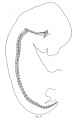
Human embryo 25 days, 19 somite pairs Scanning EM. (Carnegie stage 11)
Week 8

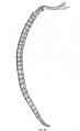
Vertebra and Spinal cord (Carnegie Stage 22)
Nucleus Pulposus


Mouse Notochord
| Mouse (E11) Notochord labeling HNF3beta{{pmid:22132119|PMID22132119}} | ||
|---|---|---|

|

|

|
|
- Links: Image 1 - E11 Notochord | Image 2 - E11 Notochord | Image 3 - E11 Notochord | Image 4 - E11 Notochord | Notochord | Mouse Development | Category:Mouse E11.0 | Image- Mouse embryo E11 and tomography | Image - Mouse embryo E11 tomography | OMIM FORKHEAD BOX A2
Molecular Factors

Abnormalities
Abnormalities include remnants of notochord that fail to regress. Locations can be along the embryonic path of the notochord and include: ecchordosis physaliphora, odontoid process of the axis, and in the coccyx. Less common locations are in the nasopharynx (Tornwaldt's cysts).
Ecchordosis physaliphora
Benign ectopic nests found along the craniospinal axis forming from notochordal remnants.{{pmid:27158576|PMID27158576}}

Brain radiography showing (A) Axial CTA (bone window); (B) Sagittal T1 MRI; (C) Sagittal T2 MRI showing EP and pontine telangiectasia; (D) CTA showing fenestrated basilar artery.{{pmid:27158576|PMID27158576}}
Tornwaldt's cysts
A rare nasopharyngeal lesion occurring in humans thought to develop from remnants of the embryonic notochord adjacent with the embryonic foregut.{{pmid:7856446|PMID7856446}}{{pmid:17315835|PMID17315835}}These cysts are covered by the nasopharynx mucous membrane. Named after Gustav Ludwig Tornwaldt (1843 - 1910) a German physician, the name is also spelled Thornwaldt.
Chordoma
Rare type of bone cancer arising from remnants of the embryonic notochord (for review see{{pmid:26363792|PMID26363792}}) Nearly all chordomas express the T-box transcription factor brachyury.
- Links: Tbx | OMIM Chordoma | chordoma foundation
References
Reviews
{{pmid:21967331}}
{{pmid:20568241}}
{{pmid:15890825}}
Articles
{{pmid:24509350}}
{{pmid:20565707}}
{{pmid:20041163}}
{{pmid:19997509}}
{{pmid:18629866}}
Search PubMed
Search NLM Online Textbooks: "Notochord" : Developmental Biology | The Cell- A molecular Approach | Molecular Biology of the Cell | Endocrinology
Search Pubmed: Notochord
Additional Images
Historic
| Historic Disclaimer - information about historic embryology pages |
|---|
|
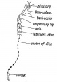
Where Remnants of the Notochord may occur in the Adult.
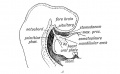
Sagittal Section showing the stomodaeum and position of the oral plate in the 3rd week
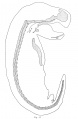
opossum embryo 12 mm
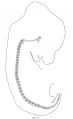
rabbit embryo 14.5 mm
cat embryo 15 mm
human embryo 32 mm


External Links
- OMIM SONIC HEDGEHOG; SHH | T BRACHYURY
Glossary Links
- Glossary: A | B | C | D | E | F | G | H | I | J | K | L | M | N | O | P | Q | R | S | T | U | V | W | X | Y | Z | Numbers | Symbols | Term Link
Cite this page: Hill, M.A. (2024, June 15) Embryology Notochord. Retrieved from https://embryology.med.unsw.edu.au/embryology/index.php/Notochord
- © Dr Mark Hill 2024, UNSW Embryology ISBN: 978 0 7334 2609 4 - UNSW CRICOS Provider Code No. 00098G