BGDB Gastrointestinal - Activity 3: Difference between revisions
mNo edit summary |
mNo edit summary |
||
| Line 127: | Line 127: | ||
* Maternal Liver function - {{#pmid:30046419}} | * Maternal Liver function - {{#pmid:30046419}} | ||
* Pancreatic Ducts | |||
** main duct identified by Wirsung in 1642, opens into the duodenum in common with the bile-duct. | |||
** accessory duct identified by Santorini in 1775, opening into the duodenum nearer the pylorus. | |||
<br> | <br> | ||
Latest revision as of 11:20, 29 April 2019
Learning Activity 3
- Describe the development of the associated organs; liver, pancreas and spleen.
- Identify the functions of these organs in the fetus.
1. Associated Organs
Liver

|
| ||||||||||||||||||||||||
| Virtual Slide Features - Stage 22 Liver | |||||||
|---|---|---|---|---|---|---|---|
|
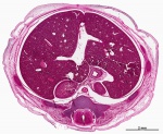
Virtual Slide - Stage 22 Liver and Ductus Venosus All Virtual Slides
The links shown in the table below are to specific features shown on the Human embryo (stage 22) Liver and Ductus Venosus virtual slide. See also notes on Liver Development Clicking the text will open the slide at a detailed view with the structure generally located in the centre of the view. The slide then can also be zoomed out from the set magnification using the controls in the upper left or the mouse. Use your browser back button to return to this table. |
You can also make your own selected feature view.
See also Permalink help | |||||
| Cardiovascular | Liver | Endocrine | Musculoskeletal | Neural | Gastrointestinal | ||

|
E3 Overview of liver region for selected high power views shown below. Note the position and size of the developing liver spanning the entire abdomen and within the liver the large central ductus venosus. |

|
E4 Central veins of liver. Radiating appearance of hepatic sinusoids. unlabeled version |

|
E5 Central vein with endothelial lining, containing nucleated erythrocytes, fetal red blood cells. The fetal liver has an important haemopoietic role. unlabeled version |

|
The Adult Liver Lobule |

Pancreas
At the foregut/midgut junction level (septum transversum) endoderm out pocketing produces 2 pancreatic buds (dorsal and ventral) that with rotation at the level of the duodenum will fuse to form the single pancreas. The dorsal bud arises first and generates most of the pancreas. The ventral bud arises beside the bile duct and forms only part of the head and uncinate process of the pancreas.
Exocrine Function - Pancreatic amylase digests starch to maltose. Postnatally, a blood test to detect amylase can be used to diagnose and monitor acute or chronic pancreatitis (pancreas inflammation).
Pancreatic Duct


The initial formation of the pancreas as two separate lobes each with their own duct that fuses leads a range of anatomical variations in the adult exocrine pancreatic duct. Pancreatic duct five variation classification: common, ansa pancreatica, branch fusion, looped, and separated. Accessory pancreatic duct (APD, of Santorini) in the embryo is the main drainage duct of the dorsal pancreatic bud emptying into the minor duodenal papilla. In the adult it has been further classified as either long-type (joins main pancreatic duct at pancreas neck portion) and short-type (joins main pancreatic duct near first inferior branch).
- Main Pancreatic Duct (MPD or Wirsung's duct) forms within the dorsal pancreatic bud and is present in the body and tail of the pancreas. Discovered by Johann Georg Wirsung (1589 - 1643) a German physician who worked as a prosector in Padua.
- Accessory Pancreatic Duct (APD or Santorini’s duct) is present mainly in the head of the pancreas. Originally dissected and delineated by Giovanni Domenico Santorini (1681 - 1737) an Italian anatomist.
- Endoscopic Retrograde Cholangiopancreatography (ERCP) is a medical procedure which allows an injected dye to display the duct system on an x ray (pancreatograms).

Human (week 8, Stage 22) pancreas
- Functions - exocrine (amylase, alpha-fetoprotein) and endocrine (pancreatic islets)
- Pancreatic buds - endoderm, covered in splanchnic mesoderm
- Pancreatic bud formation - duodenal level endoderm, splanchnic mesoderm forms dorsal and ventral mesentery, dorsal bud (larger, first), ventral bud (smaller, later)
- Duodenum growth/rotation - brings ventral and dorsal buds together, fusion of buds
- Pancreatic duct - ventral bud duct and distal part of dorsal bud, exocrine function
- Islet cells - cords of endodermal cells form ducts, which cells bud off to form islets
| Carnegie Stage | Days | Event |
|---|---|---|
| 10 | 25-27 | distal endoderm foregut |
| 12 | 29-31 | pancreatic duodenal endoderm
extra-hepatic billiard duct |
| 13 | 30-33 | pancreatic bud |
| 19 | 47 | "trunk" progenitor
"tip" progenitor |
| 23 | 8 weeks + | fetal beta cell
ductal cell |
| fetal | 14 weeks | acinar cell |
| Table data[3] Links: pancreas | exocrine pancreas | pancreas molecular timeline | timeline | ||
- Week 7 to 20 - pancreatic hormones secretion increases, small amount maternal insulin
- Week 10 - glucagon (alpha) differentiate first, somatostatin (delta), insulin (beta) cells differentiate, insulin secretion begins
- Week 15 - glucagon detectable in fetal plasma
Spleen
| The spleen is not a gastrointestinal organ, but it does develop from the late embryonic period within the dorsal mesentery.
Later the retroperitoneal position of the developing kidneys is also shown either side of the dorsal (thoracic) aorta.
Legend
|
<html5media height="450" width="300">File:Lesser sac 01.mp4</html5media> |
2. Fetal Organ Functions
Now consider that digestion is not a major function of the fetal gastrointestinal system. Then what are the fetal functions of the tract and associated organs?
At birth, the umbilical vein and the ductus venosus collapse; the portal vein becomes the only afferent vein of the liver.
Interactive Component
Additional Information
| Additional Information - Content shown under this heading is not part of the material covered in this class. It is provided for those students who would like to know about some concepts or current research in topics related to the current class page. |
- Detailed Organ pages - liver | gallbladder | pancreas | spleen
- Liver - Haematopoietic Stem Cells
- Gao X, Xu C, Asada N & Frenette PS. (2018). The hematopoietic stem cell niche: from embryo to adult. Development , 145, . PMID: 29358215 DOI.
- Ciriza J, Thompson H, Petrosian R, Manilay JO & García-Ojeda ME. (2013). The migration of hematopoietic progenitors from the fetal liver to the fetal bone marrow: lessons learned and possible clinical applications. Exp. Hematol. , 41, 411-23. PMID: 23395775 DOI.
- Gao S & Liu F. (2018). Fetal liver: an ideal niche for hematopoietic stem cell expansion. Sci China Life Sci , 61, 885-892. PMID: 29934917 DOI.
- Liver - Endocrine
- DHEA and DHEAS 16-hydroxylation by the fetal liver, then delivered to placenta, forms estrogen with estriol being the major type produced.
- Neural development - Harris RBS & Bouret SG. (2017). Development of Hypothalamic Circuits That Control Food Intake and Energy Balance. , , . PMID: 28880512 DOI.
- Maternal Liver function - Kelly C & Pericleous M. (2018). Pregnancy-associated liver disease: a curriculum-based review. Frontline Gastroenterol , 9, 170-174. PMID: 30046419 DOI.
- Pancreatic Ducts
- main duct identified by Wirsung in 1642, opens into the duodenum in common with the bile-duct.
- accessory duct identified by Santorini in 1775, opening into the duodenum nearer the pylorus.
| Gastrointestinal Tract Terms | ||
|---|---|---|
| ||
|
BGDB: Lecture - Gastrointestinal System | Practical - Gastrointestinal System | Lecture - Face and Ear | Practical - Face and Ear | Lecture - Endocrine | Lecture - Sexual Differentiation | Practical - Sexual Differentiation | Tutorial
Glossary Links
- Glossary: A | B | C | D | E | F | G | H | I | J | K | L | M | N | O | P | Q | R | S | T | U | V | W | X | Y | Z | Numbers | Symbols | Term Link
Cite this page: Hill, M.A. (2024, June 15) Embryology BGDB Gastrointestinal - Activity 3. Retrieved from https://embryology.med.unsw.edu.au/embryology/index.php/BGDB_Gastrointestinal_-_Activity_3
- © Dr Mark Hill 2024, UNSW Embryology ISBN: 978 0 7334 2609 4 - UNSW CRICOS Provider Code No. 00098G
- ↑ Godlewski G, Gaubert-Cristol R, Rouy S & Prudhomme M. (1997). Liver development in the rat and in man during the embryonic period (Carnegie stages 11-23). Microsc. Res. Tech. , 39, 314-27. PMID: 9407542 <314::AID-JEMT2>3.0.CO;2-H DOI.
- ↑ Godlewski G, Gaubert-Cristol R, Rouy S & Prudhomme M. (1997). Liver development in the rat during the embryonic period (Carnegie stages 15-23). Acta Anat (Basel) , 160, 172-8. PMID: 9718390
- ↑ Jennings RE, Berry AA, Kirkwood-Wilson R, Roberts NA, Hearn T, Salisbury RJ, Blaylock J, Piper Hanley K & Hanley NA. (2013). Development of the human pancreas from foregut to endocrine commitment. Diabetes , 62, 3514-22. PMID: 23630303 DOI.