Category:Liver: Difference between revisions
mNo edit summary |
mNo edit summary |
||
| Line 4: | Line 4: | ||
:'''Links:''' {{liver}} | {{liver histology}} | :'''Links:''' {{liver}} | {{liver histology}} | ||
{{Gastrointestinal Tract Links}} | |||
<br> | |||
{{Virtual Slide Features - Stage 22 Liver}} | {{Virtual Slide Features - Stage 22 Liver}} | ||
Revision as of 13:05, 29 April 2018
This Embryology category lists content related to liver development. Note that the liver has many different functions in addition to gastrointestinal, including fetal haemopoietic and endocrine.
- Links: liver | liver histology
| Virtual Slide Features - Stage 22 Liver | |||||||
|---|---|---|---|---|---|---|---|
|
Virtual Slide - Stage 22 Liver and Ductus Venosus All Virtual Slides
The links shown in the table below are to specific features shown on the Human embryo (stage 22) Liver and Ductus Venosus virtual slide. See also notes on Liver Development Clicking the text will open the slide at a detailed view with the structure generally located in the centre of the view. The slide then can also be zoomed out from the set magnification using the controls in the upper left or the mouse. Use your browser back button to return to this table. |
You can also make your own selected feature view.
See also Permalink help | |||||
| Cardiovascular | Liver | Endocrine | Musculoskeletal | Neural | Gastrointestinal | ||
Subcategories
This category has the following 2 subcategories, out of 2 total.
Pages in category 'Liver'
The following 66 pages are in this category, out of 66 total.
B
G
- Template:Gall bladder
- Template:Gall-bladder
- Template:Gallbladder
- Gastrointestinal Tract - Gall Bladder Development
- Gastrointestinal Tract - Gallbladder Development
- Gastrointestinal Tract - Gallbladder Histology
- Gastrointestinal Tract - Histology
- Gastrointestinal Tract - Liver Development
- Gastrointestinal Tract - Liver Histology
- Gastrointestinal Tract - Pancreas Histology
L
P
- Paper - A Contribution to the Embryology of the Liver and Vascular System in Man
- Paper - A contribution to the morphology and development of the mammalian liver
- Paper - A contribution to the morphology and development of the mammalian liver (1908)
- Paper - A Note on the Development of the Septum Transversum and the Liver
- Paper - A note on the post-natal growth of the kidney, thyroid gland and liver (1924)
- Paper - A Study of the Structural Unit of the Liver
- Paper - Congenital Anomalies of the Liver (1929)
- Paper - Functions of the liver in the embryo
- Paper - Notes on the origin of the liver (1891)
- Paper - On the relation of the liver cells to the blood-vessels and lymphatics
- Paper - Retrogressive Changes in the Fetal Vessels and the Suspensory Ligament of the Liver
- Paper - The development of the lobule of the pig's liver (1919)
- Paper - The early morphogenesis and histogenesis of the liver in Sus scrofa domesticus, including notes on the morphogenesis of the ventral pancreas
- Paper - The embryogenesis of human bile capillaries and ducts
R
- Template:Ref-Bayon1912
- Template:Ref-Bloom1926
- Template:Ref-Bradley1908
- Template:Ref-Frazer1920
- Template:Ref-Gladstone1924b
- Template:Ref-Herring1906
- Template:Ref-Hilton1903
- Template:Ref-Ingalls1908
- Template:Ref-Jackson1909b
- Template:Ref-Johnson1919
- Template:Ref-Lewis1905c
- Template:Ref-MacMahon1929
- Template:Ref-Mall1901b
- Template:Ref-Meyer1914
- Template:Ref-Scammon1915
- Template:Ref-Scammon1916
- Template:Ref-Severn1968
- Template:Ref-Severn1971
- Template:Ref-Severn1972
- Template:Ref-Thompson1914
- Template:Ref-White1939
- Template:Ref-Zorn2008
- Template:Ribavirin
Media in category 'Liver'
The following 124 files are in this category, out of 124 total.
 Adult human liver cells.jpg 900 × 1,095; 245 KB
 Bailey264.jpg 881 × 562; 77 KB
 Bailey266.jpg 731 × 913; 178 KB
 Bailey270.jpg 736 × 448; 53 KB
 Bailey272.jpg 862 × 597; 120 KB
 Bailey273.jpg 560 × 522; 58 KB
 Bailey274.jpg 558 × 442; 32 KB
 Bailey275.jpg 485 × 483; 48 KB
 Bailey276.jpg 855 × 619; 126 KB
 Bailey277.jpg 896 × 480; 112 KB
 Bailey281.jpg 865 × 1,028; 211 KB
 Dog liver portosystemic shunts.jpg 800 × 264; 19 KB
 Embryo-1951-09-01-Slide-60 Scene11-5.jpg 1,440 × 900; 268 KB
 Embryo-1951-09-01-Slide-60 Scene11-6.jpg 1,440 × 900; 220 KB
 Embryo-1951-09-01-Slide-60 Scene11-7.jpg 1,440 × 900; 163 KB
 Embryo-1951-09-01-Slide-60 Scene11-8.jpg 1,440 × 900; 125 KB
 Fetal liver erythroblasts 01.jpg 905 × 534; 69 KB
 Fetal liver weight growth graph.jpg 800 × 521; 34 KB
 Gray0475.jpg 2,042 × 1,363; 350 KB
 Gray0990.jpg 800 × 407; 60 KB
 Gray1223.png 537 × 500; 55 KB
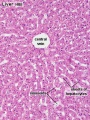
 Histology-fetal liver HEx100.jpg 1,280 × 1,024; 214 KB
 Histology-fetal liver HEx40.jpg 1,000 × 800; 281 KB
 HMB2011 Liver Histology 01.mp3 ; 1.08 MB
- HMB2011 Liver Histology 02.mp3 ; 870 KB
- HMB2011 Liver Histology 03.mp3 ; 757 KB
- HMB2011 Liver Histology 04.mp3 ; 638 KB
- HMB2011 Liver Histology 05.mp3 ; 1.04 MB
 Human liver week 9.jpg 1,200 × 991; 425 KB
 Ingalls1908 fig01.jpg 628 × 779; 35 KB
 Ingalls1908 fig02.jpg 628 × 779; 39 KB
 Ingalls1908 plate01.jpg 927 × 1,000; 204 KB
 Ingalls1908 plate02.jpg 988 × 1,000; 196 KB
 Keibel Mall 2 293.jpg 1,280 × 766; 304 KB
 Keibel Mall 2 295.jpg 1,000 × 920; 83 KB
 Keibel Mall 2 297-299.jpg 1,280 × 786; 135 KB
 Keibel Mall 2 297.jpg 354 × 359; 13 KB
 Keibel Mall 2 298.jpg 599 × 605; 40 KB
 Keibel Mall 2 299.jpg 733 × 1,034; 120 KB
 Keibel Mall 2 300-302.jpg 1,964 × 3,171; 814 KB
 Keibel Mall 2 300.jpg 1,279 × 627; 78 KB
 Keibel Mall 2 301.jpg 1,280 × 779; 140 KB
 Keibel Mall 2 302.jpg 1,278 × 803; 149 KB
 Keibel Mall 2 303-306.jpg 1,817 × 2,328; 520 KB
 Keibel Mall 2 303.jpg 659 × 833; 79 KB
 Keibel Mall 2 305.jpg 701 × 764; 77 KB
 Keibel Mall 2 306.jpg 569 × 832; 66 KB
 Keith1902 fig212.jpg 1,123 × 750; 139 KB
 Keith1902 fig213a.jpg 854 × 800; 166 KB
 Keith1902 fig213b.jpg 800 × 564; 64 KB
 Keith1902 fig214.jpg 750 × 549; 58 KB
 Keith1902 fig215.jpg 704 × 600; 56 KB
 Keith1902 fig216.jpg 1,000 × 771; 170 KB
 Keith1902 fig217.jpg 1,000 × 726; 144 KB
 Keith1902 fig218.jpg 800 × 475; 70 KB
 Keith1902 fig220.jpg 1,000 × 632; 116 KB
 Kollmann539.jpg 736 × 696; 132 KB
 Kollmann555.jpg 605 × 585; 61 KB
 Kollmann562.jpg 670 × 581; 68 KB
 Kollmann563.jpg 754 × 848; 143 KB
 Liver animated cartoon.gif 300 × 200; 239 KB
 Liver cholangiocyte tubulogenesis 01.jpg 800 × 236; 81 KB
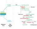 Liver development signaling.jpg 600 × 467; 45 KB
 Liver hepatocyte from stem cell.png 600 × 444; 96 KB
 Liver histology 001.jpg 400 × 533; 94 KB
 Liver histology 002.jpg 375 × 500; 54 KB
 Liver histology 003.jpg 375 × 500; 52 KB
 Liver histology 004.jpg 600 × 400; 70 KB
 Liver histology 005.jpg 800 × 664; 166 KB
 Liver histology 006.jpg 1,280 × 1,024; 664 KB
 Liver histology 007.jpg 1,280 × 1,024; 313 KB
 Liver histology 008.jpg 1,280 × 1,024; 214 KB
 Liver histology 009.jpg 1,280 × 1,024; 373 KB
 Liver histology 101.jpg 1,280 × 1,024; 410 KB
 Liver histology 102.jpg 1,280 × 1,024; 475 KB
 Liver histology 103.jpg 1,280 × 1,024; 330 KB
 Liver histology EM01.jpg 1,028 × 708; 141 KB
 Liver histology EM02.jpg 1,028 × 707; 154 KB
 Liver plasmodium infection cartoon.jpg 1,000 × 450; 78 KB
 Liver polyploidy 01.jpg 800 × 619; 119 KB
 Liver SEM01.jpg 2,000 × 1,333; 350 KB
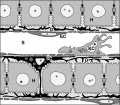
 Liver sinusoidal endothelial cell fenestrations.jpg 926 × 474; 184 KB
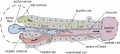 Liver structure cartoon.jpg 1,000 × 451; 78 KB
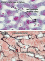 Liver- Kupffer cell and reticular fibre.jpg 600 × 800; 49 KB
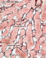 Liver-reticular fibre.jpg 700 × 875; 77 KB
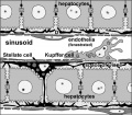
 Liver-sinusiod cartoon.jpg 600 × 523; 51 KB
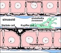 Liver-sinusoid colour cartoon.jpg 600 × 523; 64 KB
 Liver-sinusoid-label cartoon.jpg 600 × 523; 58 KB
 Mall1906-fig06.jpg 600 × 716; 111 KB
 Mall1906-fig07.jpg 503 × 753; 102 KB
 Mall1906-fig08.jpg 726 × 753; 75 KB
 Mall1906-fig09.jpg 452 × 744; 111 KB
 Mall1906-fig10.jpg 660 × 744; 71 KB
 Mall1906-fig11.jpg 681 × 735; 89 KB
 Mall1906-fig12.jpg 544 × 735; 68 KB
 Mall1906-fig13.jpg 592 × 706; 129 KB
 Mall1906-fig14.jpg 673 × 699; 50 KB
 Mall1906-fig15.jpg 887 × 1,000; 115 KB
 Mall1906-fig16.jpg 708 × 890; 155 KB
 Mall1906-fig17.jpg 705 × 881; 98 KB
 Mall1906-fig18.jpg 933 × 584; 66 KB
 Mall1906-fig19.jpg 596 × 610; 102 KB
 Mall1906-fig20.jpg 768 × 1,000; 115 KB
 Mall1906-fig21.jpg 1,000 × 763; 72 KB
 Mall1906-fig22.jpg 694 × 880; 195 KB
 Mall1906-fig23.jpg 650 × 850; 191 KB
 Mall1906-fig24.jpg 577 × 850; 176 KB
 Mall1906-fig25.jpg 1,165 × 887; 153 KB
 Mall1906-fig26.jpg 766 × 639; 78 KB
 Mall1906-fig27.jpg 890 × 800; 168 KB
 Mall1906-fig28.jpg 1,000 × 803; 146 KB
 Mall1906-fig29.jpg 780 × 700; 59 KB
 Mall1906-fig30.jpg 845 × 650; 67 KB
 Mouse hematopoietic stem cell.gif 600 × 595; 40 KB
 Stage 22 image 131.jpg 1,000 × 668; 145 KB
 Stage 22 image 180.jpg 1,000 × 668; 145 KB
 Stage 22 image 181.jpg 1,000 × 653; 263 KB
 Stage 22 image 182.jpg 1,000 × 653; 211 KB
 Thompson1908 fig01.jpg 1,134 × 865; 295 KB
 Thompson1908 fig02.jpg 1,200 × 628; 142 KB
 Thompson1908 fig03.jpg 1,209 × 600; 121 KB
 Waterston07.jpg 562 × 655; 79 KB
 West05.jpg 457 × 759; 30 KB
 Zorn2008 fig01.jpg 1,200 × 1,158; 110 KB


