Blastocyst Development: Difference between revisions
| Line 72: | Line 72: | ||
==Additional Images== | ==Additional Images== | ||
<gallery> | <gallery> | ||
Bovine blastocyst KRT18, FN1 and MYL6 expression.jpg|Bovine blastocyst KRT18, FN1 and MYL6 expression | File:Bovine blastocyst KRT18, FN1 and MYL6 expression.jpg|Bovine blastocyst KRT18, FN1 and MYL6 expression | ||
File:Bovine_blastocyst_KRT18_and_MYL6_expression.jpg|Bovine blastocyst KRT18 and MYL6 expression | File:Bovine_blastocyst_KRT18_and_MYL6_expression.jpg|Bovine blastocyst KRT18 and MYL6 expression | ||
</gallery> | </gallery> | ||
Revision as of 15:30, 14 October 2010
Introduction

(Greek, blastos = sprout + cystos = cavity) or blastula, the term used to describe the hollow cellular mass that forms in early development. The blastocyst consists of cells forming an outer trophoblast layer, an inner cell mass and a fluid-filled cavity. The blastocyst inner cell mass is the source of true embryonic stem cells capable of forming all cell types within the embryo. In humans, this stage occurs in the first and second weeks after the zygote forms a solid cellular mass morula stage) and before implantation.
- Links: Fertilization | Week 1 | Morula | Blastocyst
Some Recent Findings

|
Model Human Blastocyst Development
The following figure is from a recent study[2] using video and genetic analysis of in vitro human development during week 1 following fertilization.

- EGA - embryonic genome activation
- ESSP - embryonic stage–specific pattern, four unique embryonic stage–specific patterns (1-4)
- Links: Figure with legend
Mouse Blastocyst Gene Expression

General gene expression patterns are indicated from genomic profiling.[4]
- red - loss of maternal mRNAs
- green - activation of embryonic genome (EGA)
- purple - maternal gene activation (MGA)
- orange - continuous expression
Inner Cell Mass
A cluster of cells located and attached on one wall of the outer trophoblast layer, these cells are also called the "embryoblast", as a term to discriminate them from "trophoblast".
Trophoblast Layer
This outer layer of cells is also called the trophectoderm (TE) epithelium. A key function is for the transport of sodium (Na+) and chloride (Cl-) ions through this layer into the blastocoel.
Differentiation of of this layer is regulated by the transcription factors Tead4[5] and then Caudal-related homeobox 2 (Cdx2).
About Tead
- TEA DNA- binding domain, these factors bind to the consensus TEA/ATTS cognate binding site[6]
- TEF-3 - renamed Tead1 and Tead4
- Tead3 - is expressed in the placental syncytiotrophoblasts
- Links: Trophoblast | OMIM -Tead4 | OMIM - Cdx2
Blastocoel Formation

- trophectoderm transports of Na+ and Cl- ions through this layer into the blastocoel
- generates an osmotic gradient driving fluid across this epithelium
- distinct apical and basolateral membrane domains specific for transport
- facilitates transepithelial Na+ and fluid transport for blastocoel formation
- transport is driven by Na, K-adenosine triphosphatase (ATPase) in basolateral membranes of the trophectoderm [8]
Molecular Factors
- E-cadherin - Calcium ion-dependent cell adhesion molecule, a cell membrane adhesive protein required for morula compaction
- epithin - A type II transmembrane serine protease, identified in mouse for compaction of the morula during preimplantation embryonic development. Expressed from 8-cell stage at blastomere contacts and co-localises in the morula with E-cadherin. PMID: 15848395
- Na, K-adenosine triphosphatase - A sodium potassium pump that generates an osmotic gradient for fluid flow into the blastocoel
- Zonula occludens-1 - (ZO-1) Tight junction protein involved in morula to blastocyst transformation in the mouse PMID: 18423437
Additional Images
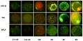
Bovine blastocyst KRT18, FN1 and MYL6 expression
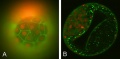
Bovine blastocyst KRT18 and MYL6 expression


References
- ↑ <pubmed>19924284</pubmed>| PMC2773928 | PLoS One
- ↑ 2.0 2.1 <pubmed>20890283</pubmed>| Nat Biotechnol.
- ↑ <pubmed>20864103</pubmed>
- ↑ <pubmed>19043080</pubmed>| Mol Hum Reprod.
- ↑ <pubmed>18083014</pubmed>
- ↑ <pubmed>8702974</pubmed>
- ↑ <pubmed>19043080</pubmed>| Mol Hum Reprod.
- ↑ <pubmed>16139691</pubmed>
Articles
<pubmed>19289087</pubmed> <pubmed>18817772</pubmed> <pubmed>18083014</pubmed> <pubmed>20157423</pubmed>
Search PubMed
Search April 2010
Search Pubmed: blastocyst development | blastocoel development | inner cell mass development | trophectoderm |
Glossary Links
- Glossary: A | B | C | D | E | F | G | H | I | J | K | L | M | N | O | P | Q | R | S | T | U | V | W | X | Y | Z | Numbers | Symbols | Term Link
Cite this page: Hill, M.A. (2024, June 17) Embryology Blastocyst Development. Retrieved from https://embryology.med.unsw.edu.au/embryology/index.php/Blastocyst_Development
- © Dr Mark Hill 2024, UNSW Embryology ISBN: 978 0 7334 2609 4 - UNSW CRICOS Provider Code No. 00098G