BGD Lecture - Gastrointestinal System Development: Difference between revisions
mNo edit summary |
mNo edit summary |
||
| Line 10: | Line 10: | ||
|} | |} | ||
:'''Links:''' [http:// | :'''Links:''' [http://php.med.unsw.edu.au/embryology/index.php?title=BGD_Lecture_-_Gastrointestinal_System_Development&printable=yes 2013 Printable version] | [http://php.med.unsw.edu.au/embryology/index.php?title=BGD_Lecture_-_Gastrointestinal_System_Development&oldid=122054 2013 Lecture] | [http://php.med.unsw.edu.au/embryology/index.php?title=BGD_Lecture_-_Gastrointestinal_System_Development&oldid=115411 2012 Lecture] | [http://emed.med.unsw.edu.au/Map.nsf/0/D5264C4F39F6E9FCCA257339000628E2?OpenDocument&login Link to Learning Activity] | [[BGDB_Practical_-_Gastrointestinal_System_Development|BGDB Practical - GIT]] | ||
| Line 27: | Line 27: | ||
|- | |- | ||
| [[File:Logo.png|80px]] | | [[File:Logo.png|80px]] | ||
| | | {{Embryo citation}} | ||
{{Gastrointestinal Tract Links}} | {{Gastrointestinal Tract Links}} | ||
|- | |- | ||
Revision as of 20:17, 5 January 2014
Introduction

|
This lecture introduces the early development of the Gastrointestinal Tract (acronym GIT).
|
- Links: 2013 Printable version | 2013 Lecture | 2012 Lecture | Link to Learning Activity | BGDB Practical - GIT
Lecture Objectives

- Understanding of germ layer contributions to the early gastrointestinal tract (GIT)
- Understanding of the folding of the GIT
- Understanding of three main GIT embryonic divisions
- Understanding of associated organ development (liver, pancreas, spleen)
- Brief understanding of mechanical changes (rotations) during GIT development
- Brief understanding of gastrointestinal abnormalities
Textbooks


Gastrointestinal Tract Movies
|
|
|
|
| |||||||||||||||||||
|
|
|
|
|
|
Week 3
(Gestational age 5 weeks)
Gastrulation

In week 3 the term "gastrulation" means "gut formation" and is the generation of the 3 germ layers.
|
Both endoderm and mesoderm will contribute to associated organs. |
Folding
| Folding of the embryonic disc then occurs ventrally around the notochord, which forms a rod-like region running rostro-caudally in the midline.
In relation to the notochord:
|
||
| Endoderm Development Movie | Amniotic Cavity Development Movie |
The ventral endoderm (shown yellow) has grown to line a space called the yolk sac. Folding of the embryonic disc "pinches off" part of this yolk sac forming the first primitive gastrointestinal tract.
Week 4
(Gestational age 6 weeks)


Coelomic Cavity
- The mesoderm initially undergoes segmentation to form paraxial, intermediate mesoderm and lateral plate mesoderm.
- Paraxial mesoderm segments into somites and lateral plate mesoderm divides into somatic and splanchnic mesoderm.
- The space forming between them is the coelomic cavity, that will form the 3 major body cavities (pericardial, pleural, peritoneal)
- Most of the gastrointestinal tract will eventually lie within the peritoneal cavity.


(only the righhand side is shown, lefthand side would be identical)
Liver Development


Endoderm and splanchnic mesoderm at the level of the transverse septum (week 4)
- Stage 11 - hepatic diverticulum development
- Stage 12 - cell differentiation, septum transversum forming liver stroma, hepatic diverticulum forming hepatic trabeculae
- Stage 13 - epithelial cord proliferation enmeshing stromal capillaries
The liver initially occupies the entire anterior body. All blood vessels enter the liver (placental, vitelline) and leave to enter the heart.
Stomach

|
|
Week 5
(Gestational age 7 weeks)
Canalization
|
|
Mesentery Development

|
Spleen
|
Week 8 - 10
(Gestational age 10-12 weeks)
Intestine Herniation


|
|
Intestine Rotation

Normal intestinal rotation (note these are gestational age weeks)[1]
Hindgut

|
|
Gastrointestinal Tract Divisions
| During the 4th week the 3 distinct portions (fore-, mid- and hind-gut) extend the length of the embryo and will contribute different components of the GIT. These 3 divisions are also later defined by the vascular (artery) supply to each of theses divisions.
|

Gastrointestinal Tract Blood Supply |
Fetal

|

|
| Small Intestine length (mm) | Liver Growth (weight grams) |
| 1 to 124 grams (birth) |
Liver
- Differentiates to form the hepatic diverticulum and hepatic primordium, generates the gall bladder then divides into right and left hepatic (liver) buds.
- Hepatic Buds - form hepatocytes, produce bile from week 13 (forms meconium of newborn)
- Left Hepatic Bud - left lobe, quadrate, caudate (both q and c anatomically Left) caudate lobe of human liver consists of 3 anatomical parts: Spiegel's lobe, caudate process, and paracaval portion.
- Right Hepatic Bud - right lobe
- Bile duct - 3 connecting stalks (cystic duct, hepatic ducts) which fuse.
- Early liver also involved in blood formation, after the yolk sac and blood islands acting as a primary site.
Pancreas

- Pancreatic buds - endoderm, covered in splanchnic mesoderm
- Pancreatic bud formation – duodenal level endoderm, splanchnic mesoderm forms dorsal and ventral mesentery, dorsal bud (larger, first), ventral bud (smaller, later)
- Duodenum growth/rotation – brings ventral and dorsal buds together, fusion of buds, exocrine function
- Pancreatic duct – ventral bud duct and distal part of dorsal bud
- Pancreatic islets - endocrine function (week 10 onwards)

Spleen
|

Gastrointestinal Tract Abnormalities

| USA Statistics | ||||||||||||||||||||||||||||||||||||||||||||||||||||||||||||||||||||||||
|---|---|---|---|---|---|---|---|---|---|---|---|---|---|---|---|---|---|---|---|---|---|---|---|---|---|---|---|---|---|---|---|---|---|---|---|---|---|---|---|---|---|---|---|---|---|---|---|---|---|---|---|---|---|---|---|---|---|---|---|---|---|---|---|---|---|---|---|---|---|---|---|---|
| ||||||||||||||||||||||||||||||||||||||||||||||||||||||||||||||||||||||||
Lumen Abnormalities
There are several types of abnormalities that impact upon the continuity of the gastrointestinal tract lumen.
Atresia
Stenosis
Duplication
|
![Gastrointestinal tract duplication sites based upon 78 clinical studies.[2]](/embryology/index.php?title=File:Gastrointestinal_tract_duplication_sites.jpg)
|
Meckel's Diverticulum
|

Meckel's Diverticulum |
Intestinal Malrotation
Presents clinically in symptomatic malrotation as:
|

Intestinal malrotation |
Intestinal Aganglionosis
(intestinal aganglionosis, Hirschsprung's disease, aganglionic colon, megacolon, congenital aganglionic megacolon, congenital megacolon)
|

|
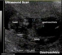
Gastroschisis
| Gastroschisis (omphalocele, paraomphalocele, laparoschisis, abdominoschisis, abdominal hernia) is a congenital abdominal wall defect which results in herniation of fetal abdominal viscera (intestines and/or organs) into the amniotic cavity.
Incidence of gastroschisis has been reported at 1.66/10,000, occuring more frequently in young mothers (less than 20 years old). By definition, it is a body wall defect, not a gastrointestinal tract defect, which in turn impacts upon GIT development. This indirect developmental effect (one system impacting upon another) occurs in several other systems.
|
|
| Gastroschisis |
Final Thoughts- After Birth
Remember that the GIT does not function until after birth consider:
- metabolic disorders discovered by neonatal diagnosis
- Neonatal feeding difficulties due to cleft lip and cleft palate.
Links: Gastrointestinal Tract - Abnormalities
Images


Terms
- allantois - An extraembryonic membrane, endoderm in origin extension from the early hindgut, then cloaca into the connecting stalk of placental animals, connected to the superior end of developing bladder. In reptiles and birds, acts as a reservoir for wastes and mediates gas exchange. In mammals is associated/incorporated with connecting stalk/placental cord fetal-maternal interface.
- amnion - An extraembryonic membrane]ectoderm and extraembryonic mesoderm in origin and forms the innermost fetal membrane, produces amniotic fluid. This fluid-filled sac initially lies above the trilaminar embryonic disc and with embryoic disc folding this sac is drawn ventrally to enclose (cover) the entire embryo, then fetus. The presence of this membane led to the description of reptiles, bird, and mammals as amniotes.
- amniotic fluid - The fluid that fills amniotic cavity totally encloses and cushions the embryo. Amniotic fluid enters both the gastrointestinal and respiratory tract following rupture of the buccopharyngeal membrane. The late fetus swallows amniotic fluid.
- buccal - (Latin, bucca = cheek) A term used to relate to the mouth (oral cavity).
- buccopharyngeal membrane - (oral membrane) (Latin, bucca = cheek) A membrane which forms the external upper membrane limit (cranial end) of the early gastrointestinal tract (GIT). This membrane develops during gastrulation by ectoderm and endoderm without a middle (intervening) layer of mesoderm. The membrane lies at the floor of the ventral depression (stomadeum) where the oral cavity will open and will breakdown to form the initial "oral opening" of the gastrointestinal tract. The equivilent membrane at the lower end of the gastrointestinal tract is the cloacal membrane.
- cloacal membrane - Forms the external lower membrane limit (caudal end) of the early gastrointestinal tract (GIT). This membrane is formed during gastrulation by ectoderm and endoderm without a middle (intervening) layer of mesoderm. The membrane breaks down to form the initial "anal opening" of the gastrointestinal tract.
- coelom - Term used to describe a space. There are extraembryonic and intraembryonic coeloms that form during vertebrate development. The single intraembryonic coelom will form the 3 major body cavities: pleural, pericardial and peritoneal.
- foregut - The first of the three part/division (foregut - midgut - hindgut) of the early forming gastrointestinal tract. The foregut runs from the buccopharyngeal membrane to the midgut and forms all the tract (esophagus and stomach) from the oral cavity to beneath the stomach. In addition, a ventral bifurcation of the foregut will also form the respiratory tract epithelium.
- gastrula - (Greek, gastrula = little stomach) A stage of an animal embryo in which the three germ layers ([E#endoderm|endoderm]/mesoderm/ectoderm) have just formed.
- gastrulation - The process of differentiation forming a gastrula. Term means literally means "to form a gut" but is more in development, as this process converts the bilaminar embryo (epiblast/hypoblast) into the trilaminar embryo ([E#endoderm endoderm]/mesoderm/ectoderm) establishing the 3 germ layers that will form all the future tissues of the entire embryo. This process also establishes the the initial body axes.
- hindgut - The last of the three part/division foregut - midgut - hindgut) of the early forming gastrointestinal tract. The hindgut forms all the tract from the distral transverse colon to the cloacal membrane and extends into the connecting stalk (placental cord) as the allantois. In addition, a ventral of the hindgut will also form the urinary tract (bladder, urethra) epithelium.
- intraembryonic coelom - The "horseshoe-shaped" space (cavity) that forms initially in the third week of development in the lateral plate mesoderm that will eventually form the 3 main body cavities: pericardial, pleural, peritoneal. The intraembryonic coelom communicates transiently with the extraembryonic coelom.
- neuralation - The general term used to describe the early formation of the nervous system. It is often used to describe the early events of differentiation of the central ectoderm region to form the neural plate, then neural groove, then neural tube. The nervous system includes the central nervous system (brain and spinal cord) from the neural tube and the peripheral nervous system (peripheral sensory and sympathetic ganglia) from neural crest. In humans, early neuralation begins in week 3 and continues through week 4.
- neural crest - region of cells at the edge of the neural plate that migrates throughout the embryo and contributes to many different tissues. In the gastrointestinal tract it contributes mainly the enteric nervous system within the wall of the gut responsible for peristalsis and secretion.
- pharynx - uppermost end of gastrointestinal and respiratory tract, in the embryo beginning at the buccopharyngeal membrane and forms a major arched cavity within the phrayngeal arches.
- somitogenesis The process of segmentation of the paraxial mesoderm within the trilaminar embryo body to form pairs of somites, or balls of mesoderm. A somite is added either side of the notochord (axial mesoderm) to form a somite pair. The segmentation does not occur in the head region, and begins cranially (head end) and extends caudally (tailward) adding a somite pair at regular time intervals. The process is sequential and therefore used to stage the age of many different species embryos based upon the number visible somite pairs. In humans, the first somite pair appears at day 20 and adds caudally at 1 somite pair/90 minutes until on average 44 pairs eventually form.
- splanchnic mesoderm - Gastrointestinal tract (endoderm) associated mesoderm formed by the separation of the lateral plate mesoderm into two separate components by a cavity, the intraembryonic coelom. Splanchnic mesoderm is the embryonic origin of the gastrointestinal tract connective tissue, smooth muscle, blood vessels and contribute to organ development (pancreas, spleen, liver). The intraembryonic coelom will form the three major body cavities including the space surrounding the gut, the peritoneal cavity. The other half of the lateral plate mesoderm (somatic mesoderm) is associated with the ectoderm of the body wall.
- stomadeum - (stomadeum) A ventral surface depression on the early embryo head surrounding the buccopharyngeal membrane, which lies at the floor of this depression. This surface depression lies between the maxillary and mandibular components of the first pharyngeal arch.
BGDB: Lecture - Gastrointestinal System | Practical - Gastrointestinal System | Lecture - Face and Ear | Practical - Face and Ear | Lecture - Endocrine | Lecture - Sexual Differentiation | Practical - Sexual Differentiation | Tutorial
Glossary Links
- Glossary: A | B | C | D | E | F | G | H | I | J | K | L | M | N | O | P | Q | R | S | T | U | V | W | X | Y | Z | Numbers | Symbols | Term Link
Cite this page: Hill, M.A. (2024, June 15) Embryology BGD Lecture - Gastrointestinal System Development. Retrieved from https://embryology.med.unsw.edu.au/embryology/index.php/BGD_Lecture_-_Gastrointestinal_System_Development
- © Dr Mark Hill 2024, UNSW Embryology ISBN: 978 0 7334 2609 4 - UNSW CRICOS Provider Code No. 00098G
- ↑ <pubmed>20549505</pubmed>| PMC2908440
- ↑ <pubmed>718292</pubmed>