File:Vein valve animation.gif
Vein_valve_animation.gif (300 × 200 pixels, file size: 54 KB, MIME type: image/gif, looped, 35 frames, 4.2 s)
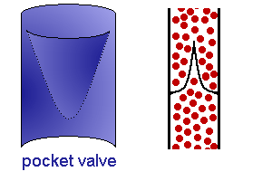
Vein Valve Animation
Animation shows how venous valves prevent the back flow of blood.
- small to medium-sized veins are characterised by the presence of valves.
- valves are formed by loose, pocket-shaped folds of the tunica intima.
- one to three pockets form the valve.
- Valves are absent in largest veins of the abdomen and thorax.
Links: Histology | Histology Stains | Blue Histology images copyright Lutz Slomianka 1998-2009. The literary and artistic works on the original Blue Histology website may be reproduced, adapted, published and distributed for non-commercial purposes. See also the page Histology Stains.
Cite this page: Hill, M.A. (2024, April 27) Embryology Vein valve animation.gif. Retrieved from https://embryology.med.unsw.edu.au/embryology/index.php/File:Vein_valve_animation.gif
- © Dr Mark Hill 2024, UNSW Embryology ISBN: 978 0 7334 2609 4 - UNSW CRICOS Provider Code No. 00098G
File history
Click on a date/time to view the file as it appeared at that time.
| Date/Time | Thumbnail | Dimensions | User | Comment | |
|---|---|---|---|---|---|
| current | 12:45, 6 July 2012 | | 300 × 200 (54 KB) | Z8600021 (talk | contribs) | ==Vein Valve Animation== {{Blue Histology}} Category:Cardiovascular Category:Blood Vessel |
You cannot overwrite this file.
File usage
The following 2 pages use this file: