Yolk Sac Development: Difference between revisions
mNo edit summary |
mNo edit summary |
||
| Line 16: | Line 16: | ||
|-bgcolor="F5FAFF" | |-bgcolor="F5FAFF" | ||
| | | | ||
* ''' | * '''The phenotypic and functional properties of {{mouse}} yolk-sac-derived embryonic macrophages'''{{#pmid:230016639|PMID30016639}} "Macrophages are well characterized as immune cells. However, in recent years, a multitude of non-immune functions have emerged many of which play essential roles in a variety of developmental processes. In adult animals, macrophages are derived from circulating monocytes originating in the bone marrow, but much of the tissue-resident population arise from erythro-myeloid progenitors (EMPs) in the extra-embryonic yolk sac, appearing around the same time as primitive erythroblasts. ...In conclusion, we have established a protocol to isolate and propagate EMs in vitro, have further defined specialized properties of yolk-sac-derived macrophages, and have identified EM-EC and EM-NSPC interactions as key inducers of EC tube formation and microglial cell maturation, respectively." | ||
* '''Expression of thyroid hormone regulator genes in the yolk sac membrane of the developing chicken embryo'''{{#pmid:28652559|PMID28652559}} "Thyroid hormones (THs) are essential for the correct development of nearly every structure in the body from the very early stages of development, yet the embryonic thyroid gland is not functional at these stages. To clarify the roles of the egg yolk as a source of THs, the TH content in the yolk and the expression of TH regulator genes in the yolk sac membrane were evaluated throughout the 21-day incubation period of chicken embryos....It is assumed that the chicken yolk sac inactivates THs contained abundantly in the yolk and supplies the hormones to the developing embryo in appropriate concentrations until the second week of incubation, while THs may be activated in the yolk sac membrane in the last week of incubation. Additionally, the yolk sac could serve as a source of iodine for the embryo." | |||
* '''New development of the yolk sac theory in diabetic embryopathy: molecular mechanism and link to structural birth defects'''{{#pmid:26432466|PMID26432466}} "Maternal diabetes mellitus is a significant risk factor for structural birth defects, including congenital heart defects and neural tube defects. ...The yolk sac vascular system is the first system to develop during embryogenesis; therefore, it is the most sensitive to hyperglycemia. The consequences of yolk sac injuries include impairment of nutrient transportation because of vasculopathy. Although the functional relationship between yolk sac vasculopathy and structural birth defects has not yet been established, a recent study reveals that the quality of yolk sac vasculature is related inversely to embryonic malformation rates." | |||
* '''Definitive Hematopoiesis in the Yolk Sac Emerges from Wnt-Responsive Hemogenic Endothelium Independently of Circulation and Arterial Identity'''{{#pmid:26418893|PMID26418893}} "Adult-repopulating hematopoietic stem cells (HSCs) emerge in low numbers in the midgestation mouse embryo from a subset of arterial endothelium, through an endothelial-to-hematopoietic transition. HSC-producing arterial hemogenic endothelium relies on the establishment of embryonic blood flow and arterial identity, and requires β-catenin signaling. Specified prior to and during the formation of these initial HSCs are thousands of yolk sac-derived erythro-myeloid progenitors (EMPs). ...In embryos lacking a functional circulation, rounded Kit(+) EMPs still fully emerge from unremodeled yolk sac vasculature. In contrast, canonical Wnt signaling appears to be a common mechanism regulating hematopoietic emergence from hemogenic endothelium. These data illustrate the heterogeneity in hematopoietic output and spatiotemporal regulation of primary embryonic hemogenic endothelium." | |||
|} | |} | ||
{| class="wikitable mw-collapsible mw-collapsed" | {| class="wikitable mw-collapsible mw-collapsed" | ||
| Line 28: | Line 34: | ||
<pubmed limit=5>Yolk Sac Development</pubmed> | <pubmed limit=5>Yolk Sac Development</pubmed> | ||
Search term: [http://www.ncbi.nlm.nih.gov/pubmed/?term=Meckel's+Diverticulum ''Meckel's Diverticulum''] | |||
<pubmed limit=5>Meckel's Diverticulum</pubmed> | |||
|} | |} | ||
{| class="wikitable mw-collapsible mw-collapsed" | {| class="wikitable mw-collapsible mw-collapsed" | ||
| Line 33: | Line 43: | ||
|- | |- | ||
| {{Older papers}} | | {{Older papers}} | ||
* '''Review - Coelomic epithelium-derived cells in visceral morphogenesis'''{{#pmid:26638186|PMID26638186}} "Coelomic cavities of vertebrates are lined by a mesothelium which develops from the lateral plate mesoderm. During development, the coelomic epithelium is a highly active cell layer, which locally is able to supply mesenchymal cells that contribute to the mesodermal elements of many organs and provide signals which are necessary for their development. ... Body wall, heart, liver, lungs, gonads, and gastrointestinal tract are populated by cells derived from the coelomic epithelium which contribute to their connective and vascular tissues, and sometimes to specialized cell types such as the stellate cells of the liver, the Cajal interstitial cells of the gut or the Sertoli cells of the testicle." | |||
* '''Embryo-fetal erythroid megaloblasts in the human coelomic cavity'''{{#pmid:20533375|PMID20533375}} "The coelomic cavity is part of the extraembryonic mesoderm, surrounding amniotic cavity, embryo, and yolk sac in the early gestation. It is now believed to represent an important transfer interface and a reservoir of nutrients for the embryo. Coelocentesis by ultrasound-guided transvaginal puncture offers an easier access to the early human embryo, from 28 days post-fertilization. However, despite some studies about its biochemical composition being reported, our knowledge about the presence of cellular elements and their quality in this compartment are still limited. Here we studied human coelomic fluids sampled from 6.6 (48 days) to 10 weeks of gestation, demonstrating the presence of functional embryonic erythroid precursors, that is, megaloblasts in the coelomic cavity." | |||
|} | |} | ||
| Line 91: | Line 104: | ||
== Terms == | == Terms == | ||
{{yolk stalk}}, {{vitelline duct}}, {{omphalomesenteric duct}} | {{yolk stalk}}, {{vitelline duct}}, {{omphalomesenteric duct}} | ||
| Line 99: | Line 110: | ||
{{Footer}} | {{Footer}} | ||
[[Category:Gastrointestinal Tract]] [[Category:Coelomic Cavity]][[Category:Endoderm]] | [[Category:Gastrointestinal Tract]] [[Category:Coelomic Cavity]][[Category:Endoderm]] | ||
Revision as of 10:25, 28 October 2018
| Embryology - 25 Apr 2024 |
|---|
| Google Translate - select your language from the list shown below (this will open a new external page) |
|
العربية | català | 中文 | 中國傳統的 | français | Deutsche | עִברִית | हिंदी | bahasa Indonesia | italiano | 日本語 | 한국어 | မြန်မာ | Pilipino | Polskie | português | ਪੰਜਾਬੀ ਦੇ | Română | русский | Español | Swahili | Svensk | ไทย | Türkçe | اردو | ייִדיש | Tiếng Việt These external translations are automated and may not be accurate. (More? About Translations) |
Introduction
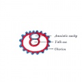
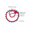
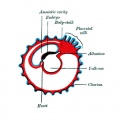
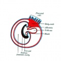
The yolk sac is an early extra-embryonic membrane which is endoderm origin and covered with extra-embryonic mesoderm. Yolk sac lies outside the embryo connected by a yolk stalk (vitelline duct, omphalomesenteric duct) to the midgut with which it forms a continuous connection. The endodermal lining is continuous with the endoderm of the gastrointestinal tract. The extra-embryonic mesoderm differentiates to form both blood and blood vessels of the vitelline system.
In reptiles and birds, the yolk sac has a function associated with nutrition. In mammals the yolk sac acts as a source of primordial germ cells and blood cells.
Note that in early human development (week 2) a transient structure called the "primitive yolk sac" forms from the hypoblast layer, this is an entirely different structure.
The yolk stalk normally degenerates around the time the midgut herniation return to the peritoneal cavity and the anterior body wall closes (week 8). Failure of complete degeneration of this structure can lead to a common intestinal abnormality, Meckel's diverticulum.
| Coelom Links: Introduction | Lecture - Week 3 Development | Lecture - Mesoderm Development | Placenta - Membranes | Category:Coelomic Cavity |
| Historic Embryology - Coelomic Cavity |
|---|
| 1891 peritoneal | 1897 human coelom | 1910 Coelom and Diaphragm | 1924 serous |
Some Recent Findings
|
| More recent papers |
|---|
 This table allows an automated computer search of the external PubMed database using the listed "Search term" text link.
More? References | Discussion Page | Journal Searches | 2019 References | 2020 References Search term: Yolk Sac Development <pubmed limit=5>Yolk Sac Development</pubmed> Search term: Meckel's Diverticulum <pubmed limit=5>Meckel's Diverticulum</pubmed> |
| Older papers |
|---|
| These papers originally appeared in the Some Recent Findings table, but as that list grew in length have now been shuffled down to this collapsible table.
See also the Discussion Page for other references listed by year and References on this current page.
|
Development Overview
Week 8
Abnormalities
Meckel's Diverticulum
ICD-11 LB15.0 Meckel diverticulum


This gastrointestinal tract abnormality is a very common (incidence of 1–2% in the general population) and results from improper closure and absorption of the omphalomesenteric duct (vitelline duct) in development. This transient developmental duct connects the yolk to the primitive gastrointestinal tract.
In addition to Meckel's diverticulum there are a range of other vitelline duct abnormalities, which depend on the degree from a completely patent duct at the umbilicus to lesser remnants (cysts, fibrous cords connecting umbilicus to distal ileum, granulation tissue at umbilicus, or umbilical hernias).
- Links: GIT Abnormalities - Meckel's Diverticulum | OMIM - Meckel's Diverticulum | Pubmed - Meckel's Diverticulum | Pubmed - omphalomesenteric duct | Pubmed - vitelline duct
References
- ↑ Too HC, Shibata M, Yayota M, Darras VM & Iwasawa A. (2017). Expression of thyroid hormone regulator genes in the yolk sac membrane of the developing chicken embryo. J. Reprod. Dev. , 63, 463-472. PMID: 28652559 DOI.
- ↑ Dong D, Reece EA, Lin X, Wu Y, AriasVillela N & Yang P. (2016). New development of the yolk sac theory in diabetic embryopathy: molecular mechanism and link to structural birth defects. Am. J. Obstet. Gynecol. , 214, 192-202. PMID: 26432466 DOI.
- ↑ Frame JM, Fegan KH, Conway SJ, McGrath KE & Palis J. (2016). Definitive Hematopoiesis in the Yolk Sac Emerges from Wnt-Responsive Hemogenic Endothelium Independently of Circulation and Arterial Identity. Stem Cells , 34, 431-44. PMID: 26418893 DOI.
- ↑ Ariza L, Carmona R, Cañete A, Cano E & Muñoz-Chápuli R. (2016). Coelomic epithelium-derived cells in visceral morphogenesis. Dev. Dyn. , 245, 307-22. PMID: 26638186 DOI.
- ↑ Renda MC, Giambona A, Fecarotta E, Leto F, Makrydimas G, Renda D, Damiani G, Jakil MC, Picciotto F, Piazza A, Valtieri M & Maggio A. (2010). Embryo-fetal erythroid megaloblasts in the human coelomic cavity. J. Cell. Physiol. , 225, 385-9. PMID: 20533375 DOI.
Reviews
Yamane T. (2018). Mouse Yolk Sac Hematopoiesis. Front Cell Dev Biol , 6, 80. PMID: 30079337 DOI.
Articles
Funayama N, Sato Y, Matsumoto K, Ogura T & Takahashi Y. (1999). Coelom formation: binary decision of the lateral plate mesoderm is controlled by the ectoderm. Development , 126, 4129-38. PMID: 10457021
Search PubMed
Search Pubmed: Coelomic Cavity Development | pericardial cavity development | pleural cavity development | peritoneal cavity development
Additional Images
Historic
| Historic Disclaimer - information about historic embryology pages |
|---|
|

Yolk sac


Terms
yolk stalk, vitelline duct, omphalomesenteric duct
Glossary Links
- Glossary: A | B | C | D | E | F | G | H | I | J | K | L | M | N | O | P | Q | R | S | T | U | V | W | X | Y | Z | Numbers | Symbols | Term Link
Cite this page: Hill, M.A. (2024, April 25) Embryology Yolk Sac Development. Retrieved from https://embryology.med.unsw.edu.au/embryology/index.php/Yolk_Sac_Development
- © Dr Mark Hill 2024, UNSW Embryology ISBN: 978 0 7334 2609 4 - UNSW CRICOS Provider Code No. 00098G