Week 3 Development Movie: Difference between revisions
No edit summary |
No edit summary |
||
| Line 2: | Line 2: | ||
{| | {| | ||
|- | |- | ||
| width=400px|<mediaplayer width='388' height='500' image="http://embryology.med.unsw.edu.au/embryology/images/d/df/Week3_folding_icon.jpg">Week3_folding.mp4</mediaplayer> | | width=400px|<mediaplayer width='388' height='500' image="http://embryology.med.unsw.edu.au/embryology/images/d/df/Week3_folding_icon.jpg">File:Week3_folding.mp4</mediaplayer> | ||
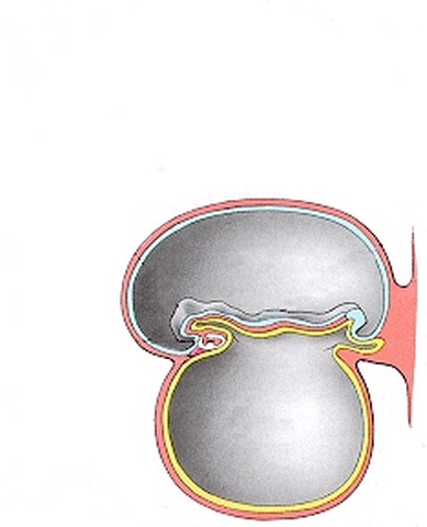
| [[File:Week3_folding icon.jpg|100px|left]] This animation shows folding of the embryonic disc beginning week 3 of development. | | [[File:Week3_folding icon.jpg|100px|left]] This animation shows folding of the embryonic disc beginning week 3 of development. | ||
| Line 28: | Line 28: | ||
Compare this sectional view of folding with the [[Development Animation - Amniotic Cavity|Amniotic Cavity]] view, filling the amnion and yolk cavities. | Compare this sectional view of folding with the [[Development Animation - Amniotic Cavity|Amniotic Cavity]] view, filling the amnion and yolk cavities. | ||
{{Amniotic Movie}} | |||
[[Category:Week 3]] | [[Category:Week 3]] | ||
Revision as of 16:21, 2 March 2013
| Embryology - 25 Apr 2024 |
|---|
| Google Translate - select your language from the list shown below (this will open a new external page) |
|
العربية | català | 中文 | 中國傳統的 | français | Deutsche | עִברִית | हिंदी | bahasa Indonesia | italiano | 日本語 | 한국어 | မြန်မာ | Pilipino | Polskie | português | ਪੰਜਾਬੀ ਦੇ | Română | русский | Español | Swahili | Svensk | ไทย | Türkçe | اردو | ייִדיש | Tiếng Việt These external translations are automated and may not be accurate. (More? About Translations) |
| <mediaplayer width='388' height='500' image="http://embryology.med.unsw.edu.au/embryology/images/d/df/Week3_folding_icon.jpg">File:Week3_folding.mp4</mediaplayer> | Embryonic disc (midline section) shown to the left and early placenta to the right. The embryonic disc dorsal (ectoderm) top and ventral (endoderm) bottom. Cranial end of disc to the left and caudal end of disc to the right. Note also the early cardiac region shown at the cranial end of disc and the allantois at the caudal end of disc extending into the connecting stalk. Folding of the embryonic disc
|
Compare this sectional view of folding with the Amniotic Cavity view, filling the amnion and yolk cavities.