User:Z5229177: Difference between revisions
| Line 12: | Line 12: | ||
==Group 3== | ==Group 3== | ||
[[User:Z5229177|Z5229177]] ([[User talk:Z5229177|talk]]) 18:21, 8 October 2018 (AEDT) | |||
The history provided a good background of Melanocytes and also the idea of Melanocytes being linked to neural crest cells. For Tissue Organ Structure and Function, it is well-organized into sub-parts such as Skin, Ears and Eyes etc. This allows readers to better understand how Melanocytes can be found in different organs and also their different specific function in each organ. | The history provided a good background of Melanocytes and also the idea of Melanocytes being linked to neural crest cells. For Tissue Organ Structure and Function, it is well-organized into sub-parts such as Skin, Ears and Eyes etc. This allows readers to better understand how Melanocytes can be found in different organs and also their different specific function in each organ. | ||
Revision as of 17:21, 8 October 2018
Peer Review:
Group 1
Z5229177 (talk) 16:26, 8 October 2018 (AEDT) The history of neural crest was well-described and detailed, considering the different discoveries made. However, it is a too lengthy and would be good to link the history of adrenal medulla to neural crest under the history section as that should be the main focus of the project.
For Developmental time course, I feel it might be good to have a timeline to show the different weeks of development and differentiation of the cortex and medulla to add on to the existing content that is already there. This might help readers to understand better.
Sections such as embryonic origins, developmental/adult function, and abnormalities/abnormal development still needs to be worked on as they are currently empty and lacking content.
Overall, the content is detailed and understandable by readers. Good job on the done parts and good use of the self-drawn figures! They are easy to understand and looks refreshing apart from the normal images or figures found online or on journal articles. Just need to brush up on those that have yet to be filled in, and also the references portion need to be listed out neatly.
Group 3
Z5229177 (talk) 18:21, 8 October 2018 (AEDT) The history provided a good background of Melanocytes and also the idea of Melanocytes being linked to neural crest cells. For Tissue Organ Structure and Function, it is well-organized into sub-parts such as Skin, Ears and Eyes etc. This allows readers to better understand how Melanocytes can be found in different organs and also their different specific function in each organ.
However, the project mostly describes about Melanocytes and its specific functions in each body part, but not much on neural crest and its relevance is being discussed under Embryonic Origins, which should be the main focus of the project.
Sections such as Development Time Course, Molecular Mechanisms/ Factors/ Genes, Abnormalities, Skin, Ears, Heart and CNS are lacking content and need to be worked on.
Overall, the function and application of Melanocytes were well-explained. However, there was not much content on how neural crest development leads to the Melanocytes being formed and its significance. Perhaps the group might want to elaborate and expand more on the Embryonic Origins section. Otherwise, the other parts of the project are understandable and done pretty well!
Group 4
Own Group
Group 5
Z5229177 (talk) 16:48, 8 October 2018 (AEDT) For the history part, your group mentioned that there is a timeline of important figures who have made contributions big or small to dorsal root ganglion being discovered. However, in the section, only 1811 Charles Bell was mentioned. Is there supposed to be more content or more names and year being discussed in this section?
For the Embryonic Origins section, the idea of the content is there. I do feel if it would be more understandable and easier to see the overview of the neural crest cells differentiating into the different types of tissues with a suitable figure or image. As neural crest cells to dorsal root ganglion is the main focus of this project, it would be good to make this section clearer especially with a suitable image or video.
For the adult function section, I understand that the content written mainly discusses about how the dorsal root ganglia contributes to the different neurons and receptors in an adult CNS and spinal cord. However, I do not see any link of how it relates to “adult function” which is the title. What type of function is your group referring to here? Are there any examples? And hence can link to how these neurons contribute to that particular function? For example, how function of running relates to the neurons being used and the function of dorsal root ganglia in this case. Or maybe the title name can be changed instead?
Overall, most of the content is well-written and explained. Good use of the self-drawn image as well. Almost all of the sections are filled with content and the glossary and referencing has been done nicely as well. I feel that the project just need to touch up on some parts and it should be good enough!
Notes:
Only can use references more than 6 months old
need to cite from original research paper (discovery) Differentiate between research & review papers
https://www.ncbi.nlm.nih.gov/pmc/articles/PMC3718324/ https://www.ncbi.nlm.nih.gov/pubmed/23723064
https://www.ncbi.nlm.nih.gov/pubmed/25662261 https://www.sciencedirect.com/science/article/pii/S0070215314000076?via=ihub
Z5229177 (talk) 11:20, 14 August 2018 (AEST)
Adding an Image
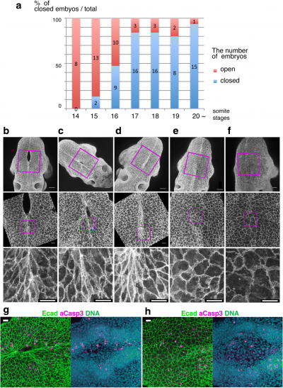
Neuropore cell shape changes[1]
Reference
PMID: 30056110
Walls ML & Hart RJ. (2018). In vitro maturation. Best Pract Res Clin Obstet Gynaecol , , . PMID: 30056110 DOI.
In vitro maturation recent article[1]
