User:Z5164785
Zygote.
Peer Reviews (Lab 10)
Group 1 (Adrenal Medulla): Perhaps describe the neural crest as a “structure” instead of a term. Great use of the student-drawn image as a guide! Overall, good simplified history.
In the normal structure and function region; good but rather brief discussion of the physiology and structure. perhaps a little bit more, and maybe an image- unless you merge it with the related anatomy component. Really love the adult adrenal gland and the effort behind it. Only complaint would be that perhaps it would have been good to have the regions of the kidney (medulla, cortex etc).
The description of the role of the adrenal medulla is very well-written; concise and seemingly accurate. Perhaps include the proper dot point structure instead of the >. The image drawn illustrating the cascade of catecholamine synthesis is also very good and I personally found it to be a helpful guide. However, it needs to be edited and correctly formatted for the webpage.
The first two sentences of the animal models section may be combined into one. Proof-reading required eg. as explained above ‘nueral’! Who is Ahonen- Is an in-text reference needed here? Perhaps this paragraph belongs in the current research section as the animal used hasn’t been mentioned. Please review this bit as the information provided is good and relevant but maybe in the wrong section.
In the current research section, the second sentence says ‘we generated’ … who are you referring to? The information here seems correct but was also quite advanced with numerous terms that I couldn’t understand- however it shows great research so well done!
Overall, great work guys! Keep it up and move along with the project consistently! Perhaps include some images from the experiments you’ve described and some more high-tech images- although the ones you have drawn are also excellent!
Group 4 (Cardiac):
Group 5 (Dorsal Root Ganglion):
Reference
PMID: 30056110
Walls ML & Hart RJ. (2018). In vitro maturation. Best Pract Res Clin Obstet Gynaecol , , . PMID: 30056110 DOI.
In vitro maturation recent article[1]
Adding an image
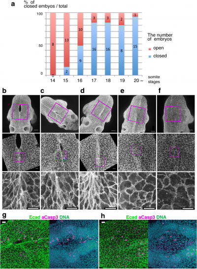
Neuropore cell shape changes[2]
- ↑ Walls ML & Hart RJ. (2018). In vitro maturation. Best Pract Res Clin Obstet Gynaecol , , . PMID: 30056110 DOI.
- ↑ Shinotsuka N, Yamaguchi Y, Nakazato K, Matsumoto Y, Mochizuki A & Miura M. (2018). Caspases and matrix metalloproteases facilitate collective behavior of non-neural ectoderm after hindbrain neuropore closure. BMC Dev. Biol. , 18, 17. PMID: 30064364 DOI.
