User:Z3465654
Online Assessment
Lab 1 Assessment
Article 1
<pubmed>24760595</pubmed> The following case study investigated the effects of hepatitis B virus (HBV) infection on sperm parameters, ovarian stimulation, and outcomes of in vitro fertilization (IVF) and embryo transfer, as the impact of HBV on human infertility was questionable. During this study, a total of 224 couples were identified, where either one or both partners were HBsAg-seropositive, and were undergoing their first IVF and embryo transfer cycle. The morphology of their sperm was analysed, as was the quality of their embryo rate, the duration of infertility and their fertilization rates, and then compared to those of 448 HBsAg-seronegative couples. In all four cases, the results of the HBsAg-seropositive couples were inferior to those of the HBsAg-seronegative couples, expressing significantly lower normal sperm morphology, top-quality embryo rate and fertilization rates, and significantly prolonged durations of infertility. It was noted however, that in regard to clinical pregnancy rates, there was no significant difference between the two groups. Based on the case study results, it was concluded that HBV infection was likely to cause infertility.
Article 2
<pubmed>24602756</pubmed> The following case study sought to investigate whether assisted reproductive technology (ART) treatments had any impact on the sex ratio of babies born. Using the United Kingdom records of women who have conceived children between 2000 and 2010 using intrauterine insemination, IVF, or intracytoplasmic sperm injection (ICSI), the records of a total of 106,066 babies born to 76,994 mothers were analysed. The results showed that each form of ART resulted in a varied sex ratio, the most significant variation occurring from IVF with 52.1% of babies born male, and the least variation occurring from ICSI embryo transfer, with 49.3% of babies being born male. It was also found that when the embryos were transferred during the blastocyst stage in ICSI and IVF, as opposed to during the early cleavage-stage ET, it resulted in approximately 6% more males being born. It was concluded however, that due to the significantly increasing number of babies born using ART treatments, more research was needed into the causes of the gender bias after such treatments.
Lab 2 Assessment
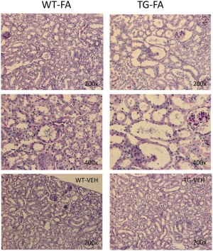
These histological views show the morphology of the kidneys of wildtype mice (left) compared to those of transgenic line A homozygous mice (right) that have been injected with Gremlin, an embryonic gene that plays a role in nephrogenesis. These images show what can occur to the morphology of the kidney if this gene is over-expressed.
<pubmed>25036148</pubmed>
Lab 3 Assessment
The kidneys first develop in the embryo by a process called nephrogenesis, in which self-renewing mesenchymal renal stem cells produce nephrons to form a simple embryonic kidney, called the pronephros. Nephrons are the main functional unit of the kidney.
<pubmed>24855634</pubmed>
An embryonic gene named gremlin (GREM1) has been found to play a key role in the formation of the kidneys and nephrogenesis in general. When fully formed, the expression of this gene is relatively low in an adult. However, it is thought that many renal diseases and their progressions are linked to an overexpression of this gremlin gene.
<pubmed>25036148</pubmed>
Nephrogenesis is stimulated by the signaling between the epithelial ureteric buds and progenitor cells, causing nephrons to develop and the ureteric buds to branch.
<pubmed>24656820</pubmed>
At birth, although the infant’s kidneys are developed enough to maintain homeostasis and are sufficient for growth and development, their functional capabilities are decreased. This is a result of the transition from depending on the placenta to maintain homeostasis of fluid and electrolyte balance while in-utero, to maturation of the neonatal glomeruli once born.
<pubmed>24781774</pubmed>
<pubmed>24623338</pubmed>
<pubmed>24488483</pubmed>
Determining nephron number is important: it can show the success/extent of nephrogenesis, and thus be used to determine if any and what genes and environmental factors may aid this process; a low nephron count has been linked to multiple cardiovascular and renal disease later in life.
<pubmed>24022365</pubmed>
<pubmed>24011574</pubmed>
The overexpression of the gremlin gene (GREM1) has been found to be a cause of renal disease.
<pubmed>25036148</pubmed> <pubmed>24500691</pubmed>
Lab 4 Assessment
Identify a paper that uses cord stem cells therapeutically and write a brief (2-3 paragraph) description of the paper's findings.
<pubmed>25130827</pubmed> A study was conducted to determine whether the combination of umbilical cord mesenchymal stem cells (UC-MSC) with haploidentical hematopoietic stem cells (haplo-HSCT) would produce a more effective outcome and positive result when transplanted into patients suffering from refractory/relapsed myeloid leukemia. Using results obtained from January 2007 to June 2013, the data of 36 patients who received such treatments were analysed with respect to the engraftment (the rate at which the stem cells are able to reproduce new cells), graft versus host disease ((GVHD) a condition in which the donor stem cells attack the recipient’s body), and their two-year overall survival.
After reviewing and analysing the results, it was determined that the average engraftment time of neutrophils was 12 days, while the average time for platelets was 14 days. The cell counts of both, however, were well below that of the normal range of a healthy individual. In terms of GVHD, 5 of the 36 patients suffered grade III to IV acute GVHD, 12 of 32 suffered chronic GVHD, 2 patients had extensive chronic GVHD, and 3 patients relapsed. Despite this, the two-year OS rate was calculated to be 76.9%, with the final assessment concluding that the combination transplantation of stem cells was a good therapeutic method, especially as an alternative to patients with high risk or unsuitable donors.
There are a number of developmental vascular "shunts" present in the embryo that are closed postnatally. Identify these shunts and their anatomical location.
<pubmed>3052747</pubmed> There are three developmental vascular ‘shunts’ present during embryo and fetal development:
• Ductus arteriosus – it connects the pulmonary artery with the descending portion of the aortic arch, and works to ‘shunt’ the majority of the output from the right ventricle away from the undeveloped lungs.
• Ductus venosus – it connects the portal sinus to the inferior vena cava, allowing oxygenated blood received from the umbilical vein to rapidly enter the central circulation by diverting around the liver.
• Foramen ovale – it connects the right atrium to the left atrium, allowing oxygenated blood from the former to enter the latter.
Lab 5 Assessment
Cystic Fibrosis
Cystic fibrosis (CF) is a hereditary abnormality affecting 1 in 2500 infants born in Australia [1]. It results from a mutation within the CF gene which is responsible for encoding a protein called cystic fibrosis transmembrane regulator (CFTR), and is located on chromosome 7 [2]. As the CFTR protein is responsible for the proper functioning of chloride channels within a cell, its defect results in an increased diffusion of salt and water across the cell, affecting the secretory glands of the body [1][2]. This causes the glands to produce increasingly salty sweat, as well as a very thick, sticky mucus, the main detriment to CF sufferers, as it causes significant impacts to several organs such as the pancreas, liver, intestines, sinuses, sex organs, and primarily the lungs [1][2][3].
The production of this thick, sticky mucus can result in blockages within the ducts and airways of the lung, causing bacteria to be trapped within. This would result in inflammation and infections capable of causing serious and permanent damage to the lungs [1][2][3]. These blockages would also result in the impaired function of digestive organs, such as the pancreas, as the enzymes produced cannot reach their destination, therefore resulting in vitamin deficiency and malnutrition [1][2].
As this abnormality is obtained genetically, both mother and father would need to be carriers for the gene, with a one-in-four chance that a child produced would inherit both copies, resulting in a positive diagnosis for CF [3]. While there is no cure for CF, there are a number of treatments available that can help to prolong their life, including salt and vitamin supplements, exercise and physiotherapy to clear lungs, and mist inhalations to open airways [1].
[1] Cystic Fibrosis Australia, 2014, About Cystic Fibrosis, [Online], Available: http://www.cysticfibrosis.org.au/all/learn/
[2] MedicineNet, 2014, Cystic Fibrosis Facts, [Online], Available: http://www.medicinenet.com/cystic_fibrosis/article.htm
[3] NHS Choices, 2014, Cystic Fibrosis – Causes, [Online], Available: http://www.nhs.uk/Conditions/cystic-fibrosis/Pages/Causes.aspx
Lab 7 Assessment
Identify and write a brief description of the findings of a recent research paper on development of one of the endocrine organs covered in today's practical.
<pubmed>24814991</pubmed> The following research article is an update to a previous discovery within the developing adrenal gland, providing additional information as to the organization of its various zones. It is well known that the adrenal cortex of an embryonic mammal will differentiate into three distinctive layers: the zona glomerulosa (zG), the zona fasciculata (zF), and the zona reticularis (zR), each of which have their own secretions. It was in 1994 however, that a fourth zone was identified located between zG and zF. This new zone was named the ‘undifferentiated cell zone (zU)’ as no significant endocrine functions were found to exist in this area. BrdU was incorporated to this zone, demonstrating that active cell division was occurring at the outer and inner regions of zU and as they proliferated, these cells migrated in two directions: towards zG and towards zF. It was proposed that these cells were stem/progenitor cells. With recent studies however, it was identified that Sonic Hedgehog existed within the cells of zU, a very important factor in embryonic development, and that these cells migrated bidirectionally as well.
Identify the embryonic layers and tissues that contribute to the developing teeth.
<pubmed>18794902</pubmed> The teeth are proposed to have originated from two main embryonic layers: the epithelium of tooth enamel is derived from the ectoderm, while the dentin and pulp of the tooth have originated from neural crest derived mesenchyme. However, the teeth are capable of being endodermal in origin, or a mixture of both endo- and ectoderm, if the oropharyngeal membrane, the membrane that separates the two layers, is broken.
Lab 8 Assessment
Provide a brief time course and overview of embryonic development of either the human testis or ovary.
Embryonic Development of the Testes The process of gonad development is one controlled by genetics. It is the presence or absence of the Y chromosome that will determine whether the gonads of the embryo will form into testis or ovaries during week 7 of the embryonic period, in particular the SRY gene located on this chromosome. This is because the presence of this gene upregulates the expression of SOX-9 [1], a transcription factor that causes the differentiation of the support cells (Sertoli cells). Once these cells are developed, they in turn begin to produce anti-Müllerian hormone (AMH) in order to promote the regression of the Müllerian duct, establishing the male phenotype [2].
The differentiation of the Sertoli cells also causes two main compartments to be formed within the developing testes: the testes cords (consist of clusters of germ cells surrounded by Sertoli cells, further surrounded by myoid cells) and the testis interstitium (includes the Leydig cells and the testis vasculature) [3].
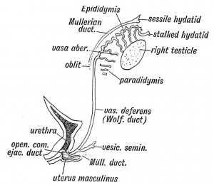
Include an image from the historic genital embryology section of the online notes in your description
Remnant of the Wolffian Body
Lab 9 Assessment
Group 1
The introduction provided good background information about the lungs and its general development, however seemed to lack any further explanation as to what else would be covered on the page (current research, abnormalities). I found most of the sentences to be short and abrupt, and more in the form of statements rather than an explanation. This is the same for the following paragraphs regarding the two zones. I would combine several of the sentences together, and restructure them so that they do not start in the same manner e.g. of the first four sentence in your introduction, three of them begin with the words ‘The respiratory system’, and over half the sentences in the entire paragraph begin with ‘The’. There are a few grammatical errors within the text that should be corrected e.g. ‘till’ of ‘until’, ‘id’ instead of ‘is’. The images used fit well, but there is no caption to explain what they are images of and what they are trying to show. This is also not indicated on the summary of the image, one of which also doesn’t include any copyright information.
The lung development stages were done very well, simplified and tabulated making it very clear. My only concern for this part is that it should be the main part of the project, the area where the development of the lungs is fully explained, yet it is the smallest section of the page. Try to expand on it maybe? Or add a picture or two to enlarge the section?
The current research and findings section seems very thorough, lots of content, good explanations. Very minor problems however; a slight tendency to over use commas in some areas, while not in others. The current models area has not been added to; make sure to fill it in, or will it be scrapped? I have also noticed a picture has been deleted so make sure to get that issue fixed if you still want to use the same image. Is the second picture under this heading part of the section? As it is after the references so I'm not sure where it lies exactly. The image should be captioned as well.
I really like the historical findings section, the information seems more concise when it is presented in bullet points. The second picture within this section is well done and very neatly labeled (I thought it was an image from the internet). The first picture though, needs a caption added as well as copyright information. The abnormalities section is very extensive which can be bother good and bad. For some of the abnormalities there is a lot of detail presented, while for others there is very little. I think maybe that as long as you mention what it is, how you get it/how it forms, some statistics and maybe an image, that should be more than enough. Also, I would remove all the sub-headings under abnormalities and have them just written in bold. Otherwise, when looking at the contents at the top of the page, it looks as though half your page is solely focused on abnormalities.
Overall, I think this page is well done and only a focus on sentence structure, a bit on grammar, and captioning pictures with correct copyright info is needed. Other than these main focus areas, one other point to make would be all the references should be at the bottom of the page.
Group 3
A good overview of the GIT, very descriptive. This section would need some referencing as most of this info isn’t exactly common knowledge. Some of the sentences seem too short for me e.g. I would reconfigure the third sentence and combine the fourth and fifth sentences into one: ‘The GIT (gastrointestinal tract) consists of three regions: the foregut, midgut and the hindgut. The majority of the organs are located in the foregut, including…..’. You also need to make sure not to use capital letters in the middle of sentences.
The timeline is sort of well organized; it’s good that you have it separated for each region so they’re not all muddled up together, but is the info in dot points under the week, or is it just written next to the week? It needs to be kept consistent. I feel like this section is a bit too spread out as well, a large portion of the left hand side has text, while the entire right side of the screen is empty. You could possible put in a picture showing these 3 regions of the gut to fill in the space? Or maybe format the info into a table, it would make it look more formal and structured. The proper referencing technique should also be used here, not added hyperlinks.
The recent findings area is a little sparse, so you should try to find a few more. The title does say findings (plural), so maybe add at least one more. The foregut section is very extensive on the information provided which is good, a lot of research has been made. Visually however, it looks a little bad as all that can be seen is a mass of text. This can be alleviated if the same thing is done as has been with the midgut and hindgut region: the use of bullet points, a small table and the use of images to offset the slabs of text. It makes it more visually appealing. Unlike the midgut portion of the page, if the images are hand drawn, make sure they are clear, legible, and with colours used that will not strain the eye. For the images drawn in that section are messy and the labels hard to read both due to the colour of the pen used and the handwriting. In both the foregut and hindgut, referencing needs to be done. There are slabs of text in both sections where no references are made.
The deformities section is good, kept simple with no extensive explanations. Are there only 2 possible deformities? If so, might be good to write a sentence mentioning that. If not, would be good to have at least 2 more deformities listed. The image drawn in this section is very neat, I like it a lot. The only problem with it is that it’s too small, making it hard to read some of the labels.
Overall, I think this page is very well done in terms of content. You have a lot of text, but I think it could do with some more pictures especially to offset some of the large slabs of texts in some areas. Make sure the pictures you have a clear and neat, and make sure you are referencing and doing it correctly.
Group 4
Just looking at the contents, if feels a little intimidating both in that it is so long as well as the use of caps. You should try and limit both; the use of all caps can be quite annoying in text and the extensive contents list can make people dread reading through your page if it looks like it’s quite long.
An introduction is recommended as it is usually a good starting point to provide the reader as sense of everything the page will cover. The system development is a little messy, but I will heed your note and pay attention to only the table. The table itself is a great idea to lay out all the events happening in the corresponding weeks, making it look neat and concise. However, the use of all caps, bold text, and two different fonts still makes this section look messy. Having both male and female events on the same table makes it look as if there is a chunk of info missing for the female side as well. I would suggest having them in separate tables next to each other, which would eliminate the empty rows in both areas. Both the image and the video (congrats on finding a video! Really good addition to the page) should be captioned.
The current research, models and findings seems well researched as there are a lot of points made. However, it is all presented in bullet points which can be visually unappealing. Some sections look incomplete as well, so an effort needs to be made to finish these areas as well as present them in an appealing manner e.g. in paragraph form with a picture next to it to both describe the text visually and offset the amount of text. The drawing of the testes should be captioned appropriately instead of the ‘alt text’ provided. It should also be enlarged, as its current size isn’t large enough to view any of the labels properly.
Historic findings looks well researched on first glance, but then I saw that only 4 sources were used to reference the section. It looks really bad when only one source is used to reference a large slab of text, which you have done twice. I suggest finding articles that state similar information and using them as references as well, to back up your current information found. Other than that, I suggest possibly formatting your section in a more appealing way; either summarize some areas in dot points, and add a picture.
The abnormalities section is nice and concise, without going into too much detail which is good. Just make sure you explain what it is, how it is formed/how you get it, some statistics and possibly an image to show what it looks like, and that’s all I believe you really need for this section.
Overall, your page is well researched with lots of info. Just make sure it looks visually appealing, is consistent in terms of font and presentation, images are used and captioned correctly, and all references are placed at the bottom of the page.
Group 5
This page seems to be done extremely well. It looks very visually appealing as multiple images are used, information is presented in tables, bullet points and very few slabs of text. The introduction is short and to the point. You could possibly add to this area a tiny bit of info concerning the embryonic development of this system, where it first started, then mention how you will expand on the fetal development. Otherwise it just seems way too short.
Explanation of the organs in this system is well done and concise. In the glands section, I would suggest not using dot points for the function of the vernix caseosa as it looks as though the dot points continue from those of the glands, therefore can be confusing when first looked at. Other than that, I would suggest that you make sure your referencing is correct and is used within the text.
The recent findings area is nicely done, but I still can’t help but feel the amount of text is just too much, even though the section is made better looking by making it purple (keep the colour, it looks awesome). The slab of text is just too much, so you should try and simplify it a bit. Historic findings are few but there is at least one for every organ which is good (more would be better). The abnormalities covered are done well, going into detail and providing a good image to describe what it looks like. I would suggest having at least 5 abnormalities, one for each organ discussed.
Overall, this page is very well done, with lots of images and colour used. The main thing I would suggest would be to make sure correct referencing is used. There were some paragraphs were no references were used at all. Also, all references should be at the bottom of the page, not within individual sections.
Group 6
At first glance, a lot of sections seem to be incomplete. On second glance, I’ve noticed that you have added all the headings used by other groups (timeline, current findings, abnormalities) as subheadings for your own project, which I think it a really smart idea. Because you have so many glands that need to be covered, writing these sections separately can be confusing with the information quickly becoming muddled up. Doing it this way eliminates that confusion.
Make sure the use of tables is appropriate, using a table for one row of info is kind of pointless (pineal gland). The timelines used should also start with the week number, otherwise it can be quite confusing trying to work out the time (e.g. try not to say times such as ‘by the second trimester’). The information presented was concise and to the point, no long-winded explanations or slabs of text which was good. The images used were relevant and captioned.
Concerning the work completed, overall it was done well. A lot more work still needs to be completed however. References should also be made in text. If you are unsure how to do this, just go into edit mode in another group’s project and see how they have done it, instead of listing all the references at the bottom of the corresponding section. Make sure all the references are also presented at the bottom of the page, not separated into sections. It would also be nice if more images are used, if not one image for every gland then at least one for every second gland mentioned (it just needs more images).
Group 7
The content looks well organized. The introduction could use a bit of work; it does a good job of introducing the CNS, but it should also mention all the other sections this page will cover regarding the topic. You might want to get rid of the using bold for brain and spinal cord, it just makes it look a little weird. Otherwise, a good embryonic developmental background is provided, it’s a good way to set the stage for when fetal development will commence from.
The information is organized well, no chunky slabs of texts. But the use of dot points is a bit extensive; almost every section of the page has dot points or makes up the complete majority of the info presented. You might want to present some of it in paragraph form e.g. the abnormalities part, as that section can still be kept quite short and not be packed with text. As long as you mention what it is, how you get it/how it forms, some statistics and use a picture, the section can be still visually appealing. The images are captioned ok, but there is a better way of doing it. In the command to input the image, continue the command with: |thumb|’whatever you want to write’], and the section in the apostrophes will be the caption under the picture (go into edit mode on another project page for a better idea, I might not have explained well).
The use of the table is well done, makes all that info easily presentable, though I see the meninges development still needs to be done. The current research models and findings looks kind of messy with just the referenced PubMed article there. It might look better if you had the article name written in bold and a couple sentences underneath each to describe what the article was trying to achieve, like what has been done under current research. A couple pictures may be included to make it all more visually appealing and colourful.
Overall, this was done well. You have a good amount of information, just try not to present it all in dot points. Make sure all your info is referenced in text, will all references displayed at the bottom of the page. Another note, try to organise your pictures in different areas of the page as well, as they are all currently on the left hand side.
Group 8
This page needs a lot of work; there are sections with little to no information, while others have just slabs of text, some of which have no references. Of those that have info presented, the topic is well covered with the large amount of content. You should use some dot points for some areas where you have a lot of info. You also need to use some images!! They will help to alleviate the slabs of content you have and add some colour to the page. Make sure you caption and reference them correctly, and add the correct copyright info.
Overall, there isn’t much I can say except add content, reference is correctly both in text and at the bottom of the page, and images and use some dot points and/or tables; don’t write everything in large slabs of text. Also, maybe get rid of that 'Muscle Gains' section, unless you actually plan to write something relevant in there haha. Otherwise, Good luck!
Lab Attendance
Lab 1 --Z3465654 (talk) 12:45, 6 August 2014 (EST)
Lab 2 --Z3465654 (talk) 11:18, 13 August 2014 (EST)
Lab 3 --Z3465654 (talk) 11:16, 20 August 2014 (EST)
Lab 4 --Z3465654 (talk) 11:06, 27 August 2014 (EST)
Lab 5 --Z3465654 (talk) 11:42, 3 September 2014 (EST)
Lab 6 --Z3465654 (talk) 11:40, 10 September 2014 (EST)
Lab 7 - Did Not Attend
Lab 8 --Z3465654 (talk) 11:08, 24 September 2014 (EST)
Lab 9 --Z3465654 (talk) 12:48, 8 October 2014 (EST)

