User:Z3465141
Attendance
Lab 1 --Z3465141 (talk) 12:45, 6 August 2014 (EST)
Lab 2 --Z3465141 (talk) 11:56, 13 August 2014 (EST)
Lab 3 --Z3465141 (talk) 11:59, 20 August 2014 (EST)
Lab 4 --Z3465141 (talk) 11:19, 27 August 2014 (EST)
Lab 5 --Z3465141 (talk) 11:41, 3 September 2014 (EST)
Lab 6 --Z3465141 (talk) 11:46, 10 September 2014 (EST)
Lab 7 --Z3465141 (talk) 11:21, 17 September 2014 (EST)
Lab 8 ----Z3465141 (talk) 13:05, 8 October 2014 (EST)
Lab 9 --Z3465141 (talk) 11:18, 15 October 2014 (EST)
Lab 10 Z3465141 (talk) 11:12, 22 October 2014 (EST)
Lab 11 --Z3465141 (talk) 12:19, 29 October 2014 (EST)
[[1]]
Assessment 1
<pubmed>25077107</pubmed> The article above aim was to determine whether vitamin D levels effects women’s clinical pregnancy rates following in vitro fertilization (IVF) treatment. A total of 173 infertile women participated in the study that met the following criteria: being in the age category of 18-41 years, follicle-stimulating hormone level 12 IU/L or lower, as well as consent.
Participants of this study were divided into two categories based on their Vitamin D via the serum 25-hydroxy-vitamin D (25[OH]D) levels. Sufficient levels were classified for women to have ≥ 75 nmol/L of vitamin D whereas insufficient levels were classed as being < 75 nmol/L vitamin D levels. Successful patients IVF cycles resulted in a clinical pregnancy, which is defined as a visible intrauterine sac upon ultrasound.
The study concluded that the womens clinical pregnancy rates were subsequently higher per IVF cycle if the patient had a sufficient level of Vitamin D. Thus forming a relationship between serum 25-hydroxy-vitamin D (25[OH]D) levels and clinical pregnancy rates.
--Mark Hill These should have been 2 references for this assessment item (3/5)
Assessment 2
<pubmed>24618008</pubmed>
--Mark Hill All information and file naming correct. Next time scale the image to fit the page better and also include a figure legend as well. (5/5)
Assessment 3
<pubmed>18631884</pubmed> <pubmed>20807610</pubmed> <pubmed>20388228</pubmed> <pubmed>21079243</pubmed>
--Mark Hill These reference are relevant. You could have also included a single sentence on why/how you selected these references. (4/5)
Assessment 4
<pubmed>23998127</pubmed>
Pre-mature ovarian failure (POF) is currently classified into two categories, these include: there are little to no remaining follicles or there is a copious quantity of follicles present in the ovaries. POF in women has been commonly treated by hormone replacement therapy, even though the treatment increases the risks of other complications including the formation of blood clots such as DVT’s and cancers such as ovarian and breast cancer. The study undertaken by Wang et. al. attempted to investigate whether Mesenchymal stem cells utilized from the human umbilical cord “umbilical cord matrix stem cells” or (UCMSCs) originating in Wharton’s Jelly has any therapeutic use for the treatment of premature ovarian failure in mice.
Wang et al. collected and isolated UCMSCs from full term umbilical cords following the drainage of the cord blood. The umbilical cords were then dissected into sections of 5-6 grams of tissue manually and treated chemically in preparation to be cultured and then harvested after 10 days. The mice were then divided into 3 categories, each consisting of 15 mice each, which included the POF and UCMSC groups. Mice in the UCMSC were intravenously injected with 1 x 10^6 hUCMSCs in 100 𝜇L PBS, whereas the mice in the POF group were exclusively injected with 100𝜇L PBS. These groups then received daily injections of intraperitoneal CTX (50mg/kg) for a total of 15 days, instigating the development of POF models of chemotherapy-induced ovarian damage.
The study concluded that following the transplantation of UCMSCs in mice in the chemotherapy treated group, the mice had a decrease in apoptosis of cumulus cells as well as restoring the normal function of the ovary. Mice treated with UCMSCs also reportedly had a significant increase in their sex hormone levels, leading to an increase in follicles present in the treated mice in comparison to the control group. In essence, the study conveyed UCMSCs could successfully restore the function of damaged ovaries as well as significantly decreasing apoptosis of granulosa cells in the developing follicles.
Vascular shunts
• Ductus venosus - connects the pulmonary artery to the proximal segment of the arotic arch allowing oxygenated blood to travel from the left umbilical vein to the inferior vena cava, thus allowing bypass of the liver. This shunt is then closed postnatally and becomes ligamentum venous.
• Foramen ovale - an opening located between the right and left atrium that directs highly oxygenated blood flow entering from the right atrium to the left atrium. This is then closed at birth and become the fossa ovalis. The remnant of a foramen ovale that had not closed after birth is known as a patent foramen ovale.
• Ductus arteriosus - connects the pulmonary artery to the proximal descending aorta. This blood vessel prevents the output of the right ventricle from entering the non-functioning and fluid filled lungs of the fetus. Ductus arteriosus then becomes the ligamentum arteriosum postnatally.
--Mark Hill Stem cell paper review is fine and your 3 shunts are correct (5/5)
Assessment 5
Gastrochisis
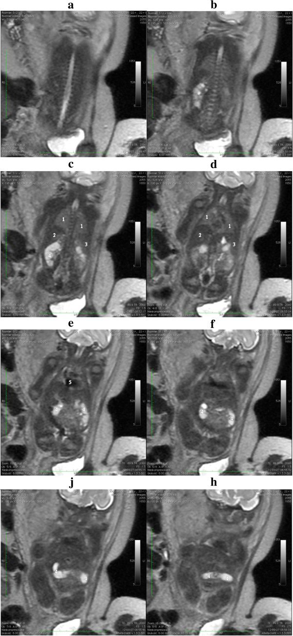
Gastrochisis is a development abnormality of the anterior abdominal wall, where the bowel protrudes without a covering sac between the developing rectus muscles, occurring slightly lateral and towards the right of the fetal umbilicus. Gastrochisis commonly occurs as an isolated malformation, occurring in approximately 2.5 in 10’000 births.
During the fourth week of normal fetal development, the lateral body of the fetus folds, moving ventrally and fusing in the midline to form the anterior body wall. In Gastrochisis it has been theorized that the incomplete fusion of the midline results in this abnormality, resulting in the abdominal viscera to protrude through the abdominal wall, herniating through the rectus muscle. This is one of many theories linked to Gastrochisis as the cause is still unclear. Other theories include, the failure of mesoderm to form in the body wall, rupture of the amnion around the umbilical ring with subsequent herniation of the bowel, abnormal involution of the right umbilical vein resulting in a weakening of the body wall and thus resulting in herniation of the bowel, and disruption of the right vitelline (yolk sac) artery with consequent body wall damage and gut herniation.
<pubmed>25059025</pubmed>
<pubmed>17230493</pubmed>
<pubmed>19419415</pubmed>
--Mark Hill this is a reasonable description of gastrochisis, please use in-text referencing so that your specific sources can be checked. (4/5)
Assessment 6
Thyroid Development
<pubmed>19389367</pubmed> The current study by Lania et. al. demonstrates that the developmental mechanisms of the endocrine gland, the thyroid, is regulated by genetic networks. Tbx1 is a prominent gene involved in the embryonic development of the thyroid gland, as well as many of the pharyngeal apparatus derivatives. In this study, the role of Tbx1 is emphasized as a key factor for regulating the size of the thyroid in early development of mice. Knockout mechanisms were preformed in mice embryos to remove the expression of Fgf8, in the mesoderm, which is regulated by Tbx1. The lack of Fgf8 thus subsequently leads to cause thyroid hypoplasia in the subjects. Thyroid sizes of the mouse embryos were measured following the removal of Fgf8, in 2 different stages of embryonic stages of development, with both stages showing a significant decrease in thyroid size. These results suggest that a Tbx1-NFgf8 pathway is a key factor in determining the size of he thyroid glands in mammalians.
In addition, mutant phenotypes were observed due to the lack of function of Tbx1. The mutant embryos presented with a hypoplastic thyroid, though the positioning of the organ was predominantly normal. Lania et. al. went on to observe a slightly larger than normal lumen present in the thyroid follicles of the mutants. These results were then supported further by Immunohistochemistry analysis of these embryos, which demonstrated that Nkx2-1 is typically expressed and that thyroglobulin is typically produced by mutant follicles.
Tooth Development
• Odontoblasts - derived from neural crest mesenchymal cells, and are differentiated under the influence of the enamel epithelium. Odontoblasts secrete predentin throughout life, which calcifies to form dentin, located under enamel.
• Ameloblasts - derived from oral epithelium of ectodermal origin. They produce enamel after the first production of dentin layer by odontoblasts and form the outermost layer of the tooth.
• Periodontal ligament - a specialised connective tissue layer that acts as an anchor for tooth in its bony socket and surrounds the tooth root coating of cementum.
--Mark Hill Yes T-Box 1 (Tbx1) appears to be important for thyroid development indirectly through its effects on the surrounding mesoderm through FGF pathway. Tooth origins are correct. (5/5)
http://www.omim.org/entry/602054
Assessment 7
Ovary Development
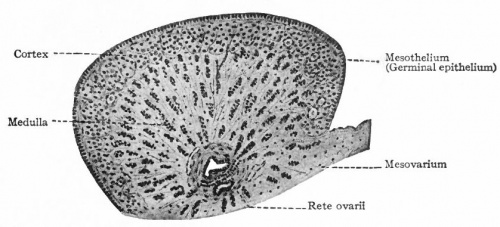
Initially, the origins of gonadal development are morphologically similar for both the testes and ovaries. It is only when the indifferent stage of sexual development occurs when the differential stages of ovary development begins. Both gonads have derivatives from the structures; Mesothelium, which lines the posterior abdominal wall, as well as underlying mesenchyme and primordial germ cells that form the earliest, undifferentiated sex cells.
During week 5 the development of ovaries begins, when a thickened area of mesothelium develops on the medial side of the mesonephros, a primitive kidney. This is then continued by the development of gonadal ridges, which results from proliferation of the mesothelium and the underlying mesenchymal tissue, as it produces a bulge on the medial mesonephros. Finger like epithelial cords then grow into the underlying mesenchyme forming the gonadal cords. In females (XX), the cortex of the indifferent gonad differentiates into an ovary, and the medulla regresses.
Primordial germ cells dwell amongst dorsal endodermal cells of the umbilical vesicle, where they first populate. They then begin to migrate to the gonadal ridge along the hindgut’s dorsal mesentery where they then migrate to the gonadal ridges during the folding process of the embryo. In week 6, the primordial germ cells enter the underlying mesenchyme and are incorporated in the gonadal cords.
Ovary development is a slow process in female embryos, and It is not until week 10, when the ovaries become histologically recognizable.
References
<pubmed>15664455</pubmed> Moore: The Developing Human Chapter 12
--Mark Hill Historic figure is fine. Ovary description is also OK, please do not cite textbook as source for assessment items, I expect you to look at the research literature. You have not provided any more detail than was covered in the lecture. (4/5)
Assessment 9
Group 1:
Firstly, great job on the layout and formatting of the project, everything is easy to find and overall, it reads well. The introduction provides great insight of what to expect on the page. However, it lacks in-text citations for the first three subheadings of the page, as well as the table of lung developmental stages. The first two images also don’t have a description when I click on it, I don’t know what I’m looking at. The “student template” is also missing for the images. I would suggest you look up the tutorial for uploading images on the pages as Mark has extensive information for the proper steps required for uploading images. Otherwise, the lung developmental stages table is informative and easy to read. I would also recommend adding an image for better visualization of the developmental process.
The historical findings and current research models have very detailed content, and look as though they have been referenced correctly using in-text citations, I’m impressed. Although, I would suggest you leave all the references to the end by simply putting </references> at the bottom of the page, as it looks neater to have them all in one place, rather than at the bottom of each sub-heading. The abnormalities section is done well and there are a wide number of abnormalities covered. The detail of the first two is more in depth than the rest, I’m unsure whether they was more information on those particular abnormalities or their still needs to be information added, but I suggest to have the same amount of information on each disease, if possible. Overall, the project is very informative and presented well. It just need a few minor edits.
Group 3:
The introduction provides a good basic outline of the overview of the GIT. Although, there are no in-text citations in the introduction and all sub-headings are not included into the overview. Be wary of spelling errors such as “GIT (Gastrointestinal Track) consist of the Fore-gut, Mid-gut and Hind-gut” that should read Gastrointestinal Tract consists of the foregut, midgut and hindgut. This section would be better it was expanded upon and images were added. The timeline provides good detail, though would benefit by better formatting and organisation of the information, maybe putting it all into a table, by week will tidy it up.
Adding images for the sections will definitely be beneficial. The images hand-drawn are great, although the colours used make it hard to read. If you plan to add anymore drawings, try and use dark colours that allow for easy readability. The images already uploaded are missing copyright, referencing and “student template” information for images such as “fetal week 10 sagittal plane”. I would suggest you look up the tutorial for uploading images on the pages as Mark has extensive information for the proper steps required for uploading images.
The deformities section should be re-titled abnormalities as per the assessment criteria and would ensure the group is following similar structure from the other projects. Again, adding an image per disease would be great. Try and do about 1-2 more abnormalities. Great job on putting all the references at the bottom of the page, it makes it very neat and accessible. Overall, a good project just needs a few edits.
Group 4:
Firstly, great job on all the contents you guys managed to present, it’s quite detailed. There seems to be no introduction though, and the page jumps straight into explaining genital development. I think if an introduction were added, it would give the whole page better structure and formatting so the reader knows what to expect when they decide if they want to read on. The dot points used for the developmental section allows for easy readability of the contents, however, the use of caps lock and arrows takes away from the overall presentation of the page. I would suggest any text you want to emphasize to make bold or underline the word. I also noticed that there was a note stating the attempt to put all the developmental information into a table, but had issues. I suggest you look at the editing basic page you can search for in the top right hand corner as it outlines a step-by-step guide into making tables etc.
In regards to referencing, there are no in-text citations for the first two subheadings. The sections were they do have citations also have a list of references at the bottom of each section. I would recommend just adding a final list of references at the bottom of the page, as it looks much neater.
I’m impressed with the level of hand-drawn diagrams uploaded. I would also recommend adding captions to the image. For example: [File: Flow Diagram of Fetal Development of External Genitalia.pptx|1000px|thumb|right|alt text]. The “alt text” should be edited to describe the caption of the drawing. This particular image seems to have a broken link though; the “alt text” also appeared in the labeled diagram of the testes. Otherwise, good job on the other images.
The current findings section seems to be untouched, with the exception of some pubmed journal article links, I’m assuming you are still in the process of adding content. The historic findings, however, is extensive and well researched. Good job.
The abnormalities section is done well. There is more than enough abnormalities listed, and they are researched well, I would just suggest adding a few more images for better visualization. Overall, great page, just needs better formatting for the mentioned sections.
Group 5:
This page has great overall structure and presentation. The introduction gives good insight of the overall contents of the page, however it is very brief and should be expanded upon.
The table included in the developmental overview serving, as a timeline is excellent, really well done. It’s easy to follow and looks very neat. I like how there is an image for each of the weeks mentioned, just don’t forget to add in-text citations for its contents. The glands sub-section is very brief and would benefit if there were more contents added. Great job on the images though. The nail section is the same, more contents needs to be added and image would look really good.
The developmental overview and historic findings sections also seems to lack in-text citations. There is also has an image with a broken link. The subsection hair seems to be well researched, however I would also suggest either bolding or underlining the words you want to emphasize such as “structure” for a neater look.
The recent findings section looks superb I love the purple background colour. Its very well researched and the link to more research papers are very helpful for readers. I would suggest you put the image at the bottom of the mentioned content though, just to avoid the big gap on the page, or even if you can manage to wrap the text around the image, it would look much better in terms of presentation.
Although disturbing, the abnormalities section I could not fault. Very well done. It is evident that it has been research well and the images allow for great visualization of the diseases mentioned.
Overall, excellent page just needs a very formatting edits and some expanded contents mentioned above. Good luck!
Group 6:
There seems to be no introduction on the page, don’t forget to add content to this section before the final submission. The overall page looks disjointed by the choice of sub-headings. I think an overall timeline is needed to know which glands/organs develop when and originate from where. It would look much neater and would be easier to follow.
The parathyroid gland and pancreas seems to be the only sections that are properly completed. Both sections have good use of images and the tables provide easy readability. The images are all properly cited, good job.
The overall referencing of the page is all over the place and lacks in-text citations. I suggest you go through the contents and add these where necessary. If you are unsure how to do this, just look at the handout Mark gave out in week 2 for further reference. Or, alternatively you could look at some of the other project pages in edit mode. I would also suggest you leave all the references to the end of the page by simply putting </references> at the bottom of the page, as it looks neater to have them all in one place, rather than at the bottom of each sub-heading.
The abnormalities section is lacking content and there is only 2 diseases listed, with no description.
Overall, the page has good content, just needs to be edited to put in-text referencing. Some sections need contents such as the placenta and adding images to the page will also improve its presentation.
Group 7:
This page is organized well, all the headings and subheadings are thought through. Although, I’m unsure while the sections brain and spinal cord are in bold? The development during fetal period image lacks the necessary “student template” at the bottom of the description summary and I was unable to open the link http://www.nichd.nih.gov/publications/pubs/acute/images/p44.gif. Otherwise, all the other images uploaded on the page look really good and are referenced correctly.
The table under the section brain development is very brief, and expansions on the content will allow for a better understanding of the content. Adding images to appear after the table will also add to the appearance of the page and give it a cleaner look.
The spinal cord and menegies development have been left untouched and the current research models have no content, just pubmed references. I understand the current research models are probably the hardest part of the assignment, but the content appears to be quite good, the formatting of the section could be improved by following the structure Mark uses. You could look at the other group projects as examples.
In regards to referencing, there are no in-text citations for the first two subheadings. I would also like to recommend just adding a final list of references at the bottom of the page, as it looks much neater.
The abnormalities section is done well. But try to minimise the use of dot points as this section lacks any structured paragraphs. It use of images are great, although there is an image that appear to have been removed and as a result, there is a broken link.
Overall, great job so far!
Group 8:
Let me start by saying, for only having two people in the group, well done. The page should have an introduction though, and this is missing. Just by simply summarizing all the information that will be covered in the page and adding it to the introduction, will improve the overall presentation significantly, you may wish to leave this to last, or edit as you go along.
The section “Making gains” is amusing, but inappropriate and should be omitted from the final submission. The timeline for the page I believe should be put into a table to save time and add to the presentation of the page, it can be easily done if you follow the steps outlined in the ‘editing basics’ page
The background information is comprehensive, however, the page is in desperate need of some images as there are just slabs of text. Images will really help break up the contents of the page and make it visually appealing.
The abnormalities section also seems to be coming along quite well. Keep up the good work.
--Mark Hill You have provided some good critical feedback. My only specific comment would be that while it is good to praise the group it should be focussed on what is actually good about the project rather than overall statements. (10/10)
Assessment 10
<pubmed>8955790</pubmed> The outer oral environment receives chemical stimuli from slender epithelial cells, which are responsible for the assembly of taste buds. To date, there have been few studies documented on the formation and morphology of prenatal human taste buds. Therefore, in this study, taste bud primordium is investigated, including its morphological changes, synaptogenesis, cell differentiation, and taste pore formation from the time of the onset of taste bud formation from approximately the 8th week until the 15th week of gestation.
Forty-two human embryonic and fetal tongues from the 6th week to the 15th week of gestation were used in the study. The tongues were then fixed in 0.1 M cacodylate buffer containing 2.5% glutaraldehyde, or 0.1 M phosphate buffer containing 2% paraformaldehyde and 1% glutaraldehyde. The samples were then contrasted overnight and prepared with uranyl acetate dissolved in 70% ethanol, which were then, washed, dehydrated and embedded in Epon. The sections of tissues were then prepared consisting of three specimens of each tongue and screened for taste bud primordial. The sections were then placed on slot grids and examined by means of transmission election microscopy.
From the results, it can be seen that the sixth to seventh postovulatory week have shown no signs of cell specialization and indicate taste bud formation, although at this stage the epithelial is 2-3 layers thick and nerves fibres begin to approach the lingual epithelium. During the 8th week of gestation, the first signs of cell differ enation responsible for the formation of taste buds were seen at the base of the taste bud primordium and the nerve fibres are able to penetrate the basal lamina. By the 9th – 11th week the lingual epithelium has thicken to compose of approximately 3-4 layers and taste bud primordial were present and resemble those that were found in earlier stages. During week 12, further differentiation of the cells can be seen, as the cells that are electron dense begin to resemble type III cells seen in human adult taste buds, as well as long, slender cells with electron-dense nuclei and electron-dense cytoplasm containing abundant mitochondria and bundles of intermediate filaments. However, the majority of taste pores do not begin to develop up until week 14-15 of gestation.
--Mark Hill This is a good summary of an early paper studying human tastebud development timeline by EM study. (5/5)
--Mark Hill Lab 11 assessment?

