User:Z3418340
--Z3418340 (talk) 12:45, 6 August 2014 (EST)
Lab Attendance
Lab 1 - --Z3418340 (talk) 12:53, 6 August 2014 (EST)
Lab 2 - --Z3418340 (talk) 11:13, 13 August 2014 (EST)
Lab 3 - --Z3418340 (talk) 13:48, 20 August 2014 (EST)
Lab 4 - --Z3418340 (talk) 12:39, 27 August 2014 (EST)
Lab 5 - --Z3418340 (talk) 11:37, 3 September 2014 (EST)
Lab 6 - --Z3418340 (talk) 11:12, 10 September 2014 (EST)
Lab 7 - -- Z3418340 (talk) 13:10, 17 September 2014 (EST)
Lab 8 - --Z3418340 (talk) 12:20, 24 September 2014 (EST)
Lab 9 - --Z3418340 (talk) 12:03, 8 October 2014 (EST)
Lab 10- --Z3418340 (talk) 11:16, 15 October 2014 (EST)
Lab 11- --Z3418340 (talk) 11:52, 22 October 2014 (EST)
Individual Assessment 1
ARTICLE 1
<pubmed>24302192</pubmed>
Inositol is an important factor with in the follicular environment, high levels have been associated with improved development of the oocyte. This particular study aims to understand the effects of treatment with inositol on oocyte quality in patients undergoing ICSI.
The researchers selected 149 patients undergoing ICSI cycles between June 2012 and May 2013, all of whom where aged under 40, had at least one previously failed attempt at ICSI and were diagnosed with polycystic ovary syndrome (PCOS). Patients were randomly divided into two groups. Group 1 consisting of 58 patients were treated with both folic acid (400 mg/day) and inositol (2000 mg/day of myo-inositol, D-chiro-inositol 400 mg/day) for 3 months prior to the ISCI cycles. Group 2 consisted of 91 patients who were treaded with folic acid (400 mg/day) alone, this group acted as the control.
(1.) The standard of IVF using ICSI was employed. Oocyte quality was routinely checked by making observations using an inverted microscope. The oocyte’s stage of maturity, size and shape, cytoplasmic characteristics and extracytoplasmic characterises were noted. (2.) Post-implantation assessments were made regarding embryo quality in light of the following parameters; number of blastomeres, degree of fragmentation and size of blastomeres. (3.) Finally at 14 days from Embryo-Transfer execution, quantitative blood detection of β-hCG is performed in order to biochemically determine pregnancy. This is followed by a final diagnosis of clinical pregnancy with ultrasound visualization.
(1.) No significant difference was found in the number of mature oocytes taken and in the number of immature oocytes taken. However the the results did show that a greater proportion of Group 1 oocytes displayed features that typical of excellent and good quality oocytes. (3.) In terms of the number of positive biochemical pregnancies there was again no statistically significant difference between Group 1 and Group 2. However Group 1 did show a statically significant increase in the number of clinical pregnancies detected.
The article is concluded discussing and highlighting the improvement in the overall quality of oocytes and parallel increase in the number of clinical pregnancies as a result of treatment with inositol.
ARTICLE 2
<pubmed>25077107 </pubmed>
The investigation was carried out on IVF patients from Mount Sinai Hospital, Toronto, Ontario. Candidates selected for the study were aged 18-41 years, with base line levels of FSH (on Day 3 of Cycle) and the ability to provide informed consent. Researchers included 173 female patients, who then underwent IVF cycles as per standard procedure.
Serum 25-hydroxy-vitamin D (25[OH]D) levels were measured one week prior to oocyte retrieval, these measurements taken in order to determine the initial Vitamin D status. Patients were either classified as having sufficient (≥ 75 nmol/L) or insufficient (< 75 nmol/L) 25(OH)D levels.
Standard IVF procedures followed; oocyte retrieval, fertilisation, growth and embryo transfer. An ultrasound was then taken 4-5 weeks after the embryo was transferred and implantation success was monitored, as indicate by the presence of a gestational sac, visible by ultrasonography. The implantation rate was calculated as the number of gestational sacs observed by ultrasonography divided by the number of embryos transferred, multiplied by 100.
With in this cohort 45.1% had sufficient levels of 25(OH)D, while 54.9% had insufficient levels. The study found a 52.5% clinical pregnancy rate per IVF cycle among women with sufficient levels of 25(OH)D levels. This was significantly higher than compare to a rate of 34.7% the among women with insufficient levels of 25(OH)D.
--Mark Hill Your summaries of these 2 selected articles are concise and accurate. You might want to also think about exploring the discussion part of the papers for additional information about the future direction of the research. (5/5)
Individual Assessment 2
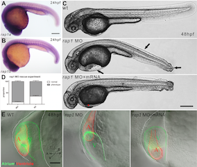
Image:Abnormal heart and caudal fin development in zebrafish due to Rap 1 knock down[1]
--Mark Hill All required information is here. You might in future think about using a slightly larger image for uploading (up to 1000px wide) as it is quite difficult to see detail in this figure at the current size. (5/5)
Individual Assessment 3
Fetal Development - Time Line
Historic Findings
[1] [2] https://www.youtube.com/watch?v=WXLPxjJszio
--Mark Hill Good references (5/5)
Individual Assessment 4
PART 1 - Identify a paper that uses cord stem cells therapeutically and write a brief (2-3 paragraph) description of the paper's findings.
Context
The use of umbilical chord derived stem cells for therapeutic purposes is certainly widespread and has served as an effective tool for the treatment of cancers, heart disease and many other conditions.
This particular study aims to further investigate the potential use of Mesenchymal stem cells (MSC)derived from chord blood, this time looking at potential use as a promoter of wound healing in diabetic patients. In these patients incomplete healing of wounds is primarily associated with poor revascularization and decreased production of growth factors in the damaged area. Since MSCs are multipotent they hold a great promise for tissue regenration and .
The main advantage of investigation into such therapies lies in the fact that MSCs can be easily isolated and refined from chord blood as oppose to any other sources.
Study and Findings
This study was conducted on genetically diabetic mice who showed delayed would healing. A wound was induced followed by subcutaneous injection of Conditioned-MSCs (CM-MSC), UC-MSC (Umbilical cord derived MSCs) or control PBS (Phosphate buffer solution).
Wound healing was reported as a percentage of the initial would that had reepitheliasized. Time taken for complete reepitheliasized was accelerated from 14 days in the PBS group, down to 4 days; following the initial injection of CM-MSC. Histological examination of would margins at 14 days revealed that CM-MSC treated wounds had relatively enhanced repiehtelizsation, a thiner layer of dense granulation tissue and increased vascularisation. Further more Immunohistological staining also showed higher capillary density in CM-MSC treated wounds compared to both the UC-MSC and PBS treated groups. Finally the PCR analysis of RNA was extracted from the CM-MSC treated mice revealed significantly higher levels of factors promoting aniogenesis such as; VEGF, PDGF, and KGF.
It was concluded that both the transplantation of UC-MSCs and CM-MSCs accelerates wound closure, increases angiogenesis and directly stimulates transrciption of vascular growth factors (VEGF, PDGF, and KGF).
Reference
<pubmed>3781996</pubmed>
PART 2 - Identify the developmental vascular "shunts" present in the embryo shunts and their anatomical location.
The three "shunts" are alternate paths for blood flow with in the circulatory system of the embryo.
Two of these shunts have the role of diverting blood from the pulmonary to the systemic circuit. The third connects the umbilical vein to the inferior vena cava.
1. The foramen ovale - An opening in the interatrial septum that allows blood to flow from the right atrium to the left atrium. Closes to leave the fossa ovale 2. The ductus arteriosus - A short, muscular vessel that connects the pulmonary trunk to the aorta. Degenrates to from the ligamentum arteriosum. 3. The ductus venosus - A temporary blood vessel that branches from the umbilical vein, allowing much of the freshly oxygenated blood from the placenta—the organ of gas exchange between the mother and fetus—to bypass the fetal liver and go directly to the fetal heart. Degenerates to become the ligamentum venosum.
--Mark Hill Very good (5/5)
Individual Assessment 5
Normal Pancreatic Development - During the fifth week of gestation one dorsal and two ventral evaginations (buds) appear on the wall of the developing foregut. Selective expansion of the duodenum causes fusion of the two ventral buds. During the seventh week of development the gut undergoes rotation and the ventral bud rotates with the gut. As the ventral bud passes behind the duodenum it fuses with the dorsal bud.
Annular pancreas (AP) results when ventral bud fails to rotate with with the duodenum. As a result the ventral bud essentially envelopes the rotating duodenum and forms a ring of pancreatic tissue surrounding the duodenum, known as a annulus.
Complete annular pancreas - pancreatic parenchyma or annular duct is seen to completely surround the 2nd part of duodenum
Incomplete annular pancreas - annulus does not surround the duodenum completely, giving a 'crocodile jaw' appearance Annular pancreas is associated with excess amniotic fluid during pregnancy (polyhydramniosis) and is often coupled with other congenital abnormalities of the gastrointestinal tract.
<pubmed>21386643</pubmed>
--Mark Hill (5/5)
Individual Assessment 6 - Lab 7
1.) Identify and write a brief description of the findings of a recent research paper on development of one of the endocrine organs covered in today's practical.
It is suspected that exposure to Endocrine Disrupting Chemicals (EDCs) disrupts normal thyroid development. This study investigates the effects of pre-conceptual, gestational, and continuous maternal exposure to sewage sludge on fetal thyroid gland development and the levels of circulating thyroid hormones in the exposed ovine fetus.
Pre-conceptual and gestational exposure in the fetuses; the first exposed throughout their lives prior and post mating (TT).
Exposure only until mating, but not thereafter (TC) resulting in exclusively pre-conceptual exposure.
Exclusively gestational exposure was achieved by exposure between mating and euthanasia (CT).
Hormone Level
Maternal and fetal blood samples were obtained and concentrations of T3,T4 and TSH were determined. Hormone level analysis revealed that treatment had no significant effect on maternal t3/T4 ratios. There was also no change in plasma levels of these hormones in maternal blood compare to the control group. Furthermore, no significant correlation of any kind was found between maternal TSH and fetal thyroid hormone levels.
Morphometric Analysis
Thyroid tissue sections were analysed and the follicle number, size and epithelial height were determined. All groups showed inter-animal variability, the phenomenon is most pronounced in the TT group . Treatment groups presented with a lower follicle count, and both groups with preconceptual exposure showed a higher percentage distribution of medium-sized follicles. The height of the follicular epithelium was unchanged and no changes in follicular resorption vacuoles was observed.
In addition morphological analysis shows a change in the relative distribution of small and large blood vessels. Smaller blood vessels representing more than 80% of all blood vessels in the thyroid tissues were predominantly affected. Female fetuses consistently show a reduction in the percentage of small blood vessel after pre-conceptual exposure and an exclusive and significant increase in thyroid cell proliferation in both groups CT and TC. Male fetuses showed significantly reduced follicle counts in both cross-over groups (CT, TC) and revealed highest thyroid cell proliferative activity in the TT group.
<pubmed>23291342</pubmed>
2.) Identify the embryonic layers and tissues that contribute to the developing teeth
Tooth development begins when dental lamina proliferates to form two horseshoe-shaped structures corresponding to the future dental arcades, this structure originates from the ectodermal layer. Enamel organs develop in the dental lamina; each swelling is the future site of a single tooth. The enamel organ exerts an organizing influence over the development of the mesodermal portions of the tooth. Gradually becoming cup-shaped, the enamel organ partially encloses an adjacent mesodermal structure which goes on to from the the dental papilla. Unenclosed mesoderm of the dental papilla contributes surrounding structures.
Source: http://www.britannica.com/EBchecked/topic/1512077/tooth-germ <pubmed></pubmed>
--Mark Hill (5/5)
Individual Assessment 7 - Lab 8
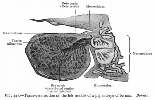
Provide a brief time course and overview of embryonic development of either the human testis or ovary. (2-3 paragraphs) Include an image from the historic genital embryology section of the online notes in your description.
The the male testis is derived from three embryonic cell layers (origins).
- The first layer in the mesothelium, linked to the posterior abdominal wall.
- The next is the underlying layer of mesencyme, which develops into embryonic connective tissue.
- Finally the primordial germ cells, which are the earliest undifferntiated sex cells.
During week 5 of embryonic development, a thickened area develops on the medial side of the mesonephros due to proliferation of epithelium and the underlying mesencyme layers - genital ridge. Epithelial chords from the mesothelium project then into the underlying mesencyme - genital chords. The gonad is now established, consisting of an external cortex and an inner medulla.In male embryos with an XY sex chromosome complex, the medulla differntiates, giving rise to the testi while the cortex degenerates.
In conjunction with the formation of genital ridges, primordial germ cells are developing in the umbilical vessicle at week 4 of embryonic development. During the folding phase, these umbilical vessicles are incorporated into the embryo (hind gut region).The primordial germs cells are now able to migrate along the dorsal mesentery from the hind gut into the genial ridges. By week 6 the germ cells have been incorporated the mesencyme via the genial chords.
The embryonic development of the early genital system beings is identical for males and females until week 7, when the of morphological characteristics begin developing. The male Y chromosome has a sex-determining region known as the SRY gene which stimulates production of Testes-Determining Factor, determines testicular differentiation. Organisation factors and TDF stimulate differentiation of gonadal chords and into seminiferious chords. At this point a thick fibrous cap envelops the developing tubules - tunica albuginea . As testis gradually enlarges, it looses its connection to the mesonephros and is suspended by the mesochorium.
The seminiferous chords continue to develop, giving rise to the Leydige cells and Sertoli cells by week 8 of embryonic development. Leydig cells being producing androgens such as Testosterone. While Sertoli cells are producing Anti-Mullerian Hormone (AMH) which prevents simultaneous development of female internal genial tract.
Week 4 - Primordial Germ Cells - differentiated Week 5 - Formation of Genital Ridges and Genital Chords - establish gonad stucture Week 6 - Primordial Germ Cells migrate into the Genial Ridges and are incorporated into the mesncyme Week 7 - Expression of the SRY gene- Increased TDF Week 8 - Differentiation of Leydig and Sertoli cells - Coupled with production of Testosterone and AMH
--Mark Hill Good summary of testis, I prefer textbooks not to be used as source material for these assessment items. (4/5)
Individual Assessment 8 - Peer Review
Group 1 - Respiratory
In this review I intend to highlight the merits of your project as well as provide some constructive criticism in light of the marking criteria of this task.
The page is well structured and provides perfect balance between written text and images. However some of the included images do not compliment the text. I suggest adding labels or descriptive annotations to these images using paint. Alternatively you could refer to these images in your text e.g “ as seen in Figure 4a” and use them to make the descriptive content easier to visualise. You could also include a simple written description of what each image showing in the image link. I found the table on the stages of lung development a really effective way of organising the content and I was able to understand much of it in a quick glimpse! I like how the text is summarised and highlights the main developmental changes that are occurring at each stage. Just to make it more engaging, perhaps you could include matching images in a another column.
Under the section of current findings, I believe that most of the information included is relevant and incredibly appropriate articles have been selected. I think its good that this section is delving into the area of molecular signalling underlying the morphological changes that we see. I believe your project would greatly benefit if there was more material discussing the biochemical signalling and recent findings in relation to this. However, I am not sure if the details on cell type should be in this section, this section might need some re-organising.
I understand that the history is a difficult topic to research. The information on our understanding of surfactant is appropriate, detailed and very informative. However I think you need to include more information on our understanding of stages in fetal lung development. Explore the transition in research focus investigating morphology to molecular changes. Perhaps use the library database to find relevant historic journal articles in the database. It was good to see the use of relevant historic images.
A number of abnormalities have been identified and described, I think its great that each section includes a description of the abnormality, and goes on to discuss the cause and implications of each disease. I would only recommend including images to make the content easy to visualise. Great Work!
Overall the project is coming along really well ! Just ensure that you proof read and review before the final submission. Also include in-text references and compile all your references to one section at the end of the page. Good Luck!!
Group 2 - Renal
In this review I will attempt to highlight the strengths of your project and identify some areas for improvement, in light of the criteria provided.
I believe the developmental timeline is a great way to summarise the major events at each stage in fetal development and serves as a simple introduction to the project. However I think it would be best if you presented this information in a tabulated format, and perhaps you should include a little more detail for each developmental stage. For instance “Week 8 – Mature kidney is formed” you could also mention some structures features that allow us to recognise that it is a mature kidney (hallmarks of a mature kidney)
I think the current research section delves into a number in interesting areas, mentioning studies investigating treatment options for congenital renal abnormalities. I think another interesting area that you could address is the molecular signalling and gene expression process that drives the underlying differentiation and development of the renal system.
The abnormalities associated with renal development in the feral period have been well researched and the information provided is well structured. However this section seems incomplete. I see a number of additional links to interesting scholarly articles. I think you should discuss some more abnormalities and divide them up into abnormalities arising in the early and late stages of fetal development. I also suggest including images or diagrams to break up the text and make the descriptive text easy to visualise.
There is has been little information added on the historic findings. This is an essential component of the project. I suggest looking at text books in the library or searching the UNSW database to find information for this section.
I really like how you have selected labeled diagrams to compliment and break up the text. Each image is relevant to the topic being discussed and the small description attached really help the reader orient them selves. Overall this project is coming along nicely. Just ensure that you are making progress on all the sections. Also only include relevant references. Finally proof read and review your work before the final submission.
Group 3 - Gastrointestinal
In this review I intend to highlight the positive features of your project while pointing out some areas that need improvement, in light of the marking criteria provided.
I really like the overview on of the topic, it is clear and succinct. I think a developmental time line is a great way to summaries all the information. I would also like to mention that this summary is very well referenced and gives an over view of the significant event is GIT development. However I think that this information would be best presented in a tabulated form. Perhaps you could use the following layout: Column1: Week, Column 2: Foregut, Column 3: Mid-gut, Column 4: Hind-gut. It would also be a good idea to include images or diagrams. I particularly like the hand drawn diagrams, they really compliment the text and help visualise the different stages of development.
However are two issues with this project, there is little information on current research. I suggest looking up emerging technologies, drugs, treatments for congenital abnormalities in relation to GIT development. You also need to address the topic of Historic Findings, I suggest using textbooks from the library, the UNSW library database and UNSW embryology page to discover how our understanding of GIT development began and how it has changed.
A great start to the project. Good luck!!
Group 4 - Genital
In this review I will attempt to highlight the merits of your project and provide some constructive criticisms in light of the marking criteria.
Great work on system development, a lot of research has been done and the page seems well organised. I suggest using the information you have collected to write up succinct paragraphs, with forget in-text referencing. Furthermore, I find that that the table is a really effective means of summarising everything, you’ve made good progress so far. I also feel that the diagrams and video really support the text and have been appropriately selected. The current research section is a looking good, it’s great that you are exploring the molecular signals driving genital development, with references to FGFs, SHH and BMPs. I think this area needs to be addressed in further depthg. I also suggest including relevant studies, methods and findings. Finally don’t forget to include references!
I see that a significant amount of research has been conducted on the historical understanding of genital system development. Your project provides a particularly interesting insight into the debate on mechanisms of testicular decent. To make this section more interactive and engaging I would suggest the inclusion of historic illustrations and diagrams. There are many images available on both the UNSW embryology database and the UNSW library database. I also suggest that further research of the female genital system. Finally use in text referencing to support your data.
The section final section of your project investigates a number of male and female genital abnormalities. The diagram on abnormalities of the vagina and uterus is particularly interesting and certainly assists my understanding of these abnormalities. I simply suggest that you provide a little more depth on each abnormality. Ensure that you address the following areas are addressed: Cause; Description; Treatments. The page is well structured and incredibly cohesive. The references are well organised. Finally I’m really impressed by the drawing and diagrams. Great work so far! Just make sure you include that introduction in the end and add all the diagrams and images you plan to.
Group 6 - Endocrine
In this review I intend to highlight the merits of your project and suggest some areas for improvement in light of the marking criterial provided.
I believe that an organ-by-organ approach to this section is great. This really helps organise the information. This layout also makes the page easy to navigate allowing students to directly refer to the section that they want to learn about. However by doing so I think you may have neglected some of the areas.
Each endocrine organ has a great introduction describing the structural features and nature of the organ. I suggest including an image or a hand-drawn diagram of each gland and location, as this would really aid understanding The time line is a great way to summaries the major stages in development, I feel that this section has been completed with sufficient research and detail.
The section on abnormalities needs to be completed, even if only one abnormality is addressed make sure you include information on the following areas. Epidemiology; Description; Cause and Treatment. Furthermore ensure that the section on current research and historical findings is researched and addressed addressed.
I feel that your project is incredibly cohesive and attempts to provide a through summary of all the main endocrine organs. However a number of sections are yet to be completed. You have a great template right now. If all these areas are completed the project will be a success. In addition; I suggest placing all the references at the end of your project page, under one heading. Good Luck!
Group 7 - Neural
In this review I intend to highlight the merits of your project and suggest some areas for improvement in light of the marking criterial provided.
The introduction provides the perfect preface for your project, it serves to summarise the topic and highlight the areas that you will be addressing.
In the first section you have discussed fetal development of the neural system in great detail. I feel that a lot of research has gone into the collection and presentation of this date. The diagrams have been appropriately selected. Each image really ties in with the content and helps explain that stage development; I particularly like the diagram summarising the cell migration. In addition the images are well referenced. In the link you provide a brief description of the image and effectively explain the meaning of all the abbreviations.
The topics addressed under the heading of current seem quite interesting. The project really succeeds in providing insight into this new MIR technology, a technology that will certainly allow us to build on current knowledge of fetal neural development. I see that the heading of future research has not been completed. However I feel that this is a very interesting sub heading and shows a clear aspiration to go beyond the scope of the course.
A number of abnormalities have been addressed. I only suggest that you ensure that each of these subheading is addressed for each abnormality. Description; Epidemiology; Cause and possible Treatments, an image would be good too.
All the content on this page is well written. I feel that all the subheadings are relevant, though some sections are not complete. The only major drawback of your project is that, at this point the area of historic findings has not been addressed at all. Make sure you address this area.
Group 8 - Musculoskeletal
In this review I hope to highlight the merits of your project and suggest some areas for improvement in line with the marking criteria.
I see that you have conducted a great amount of research on the fetal development of the musculoskeletal system. The content clearly goes beyond the material covered in the lectures. It was interesting to read about the different transcriptions factors involved in induction and regulation of myoblast differentiation. I think it will be good to see a summary of all this information in a timeline format. I suggest simply highlighting the main developments at each stage.
You have made a good start on abnormalities. I suggest that you begin by selecting one abnormality include Description; Epidemiology; Cause and Treatment. You can add more later.
The page needs a little more structure. Make sure you include appropriate sub-heading and organise the information before you submit the project. Remember we were asked specifically to address the topics of current research and historic findings.
Finally it would be good see some images to support the text. Perhaps diagrams on tendon development would help summarise the process.
Great work so far!! Hope this feed back helps.
--Mark Hill Excellent (10/10)
Individual Assessment 10
Identify a recent research paper on sensory development (not hearing) and write a brief summary (several paragraphs) of the research methods and findings. Include at the ned a link to the relevant wiki sensory notes page
The Semaphorin 3 (Sema3) family of genes known to play a role in the development of the Central Nervous System and Visual System development. This investigation aims to elucidate the role of these Sema3 timing of the developing mammalian visual system gene expression using a rat model.
Method
Embryonic Wistar rats at stages E16-E19 of development were obtained by cesarean section of mother rats. Eyes and Superior Colliculus (SC) were dissected out of the foetus. The retina was then isolated from surrounding tunica and viterious, while the SC was isolate from the surrounding meninges. Both structures then placed in storage in ice cold media.
RNA was extracted using Tri-Reagent, followed by treatment with recombinant DNAeI. cDNA was then synthesised from the RNA. Previously validated primers were used to quanity RNA transcript expression of Sema33a-f and Plxna1-4a, Nrtp1-2 and L1cam. The qPCR technique was used to determine the expression levels for each specimen and the values were standardised.
Results
Developmental expression profiles were developed for the for the Sema3s taking into account statically significant data.
Expression levels in the retina could be separated into three qualitative groups: relatively high expression of Sema3f and Plxna2; moderate expression of Nrp1 and Plxna1; and relatively low expression of the rest. There were statistically significant changes in the level of expression of all Sema3 RNAs in the retina. In addition, of all the other receptors studied Nrp2, Plxna2, Plxna3, and Plxna4a showed statistically significant changes.
Sema3a transcript expression levels were significantly increased at P14 and in the adult.Sema3c RNA expression was also relatively steady through to P0, in- creasing significantly through to P21, and remaining at that level into the adult. Sema3e RNA levels appeared to increase gradually with retinal maturation and were significantly higher than E16-P7 levels at P21 and in adult rats. Sema3f transcription was temporarily greater at P0 and then increased again at P21 and beyond.
Many of the significant peaks in transcript expression occurred after the main developmental epochs. However, the changes that were quantified in the retina before P21 occurred during periods of RGC apoptosis, and synapse generation and maturation.
<pubmed>25283545</pubmed>
External Link - Development of the Visual System https://embryology.med.unsw.edu.au/embryology/index.php/Sensory_-_Vision_Development
--Mark Hill (5/5)
Individual Assessment 11 - Week 13
<pubmed>4041046</pubmed>
Pub Med
http://www.ncbi.nlm.nih.gov/pubmed/25084016
<pubmed>25084016</pubmed>

