User:Z3416697: Difference between revisions
No edit summary |
No edit summary |
||
| Line 87: | Line 87: | ||
==Lab attendance week 8== | ==Lab attendance week 8== | ||
--[[User:Z3416697|Z3416697]] ([[User talk:Z3416697|talk]]) 11:54, 17 September 2014 (EST) | --[[User:Z3416697|Z3416697]] ([[User talk:Z3416697|talk]]) 11:54, 17 September 2014 (EST) | ||
==Lab Report- week 8== | |||
==lab Attendance WEEK 9== | |||
Revision as of 11:09, 24 September 2014
--Z3416697 (talk) 12:45, 6 August 2014 (EST)
lab attendance
Lab 1 http://www.ncbi.nlm.nih.gov/pubmed
<pubmed>2508416</pubmed>
Lab Assessment 1
ARTICLE 1
<pubmed>25071849</pubmed>
In Vitro Fertilization (IVF) is a popular method of Assisted Reproduction which allows for fertilization to occur under optimal conditions (which may not be achieved in those that are having trouble conceiving), with monitored hormone levels and calculated growth of the embryo so as to maximize implantation, and ideally, successful pregnancy. This research article endeavors to assess the different variables available in IVF to determine which method yields the most successful amount of pregnancies. The variables in question are whether the embryo should be fresh (within a hours of fertilization), or frozen-thawed (embryo frozen a few days after fertilization and thawed when the mother is ready to receive an embryo). Another variable considered is the developmental stage at which the embryo is implanted; some embryos are implanted at the cleavage stage (hours after fertilization) and others are implanted at the blastocyst stage (5 days after fertilization), this study also considered implants at the cleavage stage extended blastocyst stage however it did not clarify what this term means, and I was unable to find elsewhere what this means.
This study observed IVF cycles of 1891 women at the Wuhan Union Hospital between January and December of 2012. Of the 1891 women observed, 1150 had fresh embryo transfers and 741 had frozen thawed embryo transfers. Of the Fresh embryo transfers 993 were implanted at the cleavage stage (799 of them women were less than 35 years old and 194 were greater than or equal to 35 years old), and 157 were implanted at the blastocyst stage (131 were less than 35 years old, 26 greater than or equal to 35 years old). Of the 741 women with frozen thawed embryo transfers, 212 were implanted at the cleavage stage of embryonic development (159 women were less than 35 and 53 were greater than or equal to 35), 137 were implanted at the cleavage stage extended blastocyst stage (111 were less than 35, 26 were greater than or equal to 35) and 328 were implanted at the blastocyst stage (276 were less than 35, 52 were greater than or equal to 35). All of these women underwent traditional methods of IVF- that is they were initially treated with Gonadotropin Releasing Hormone to stimulate follicular development. When two or more follicles were greater than 18mm Human Chorionic Gonadotropin was injected to assist follicular maturation. 24-26 hours later the ova were collected (Ovum Pick Up/OPU) and 4-6 hours after OPU IVF or Intracytoplasmic Sperm Injection (ICSI) were performed on the ova. The embryos were assessed by morphology and rate of development and at day three they are transferred onto a blastocyst medium and cultured for 2-3 days until it forms a blastocyst. The blastocysts are socred according to the Gardner standard and usually 1-2 good embryos are implanted. The remaining viable embryos are cryopreserved via vitrification and may be used if the current IVF cycle is unsuccessful. The Estrogen and Progesterone levels are monitored and regulated throughout the process according to the characteristics of the patient. Clinical pregnancy was defined by the presence of a gestational sac with or without a heart beat 30 days after implantation.
From this experiment it was found that there was a greater amount of successful pregnancies in women less than 35 years old resulting from implantation of fresh embryos at the cleavage stage (52.7% success) as opposed to the blastocyst stage (35.88%). There was no statistically significant differences clinically between the two types of implantation [ie- multiple pregnancies, abortion or ectopic pregnancy]. There were also a significantly higher number of pregnancies in the cleavage stage [41.24%] vs the blastocyst stage [26.92%]. However, this was not that case for those whom has frozen-thawed embryo transfers, as there was generally a much greater success rate for pregnancy in those who had blastocyst stage transfers than those who had cleavage stage transfers. Furthermore, there is a significantly greater pregnancy rate between in fresh embryo transfers compared to frozen thawed embryo transfers at the cleavage stage. Overall, there was a greater incidence of clinical pregnancy resulting from fresh cleavage stage embryo transfers compared to any other type of transfer [ie fresh blastocyst stage, frozen thawed cleavage stage and frozen thawed blastocyst stage]. These researchers concluded that this type of implantation should be used under normal conditions, and other methods should only be considered if the mother is of compromised health (eg has Ovarian Hyper Stimulation Syndrome), in which case as fresh blastocyst stage embryo is used.
ARTICLE 2
<pubmed>25017405 </pubmed> This article aimed to assess the success rate of clinical pregnancy of women at differing age groups whom have undergone assisted reproductive methods such as In-Vitro Fertilzation and Intracytoplasmic Sperm Injection. Based on previous literature it was expected that the success rate would decline as the woman ages, as typically the most successful IVF cycles are seen in women whom are 25-30 years old. Also based on natural conception rates, spontaneous conception rates begin to decrease around 31-35 years old, and women tend to have difficulty conceiving around the ages of 35-39. General fecundabilty (probability of released ovum becoming fertilized and resulting in a successful pregnancy in one menstrual cycle) tend t decrease as the woman reaches menopause (around the early fifties) and at 5-10 years prior to this (40-44 years old), half of the women have reduced reproductive capacity.
This study observed 2,900 women undergoing IVF at the KK Women and Children’s hospital, of which yielded 3,412 fresh IVF cycles. The women were classifies into sub groups based only on age [< 30 years; 30–35 years; 36–37 years; 38 years; 39 years; 40–44 years; and ≥ 45 years]. The IVF cycles were monitored and the average number of occytes, average duration of stimulation and fertilization rates were observed. Furthermore, the number of cycles until successful embryo transfer, clinically successfully pregnancy rates, miscarriage rates and multiple pregnancy rates were also reported. In this study, most of the patients undergoing IVF were between the ages of 30-35 and 36-37, and the least amount of patients belonged to the ≥ 45 years group. Of these, it was found that there was a 15% miscarriage rate in those younger than 30 years old, but this figure doubled to 30% at the age of 38 and among women aged 40-44, the miscarriage rate was as high as 55%. Only half of the women aged greater than 45 reached embryo transfer, and of those, none proceeded to successful clinical pregnancy. Furthermore, compared to other age groups, these women had the lowest amount of oocytes collected and the lowest fertilization rate with only 50% of oocytes proceeding to fertilization, compared to 95% in women less than 30 years old. However, it was found that there was no differences in mean duration of ovarian stimulation prior to ovum collection based on age. Finally, it was found that there was a general decreasing trend of successful clinical pregnancies as age increased in women undergoing IVF, as the rate of live births was highest in women who were less than 30 years old. The occurence of multiple births was highest in this age group also.
Overall it was found that the age of the woman undoubtedly has an effect on the reproductive capacity of a woman undergoing IVF. The researchers hypothesized that the general decreased success of an aging woman may be due to decreasing ovarian reserve, poorer oocyte quality, lower embryo implantation rates, altered hormonal environment resulting in ovulatory dysfunction and uterine problems. Male factors were also considered to be causative.
Lab Assessment 2
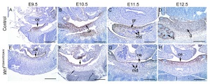
Mutation of Gene Wt1 Causes Aberrant Gonadal Development.jpeg[1]
--Z3416697 (talk) 11:08, 20 August 2014 (EST)
Lab Attendance: LAB 2
--Z3416697 (talk) 12:56, 20 August 2014 (EST) Forgot to do my attendance at the lab of week 3 given by guest Speakers Hayden Homer and Rob Gilchrist about meiosis, oocyte quality and various technologies in assisted reproduction
Lab Attendance week 4
--Z3416697 (talk) 11:39, 20 August 2014 (EST)
Lab three Online Assessment
Genital system development is an extremely interesting area of embryology as it is not until the later stages of embryogenesis (around week 4-6) that sexual differentiation occurs in the fetus, and the sexual organs actually look very similar up until this point, and the formation of the correct sex organs depend really on whether the genital ridge releases Testosterone or oestrogen <pubmed>24240231</pubmed> <pubmed>24928207</pubmed> <pubmed>24741072</pubmed> --Z3416697 (talk) 20:10, 26 August 2014 (EST)
Lab 4 Attendance
--Z3416697 (talk) 11:07, 27 August 2014 (EST)
Lab 4 Online Assessment
--Z3416697 (talk) 14:23, 2 September 2014 (EST)
<pubmed>25137413</pubmed>
Hepatocellular carcinoma and other degenerative liver diseases often result in hepatic dysfunction and ultimately organ failure. To avoid this otherwise fatal diagnosis, bio-artificial livers and hepatocyte transplantation provides some leg- room whilst waiting on a full-organ transplant. However liver transplants, much like many other organs, are often hard to come by and are usually sensitive to the immune response of the host. As a result, researchers are looking towards stem cells as an answer to this problem. Mesenchymal stem cells derived from the Wharton’s Jelly of the umbilical Chord are often chosen because they have been found to have a slightly higher multipotency and immunogenicity. The umbilical stem cells were selected because they are more primitive compared to other Mesenchymal stem cells and do not express the major histocompatibility complex class II antigens- a key determinant in illiciting an immune response form the host. However, hepatocyte like cells derived from stem cells are not clinically used because they do not express enough functional proteins and do not exhibit a full level of metabolic activity.
This study aimed to identify and observe the transcription factors involved in hepatocyte differentiation from Human umbilical cord Mesenchymal Stem Cells (HuMSCs), they were particularly interested in Hepatocyte Nuclear Factor 4-alpha (HNF-4alpha) which is believed to behave like a “master gene” in driving hepatocyte differentiation and maturation. This study continued to expose the differentiating HuMSCs to excess amounts of HNF-4alpha and hypothesized a significant improvement in the differentiation status of the hepatocyte-like cells, providing a basis for future clinical application of HuMSCs in the treatment and management of liver diseases.
The research initially confirmed that the HuMSCs are of low immunogenicity- a critical issue with organ transplantation. Once this was confirmed, they observed that there was an up-regulation of functional hepatic enzymes once the differentiating hepatocyte-like stem cells were exposed to HNF-4alpha. An important finding they discovered that the time at which the stem cells were exposed to HNF-4alpha were integral in ensuring correct hepatocyte differentiation. Ultimately they concluded that the differentiation of HuMSCs could be improved by exposure to high levels of HNF-4alpha at specific times in the development of hepatocyte-like stem cells, an important discovery for the progression of the therapeutic application of stem cells.
The three shunts which are present in fetal development but are closed postnatally are:
• ductus arteriosis → channel between the pulmonary artery and aorta in the fetus which bypasses the lungs to distribute oxygenated blood from the placenta derived from the mothers circulation.
• ductus venosus → shunts a portion of the left umbilical vein blood flow which would flow directly to the inferior vena cava- allows b=oxygenated blood to bypass the liver.
• foramen ovale → allows passageway of blood from left atrium to right atrium in the fetus.
Lab 5 Attendance
--Z3416697 (talk) 11:54, 3 September 2014 (EST)
Lab 5 Online Assessment
--Z3416697 (talk) 14:10, 16 September 2014 (EST)
Congenital Lobar emphysema (CLE) is a condition of the respiratory system which results in hyperinflation of one or more pulmonary lobes and is usually diagnosed postnatally, usually in the neonatal period- however there have been cases where CLE is not diagnosed until 10 years after birth. The clinical symptoms of CLE are often at birth as dyspnea, cyanosis and recurrent respiratory tract infections, as well as generalized neonatal respiratory distress and hyperinflation and hyperaeration of the pulmonary lobes- often seen as visible enlargements or imaged through CT. CLE is a rare disorder affecting only 1 in 20,000 to 30, 000 newborns, and appears to have a slight male predominance [3:1 male to female ratio], although the cause of this gender predilection is not fully understood.
The developmental causes of CLE are often as final result of a number of bronchopulmonary disturbances during development. These result from abnormal interactions between embryonic endodermal and mesodermal components of the lung which may result in abnormalities in airway or alveoli number and size, however the exact pathogenesis of CLE is not able to be determined in approximately half of cases. Another frequently observed cause of CLE is an obstruction of the developing airway, which creates a “ball valve” resulting in an uneven distribution of air- favoring greater airflow to the affected lobe during inspiration than is able to be cleared in expiration- resulting in air trapping. There may also be vascular abnormalities which produce compression, bronchial stenosis, bronchogenic cysts and congenital cytomegaloviral infections observed.
<pubmed>PMC2141574</pubmed>
Lab attendance week 8
--Z3416697 (talk) 11:54, 17 September 2014 (EST)
