User:Z3415141
Welcome to the 2014 Embryology Course!
- Links: Timetable | How to work online | One page Wiki Reference Card | Moodle
- Each week the individual assessment questions will be displayed in the practical class pages and also added here.
- Copy the assessment items to your own page and provide your answer.
- Note - Some guest assessments may require completion of a worksheet that will be handed in in class with your student name and ID.
| Individual Lab Assessment |
|---|
|
| Lab 12 - Stem Cell Presentation Assessment | More Info | |
|---|---|---|
| Group | Comment | Mark (10) |
| 1/8 |
|
7 |
| 2 |
|
7.5 |
| 3 |
|
7.5 |
| 4 |
|
8.5 |
| 5 |
|
8.5 |
| 6 |
|
8.5 |
| 7 |
|
7.5 |
Lab Attendance
Lab 1--Z3415141 (talk) 12:48, 6 August 2014 (EST)
Lab 2--Z3415141 (talk) 11:15, 13 August 2014 (EST)
Lab 3--Z3415141 (talk) 11:13, 20 August 2014 (EST)
Lab 4--Z3415141 (talk) 13:01, 27 August 2014 (EST)
Online Assessment 1
<pubmed>25100106</pubmed>
Some women for personal reasons prefer to take subcutaneous progesterone for luteal phase support when undergoing in vitro fertilization (IVF) in an attempt to avoid some of the side effects of vaginal progesterone. In attempting to work out whether the luteal phase of IVF better supports the ongoing pregnancy rate with the use of either the new aqueous preparation of subcutaneous progesterone or vaginal progesterone 800 random women aged between 18-42 years undergoing IVF received an embryo transfer where 3 oocytes were randomly placed in either a preparation progesterone administered subcutaneously or vaginal progesterone. If a viable pregnancy was to occur, then progesterone treatment continued for up to 12 weeks of the gestation process.
It was found that the pregnancy rate for vaginal progesterone (44.4%) in comparison to subcutaneous progesterone (41.6%) was consistently higher. While luteal phase support has clearly shown to improve pregnancy rates in women in the past this research shows that those women whom chose to go with the subcutaneous progesterone preparations are less likely to have a successful pregnancy than those who go with the traditional vaginal progesterone in supporting the luteal phase of IVF. It is important to note though due to the relatively small sample and dosage sizes of this study and the fairly small difference between the two groups shows that more testing should be completed before the results of this study should be considered fully conclusive.
<pubmed>25105102</pubmed>
A women’s worst nightmare is the sign of vaginal bleeding symbolism what may be a miscarriage. This study examined the effects of progesterone supplementation during the luteal phase on the possibility of miscarriage and live birth rate in natural frozen-thawed embryo transfer (FTET) cycles 228 women who underwent FTET cycles over a period between 2009- 2012 were examined.
The women were split up into two groups where one would receive progesterone support during the luteal phase and another that did not receive any progesterone support during the pregnancy. Overall there were no major differences in the women’s characteristics. This included the number of oocytes received, matured and fertilised. However, miscarriage rate was significantly lower in the group that received the progesterone as well as the live birth rate in comparison to those that did not receive any progesterone. The implications of this study loom large for women undergoing FTET cycles as it shows that if the luteal phase is supported with progesterone than the probability of a women’s worst nightmare can be decreased.
Online Assessment 2
Online Assessment 3
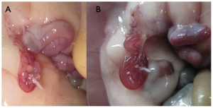
Midgut Formation during the development of the human fetus
1. <pubmed>18606147</pubmed> 2. <pubmed>14745932</pubmed> 3. <pubmed>24414177</pubmed>
Online Assessment 4
1. Identify a paper that uses cord stem cells therapeutically and write a brief (2-3 paragraph) description of the paper's findings.
The neuroprotective mechanisms and effects of implanted cord-derived msenchymal stem cells from a human umbilical cord(hUC-MSCs) were examined in ischemic stroke. An improvement in infarct volume and neurobehavioral function in the hUC-MSCs was discoverewd.
To do so the hUC-MSCs were cultured after they were isolated from the subendothetelial/endothelial layers of the umbilical cord of a human. Twenty days after the of induction of in vitro neuronal differentiation the cultured hUC-MSCs showed morphological features similar to those neurons. However there was no detection of functional neuron type channels.
From the discovery that the morphological differences meant that the cells were now able to produce neurotrophic factors but had not become active neuronal cells it was concluded that this may be related to the neuroprotective effects of hUC-MSCs.
<pubmed>18634757</pubmed>
2. There are a number of developmental vascular "shunts" present in the embryo, that are closed postnatally. Identify these shunts and their anatomical location.
Ductus Venosus: Joins the descending aorta and left pulmonary artery.
Ductus Arteriosus: connects the pulmonary artery directly to the ascending aorta.
Foamen Ovale: An opening between the septum of the left and right atria of the heart.
