User:Z3374215
Lab Attendance
Lab 1 --Z3374215 11:49, 25 July 2012 (EST)
Lab 2 --Z3374215 10:06, 1 August 2012 (EST)
Lab 1 Assessment
1) Identify the origin of In Vitro Fertilization and the 2010 nobel prize winner associated with this technique and add a correctly formatted link to the Nobel page.
The Nobel Prize for physiology or medicine in 2010 was awarded to Robert G. Edwards for his efforts in the development of In Vitro fertilization. Robert G. Edwards developed the idea of In Vitro fertilization since the 1950s. He first made fundamental discoveries in the life cycles of human eggs and the optimal time for fertilization before pairing with a gynecologist, Patrick Steptoe, and eventually seeing to the successful birth of an IVF baby in 1978. [1].
2) Identify and add a PubMed reference link to a recent paper on fertilisation and describe its key findings (1-2 paragraphs). "The relative contributions of propulsive forces and receptor-ligand binding forces during early contact between spermatozoa and zona pellucida of oocyte" was published by the Journal of Theoretical Biology in Nov. 2011 [2]. This report discusses the two main ways in which spermatozoa penetrate the zona pellucida of oocytes. The sperm utilize propulsive forces to assist in penetration. This is achieved through the motion of the flagella. The other factor important to penetration is the binding of sperm to ligands on the surface of the zona pellucida of the oocyte (ZP3). The report addresses the question of which of the cofactors is most imperative to the successful fertilization of the oocyte. A biomechanical model of the sperm-oocyte process was developed. It predicted that during early penetration the propulsive forces were stronger than the biochemical ligand binding. It was also predicted that the constant movement and overpowering force of the propulsion of sperm would make binding to ZP3 ligands difficult, making the large number of ZP3 receptors on the head of the sperm significantly important at this early stage.
References
- ↑ http://www.nobelprize.org/nobel_prizes/medicine/laureates/2010/press.html
- ↑ <pubmed>22100500</pubmed>
Lab 2 Assessment
1) Upload an image from a journal source relating to fertilization or the first 2 weeks of development as demonstrated in the practical class. Including in the image “Summary” window: An image name as a section heading, Any further description of what the image shows, A subsection labeled “Reference” and under this the original image source, appropriate reference and all copyright information and finally a template indicating that this is a student image.
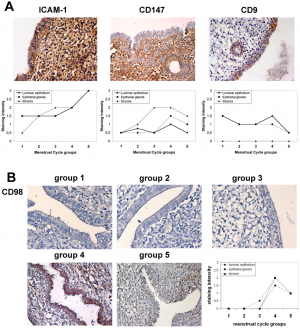
Expression of Endometrial CD98 in Implantation
A. Immunolocalization of ICAM-1 (HU5/3 mAb), CD147 (VJ1/9 mAb) and CD9 (VJ1/20 mAb) in human endometrium throughout the menstrual cycle. The micrographs show representative immunohistochemical stains of luminal epithelium in group 4 samples (day 20), corresponding to the implantation window. Images correspond to a 200× magnification. The charts plot expression based on semi-quantitative staining analysis throughout the menstrual cycle in three to five endometrial samples per group in epithelial glands (▪), luminar epithelium (♦) or stroma (▴). B. Immunolocalization of CD98 (anti-CD98 pAb) in human endometrium throughout the menstrual cycle. Micrographs depict the luminar epithelium in the 5 menstrual cycle groups in sequence: group1, = day 5, group 2 = day9, group 3 = day 15, group 4 = day 20, and group 5 = day 25. Images correspond to a 200× magnification. The chart plots semi-quantitative analysis of the stainings throughout the menstrual cycle in three to five endometrial samples per group as in A. [1] Student Number: 3374215
Reference
- ↑ <pubmed>20976164</pubmed>
http://www.plosone.org/article/info%3Adoi%2F10.1371%2Fjournal.pone.0013380 Copyright: © 2010 Domínguez et al. This is an open-access article distributed under the terms of the Creative Commons Attribution License, which permits unrestricted use, distribution, and reproduction in any medium, provided the original author and source are credited.
- Note - This image was originally uploaded as part of an undergraduate science student project and may contain inaccuracies in either description or acknowledgements. Students have been advised in writing concerning the reuse of content and may accidentally have misunderstood the original terms of use. If image reuse on this non-commercial educational site infringes your existing copyright, please contact the site editor for immediate removal.
2) Identify a protein associated with the implantation process, including a brief description of the protein's role (1-2 paragraphs).
A study has identified trophinin as a protein important to the adhesion implantation process. It is believed to be a single intrinsic protein that spans the membrane due to hydrophobic tendencies. This molecule can adhere without the aid of calcium unlike many cell adhesion molecules. Trophinin molecules bind with other trophinin molecule in trans structure on the cell surface. Immunostaining showed that antigens specific to the trophinin molecule can be found in both trophoblast cells and in the maternal epithelium near implantation sites of the embryo. The protein has been found to be encoded in the short arm of the X chromosome. It is also present in the mouse, sheep and bovine, along with monotremes and marsupials. It appears that the binding of the trophectoderm (consists of trophoblasts and is the connection between the blastocyst and the maternal cells) is essential to invasion and proliferation of cells. In embryonic cells trophinin induces and promotes invasion and proliferation. In maternal cells the same protein promotes apoptosis (controlled cell death) so as to allow the acceptance of the embryo. Therefore it is a dual signalling molecule. [1]
Reference
- ↑ <pubmed>22717627</pubmed>
