User:Z3374215: Difference between revisions
No edit summary |
|||
| Line 14: | Line 14: | ||
Lab 7 --[[User:Z3374215|Z3374215]] 10:14, 12 September 2012 (EST) | Lab 7 --[[User:Z3374215|Z3374215]] 10:14, 12 September 2012 (EST) | ||
Lab 8 --[[User:Z3374215|Z3374215]] 11:34, 19 September 2012 (EST) | |||
==Lab 1 Assessment== | ==Lab 1 Assessment== | ||
Revision as of 11:34, 19 September 2012
Lab Attendance
Lab 1 --Z3374215 11:49, 25 July 2012 (EST)
Lab 2 --Z3374215 10:06, 1 August 2012 (EST)
Lab 3 --Z3374215 10:06, 8 August 2012 (EST)
Lab 4 --Z3374215 12:01, 15 August 2012 (EST)
Lab 5 --Z3374215 10:05, 22 August 2012 (EST)
Lab 6 --Z3374215 10:08, 29 August 2012 (EST)
Lab 7 --Z3374215 10:14, 12 September 2012 (EST)
Lab 8 --Z3374215 11:34, 19 September 2012 (EST)
Lab 1 Assessment
1) Identify the origin of In Vitro Fertilization and the 2010 nobel prize winner associated with this technique and add a correctly formatted link to the Nobel page.
The Nobel Prize for physiology or medicine in 2010 was awarded to Robert G. Edwards for his efforts in the development of In Vitro fertilization. Robert G. Edwards developed the idea of In Vitro fertilization since the 1950s. He first made fundamental discoveries in the life cycles of human eggs and the optimal time for fertilization before pairing with a gynecologist, Patrick Steptoe, and eventually seeing to the successful birth of an IVF baby in 1978. [1].
2) Identify and add a PubMed reference link to a recent paper on fertilisation and describe its key findings (1-2 paragraphs).
"The relative contributions of propulsive forces and receptor-ligand binding forces during early contact between spermatozoa and zona pellucida of oocyte" was published by the Journal of Theoretical Biology in Nov. 2011 [2]. This report discusses the two main ways in which spermatozoa penetrate the zona pellucida of oocytes. The sperm utilize propulsive forces to assist in penetration. This is achieved through the motion of the flagella. The other factor important to penetration is the binding of sperm to ligands on the surface of the zona pellucida of the oocyte (ZP3). The report addresses the question of which of the cofactors is most imperative to the successful fertilization of the oocyte. A biomechanical model of the sperm-oocyte process was developed. It predicted that during early penetration the propulsive forces were stronger than the biochemical ligand binding. It was also predicted that the constant movement and overpowering force of the propulsion of sperm would make binding to ZP3 ligands difficult, making the large number of ZP3 receptors on the head of the sperm significantly important at this early stage.
References
- ↑ http://www.nobelprize.org/nobel_prizes/medicine/laureates/2010/press.html
- ↑ <pubmed>22100500</pubmed>
Lab 2 Assessment
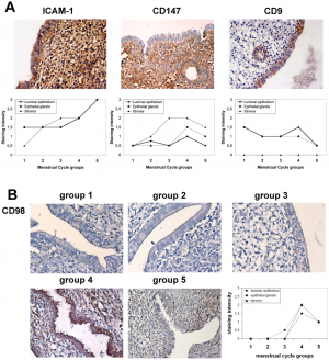
1) Upload an image from a journal source relating to fertilization or the first 2 weeks of development as demonstrated in the practical class. Including in the image “Summary” window: An image name as a section heading, Any further description of what the image shows, A subsection labeled “Reference” and under this the original image source, appropriate reference and all copyright information and finally a template indicating that this is a student image.
Image: Expression of Endometrial CD98 in implantation[1]
2) Identify a protein associated with the implantation process, including a brief description of the protein's role (1-2 paragraphs).
A study has identified trophinin as a protein important to the adhesion implantation process. It is believed to be a single intrinsic protein that spans the membrane due to hydrophobic tendencies. This molecule can adhere without the aid of calcium unlike many cell adhesion molecules. Trophinin molecules bind with other trophinin molecule in trans structure on the cell surface. Immunostaining showed that antigens specific to the trophinin molecule can be found in both trophoblast cells and in the maternal epithelium near implantation sites of the embryo. The protein has been found to be encoded in the short arm of the X chromosome. It is also present in the mouse, sheep and bovine, along with monotremes and marsupials. It appears that the binding of the trophectoderm (consists of trophoblasts and is the connection between the blastocyst and the maternal cells) is essential to invasion and proliferation of cells. In embryonic cells trophinin induces and promotes invasion and proliferation. In maternal cells the same protein promotes apoptosis (controlled cell death) so as to allow the acceptance of the embryo. Therefore it is a dual signalling molecule. [2]
Reference
Lab 3 Assessment
1) Identify the difference between "gestational age" and "post-fertilisation age" and explain why clinically "gestational age" is used in describing human development.
The gestational age refers to the time since the last normal menstruation period[1]. Whereas post-fertilisation age is calculated from the time of fertilization. There can be confusion between the terms espcially as gestational age is two weeks longer than post-fertilisation age[2]. Although in itself the term gestation age is confusing as there is no actual conceptus in until fertilisation but it is accepted by clinicians through widespread use[3]. As exact post-fetilisation age would be difficult to determine gestational age is used clinically. In assisted reproduction cases post-fertilisation age can be accurately determined but 2 weeks are generally added to age for ease of understanding[4].
2)Identify using histological descriptions at least 3 different types of tissues formed from somites
Somites form the dermis of the dorsal epithelium, skeletal muscles and some connective tissue, specifically, the vertebrae and ribs.[5]
References
- ↑ Moore, K.L., 2011 The Developing Human 9th ed. W.B. Saunders
- ↑ <pubmed>15520122,</pubmed>
- ↑ <pubmed>16006453</pubmed>
- ↑ <pubmed>15520122,</pubmed>
- ↑ Gilbert SF. Developmental Biology. 6th edition. Sunderland (MA): Sinauer Associates; 2000. Paraxial Mesoderm: The Somites and Their Derivatives. Available from: http://www.ncbi.nlm.nih.gov/books/NBK10085/
Lab 4 Assessment
1) Identify the 2 invasive prenatal diagnostic techniques related to the placenta and 2 abnormalities that can be identified with these techniques. Prenatal placental biopsy an invasive diagnostic technique for genetic abnormalities (such as trisomy 21) in the fetus. A karyotype is constructed allowing analysis of the chromosomes. It is used in the second and third trimester of pregnancy to confirm suspected malformations. Placental biopsies are sonographically guided[1]. Chorionic villus testing is another invasive technique carried out transcervically in the first trimester to detect inherited disorders such as haemophilia [2]
2) Identify a paper that uses cord stem cells therapeutically and write a brief (2-3 paragraph) description of the paper's findings.
Mesenchymal stem cells derived from the human umbilical cord have been used as a therapeutic treatment for neuromyelitis optica. Neuromyelitis optica is an autoimmune inflammatory disease that effects the optic nerve and spinal cord. Stem cells have been seen to provide differentiation potential to neural cells, secrete necessary factors and help regulate immunological function.
Five patients were treated with stem cell injections and then monitored for 18 months to analyse the effects both adverse and any improvements. Four out of the five patients gained some relief following treatment. Signs and symptoms decreased and the frequency of relapse was lessened. The neurological lesions also decreased in volume and severity as seen by MRI. The paper summarised that human umbilical cord stem cells were an appropriate therapy technique[3].
References
Lab 7 Assessment
1. (a) Provide a one sentence definition of a muscle satellite cell
Muscle satellite cells are progenitor cells and are involved in muscle growth and repair as they can induce regenerated muscle and additional satellite cells[1]
(b) In one paragraph, briefly discuss two examples of when satellite cells are activated.
A study investigating exercised induced satellite cell activation in skeletal muscle of growing and mature rats concluded that satellite cells are activated by acute sessions of prolonged eccentric exercise. It also concluded that exercise affected the proliferation of young mitotically active satellite cells[2]. Satellite cells are also activated when damage occurs. A study indicated that two variants of the IGF-I gene are necessary for activation of satellite cells. The study examined induced lesions to the anterior tibialis muscle of rats. The results showed that one variant of the gene which gives rise to a growth factor, MGF, is initially produced after injury and it activates satellite cells then IGF-IEa is expressed to maintain the repair process [3]
2. In one brief paragraph, describe what happens to skeletal muscle fibre type and size when the innervating motor nerve sustains long term damage such as in spinal cord injury.
In a study involving 12 human patients suffering from spinal cord injuries a section of the vastus lateralis muscle was biopsied at 3 intervals within the first 6month following injury. From 6-24 weeks after injury they showed 27-56% atrophy of Type I, IIa and IIax+IIx fibers. There was increased conversion between muscle types, type IIa decreased and type IIax+IIx increased. However there was little change in proportion of tpye I fibers during this period[4]
