User:Z3372817: Difference between revisions
No edit summary |
|||
| Line 225: | Line 225: | ||
==Lab Assessment 9== | ==Lab Assessment 9== | ||
Group 2 | |||
Group 3 | |||
*References are missing from the overview section. Although it serves as an introduction, you can still include references to support what you're saying. Also, maybe the language of this section should be edited to be a bit more formal, like the 2nd sentence in particular | |||
*GIT = Gastrointestinal tract, not track | |||
*The hyphens between "foregut" etc are not needed | |||
*The timeline is a good idea! Everything was simplified. Maybe look to see if you can add some images to this section | |||
*Week 6 of timeline: I don't think a liver can "obtain" a colour. Look to change the wording | |||
*Maybe to simplify the timeline section better, tabulate the findings according to time (weeks), rather than dividing it by the midgut, foregut and hind gut section. It makes it hard to follow | |||
*Need some more work on the recent findings section. Just some tips, when researching on pubmed, there's an option to look at recent articles by customising dates to say 2012-onwards | |||
*Many potentials for adding images to the "foregut" section. If you find that copyright is too difficult to get around, then you can sketch or trace images from textbooks and upload them | |||
*Great effort with the drawn images in the "midgut" section! Be wary of colour choice though, as the green highlighter and blue pen can be a bit difficult to see. Otherwise think of adjusting contrast on the images to make the diagram stand out more | |||
*Maybe think of adding a video from YouTube to show some features of GIT fetal development, like the rotations. If you do that, be sure to include the 11-digit cache code as your reference point | |||
Revision as of 18:15, 11 October 2014
Lab Attendance
Lab 1 --Z3372817 (talk) 12:45, 6 August 2014 (EST)
Lab 2 --Z3372817 (talk) 11:10, 13 August 2014 (EST)
Lab 3 --Z3372817 (talk) 11:13, 20 August 2014 (EST)
Lab 4 -- Absent
Lab 5 --Z3372817 (talk) 11:53, 3 September 2014 (EST)
Lab 6 --Z3372817 (talk) 11:01, 10 September 2014 (EST)
Lab 7 -- Absent
Lab 8 --Z3372817 (talk) 11:05, 24 September 2014 (EST)
Lab 9 --Z3372817 (talk) 11:28, 8 October 2014 (EST)
http://www.ncbi.nlm.nih.gov/pubmed
Lab Assessment 1
Research Article 1:
<pubmed>24992752</pubmed> This article tests the effect of traditional Chinese herbs on infertile women. The method employed was to conduct tests on 433 infertile women below the age of 42 and dividing the groups into test subjects, those who will be administered Chinese herbs, against the control. The groups were made up of 216 people and 217 in the respective groups. All subjects were given 1 out of 4 options of ultra-ovulation-promoting therapy to assist in the in-vitro fertilisation.
The main categories of measurement and the subsequent findings were:
| Category | Result |
|---|---|
| Endometrium thickness | Higher than control |
| Number of acquired eggs | No difference with control |
| Rates of normal fertility | Higher than control |
| High quality embryos | Higher than control |
| Biochemical and clinical pregnancy rate of subjects | Higher than control |
The findings showed an overall improved quality of fertility in these otherwise infertile women of the intervention group. The embryos also exhibited increased quality. This finding then suggested an improved success rate of IVF-embryo transplantation cycles and increased outcomes and safety of assisted reproductive technology.
Research Article 2:
<pubmed>23835722</pubmed> The effect of two different oxygen levels on embryo development was tested. The female gametes (oocyte) of 258 women were divided in a randomised study into 2 different groups; incubator of 5% oxygen concentration versus an incubator of 20% oxygen concentration. The purpose of the incubator is to ensure oxygen concentration is constant throughout the course of the experiment.
The matters of interest along with the clinical outcomes are as follows:
| Category | Result |
|---|---|
| Fertilisation | Same between groups |
| Cleavage | Same between groups |
| Embryo quality | Higher in 5% conc. group
(more blastomeres, more cycles of favourable embryos) |
| Blastocyst formation | Higher in 5% conc. group |
| Implantation | Higher in 5% conc. group |
| Pregnancy | Higher in 5% conc. group |
| Live birth rates | Higher in 5% conc. group |
The findings showed higher, greater quality embryos were seen in test subjects of the 5% oxygen concentration group. Smaller oxygen levels in incubation during embryo development was more favourable.
--Mark Hill Very good. (5/5)
Lab Assessment 2
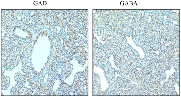
IHC image of mice fetal lung tissue showing the role of GAD and GABA in respiratory fetal development
Reference
Chintagari NR, Jin N, Gao L, Wang Y, Xi D, et al. (2010) Role of GABA Receptors in Fetal Lung Development in Rats. PLoS ONE 5(11): e14171. doi:10.1371/journal.pone.0014171 | PLoS One: Role of GABA Receptors in Fetal Lung Development in Rats
© 2010 Chintagari et al. This is an open-access article distributed under the terms of the Creative Commons Attribution License, which permits unrestricted use, distribution, and reproduction in any medium, provided the original author and source are credited.
--Mark Hill This is the correct reference link shown below. you do not need to include the student image template here (I have deleted), only with the uploaded file information. (4/5)
<pubmed>21152393</pubmed>
Lab Assessment 3
1. <pubmed>23431607</pubmed> Comparison between historical and current literature in regards to the development of the respiratory system
2. Developmental Biology, 6th edition By Scott F Gilbert. Swarthmore College Sunderland (MA): Sinauer Associates; 2000. ISBN-10: 0-87893-243-7
- Links: | Developmental Biology
Comparative embryology with detail on historical understandings of early respiratory development observed in various species. Accessible through PubMed.
3. Human Embryology and Morphology, 1902 By Arthur Keith London: Edward Arnold.
Historical images of past understandings on respiratory development
4. YouTube Video explaining early respiratory development
--Mark Hill I would have liked to have seen references initially not from textbooks or the current website, but from the research literature. (4/5)
Lab Assessment 4
(1) Paper on cord stem cells
<pubmed>23978163</pubmed>
The neurodevelopmental disorder of autism is poorly understood and therapy is currently dependent on the study of behaviour of the individuals in which the disorder manifests itself in. The utilisation of stem cells in treatment of autism is innovative, which this study outlines. The focus of the investigation is concerned with the combined transplantation of human cord blood mononuclear cells (CBMNCs) and umbilical cord-derived mesenchymal stem cells (UCMSCs) in treating children with autism.
The study does this through non-randomized, open-label, single center phase I/II trial investigations of 37 subjects diagnosed with autism. These subjects were then divided into three groups:
- Group 1 (14 subjects): received CBMNC transplantation and rehabilitation therapy
- Group 2 (9 subjects): received transplantation of both CBMNC and UCMSC as well as rehabilitation therapy
- Group 3 (14 subjects): received only rehabilitation therapy
Group 3 was used as the control for the trial.
Transplantations were performed by 4 separate stem cell infusion injections once a week. The Childhood Autism Rating Scale (CARS), Clinical Global Impression (CGI) scale and Aberrant Behavior Checklist (ABC) were used to comparatively assess between the therapeutic efficacy preceding and following treatment. Conclusions made to the study found that Group 2 combination treatment showed the greatest therapeutic effect for autism.
(2) Developmental vascular shunts
There are 3 development vascular shunts present in the embryo which later close postnatally. They are:
Foramen ovale: anatomical location is between the right and left atrium of the heart.
Ductus arteriosus: anatomical location between the descending aorta and the pulmonary artery of the heart.
Ductus venosus: anatomical location is within the liver and the veins in connection with it. The source of blood passing through this shunt is from the umbilical vein, which then drains into the IVC.
Lab Assessment 5
Causes for Meconium plug syndrome
This abnormality of gastrointestinal (GIT) development is characterised by the failure of the newborn to pass the meconium from its GIT system within 24-48 hours of being born. The aetiology is somewhat unclear, but there are a number of commonly associated factors that are related to the manifestation of this abnormality in neonates. These are:
(1) Prematurity: The condition is substantially prominent in premature neonates along with a variation of other factors [1]. The approximate incidence for its occurrence in newborns is estimated to range from 1 in every 500 to 1 in every 1,000 neonates.
(2) A thickened immobile meconium: The abnormality is of a transient nature where it is most commonly associated to the presence of a thickened and immobile meconium that obstructs the distal colon or rectum. The condition is somewhat alleviated when the infant passes the meconium plug, with normal bowel movements following this. Some newborns may require some form of rectal stimulation in order to relieve them from the plug obstructing the normal passage, such as the administration of saline enemas [2].
(3) Hirschsprung's disease: In a study conducted to determine the current significance of meconium plug syndrome, it was concluded that 13 per cent of patients who were found to have a meconium plug were followed up after the passing of the plug and later found to be diagnosed with Hirschsprung's disease [3].
(4) Location: As the abnormality is a benign condition, this means it is often restricted to the distal colon or rectum, unlike other plugs such as the ileal meconium plug [4].
(5) Colon aganglionosis: The loss of normal ganglion-cell content along the wall of the bowel has been found to be a factor that is also associated with the abnormality [5]
References
Lab Assessment 7
(1) Recent findings on pancreatic development
<pubmed>24265565</pubmed>
Congenital anomalies of the pancreas and pancreatic ducts may go undetected until adulthood and only discovered randomly upon unintentional discovery such as during surgery. Imaging is highly recommended for adults who experience persistent signs and symptoms of abdominal pain. This paper outlines two pioneering imaging technologies - MRCP and MDCT - which allows for early detection of ductal anatomic variants and congenital anomalies of the pancreas juxtaposed to normal pancreatic embryology. These techniques are a breakthrough in pancreatic-related pathology and diagnosis.
Magnetic resonance cholangiopancreaticography (MRCP) is increasing in its use as it can detect deviations from the norm in the anatomy of the biliary tree and pancreatic duct in a non-invasive manner. It identifies the course and drainage patterns of the ducts to diagnose developmental anomalies. Improvements in multidetected computed tomography (MDCT) technology allows scanning of the biliary tree and pancreas. It produces high resolution images that allow the identification of the optimum planes for viewing to be selected to provide more accurate results in diagnosis.
(2) Embryonic layers and tissues contributing to teeth development
- Epithelial/mesenchymal interactions are important during the course of teeth development:
- Ectoderm from the first overlying pharyngeal arch
- Neural crest cell contribution: NCCs have an inductive influence with the overlying ectoderm
- Ectomesenchymal cells
- Odontoblasts: Mesenchymal cells derived from NCCs which differentiate under the influence of enamel epithelium. It forms predentin which calcifies to form dentin
- Ameloblasts: Produce enamel which lead to teeth growth within the ossifying mandible (jaw)
Lab Assessment 8
(1) Time course of embryonic development human testis
image from the historic genital embryology section
Lab Assessment 9
Group 2
Group 3
- References are missing from the overview section. Although it serves as an introduction, you can still include references to support what you're saying. Also, maybe the language of this section should be edited to be a bit more formal, like the 2nd sentence in particular
- GIT = Gastrointestinal tract, not track
- The hyphens between "foregut" etc are not needed
- The timeline is a good idea! Everything was simplified. Maybe look to see if you can add some images to this section
- Week 6 of timeline: I don't think a liver can "obtain" a colour. Look to change the wording
- Maybe to simplify the timeline section better, tabulate the findings according to time (weeks), rather than dividing it by the midgut, foregut and hind gut section. It makes it hard to follow
- Need some more work on the recent findings section. Just some tips, when researching on pubmed, there's an option to look at recent articles by customising dates to say 2012-onwards
- Many potentials for adding images to the "foregut" section. If you find that copyright is too difficult to get around, then you can sketch or trace images from textbooks and upload them
- Great effort with the drawn images in the "midgut" section! Be wary of colour choice though, as the green highlighter and blue pen can be a bit difficult to see. Otherwise think of adjusting contrast on the images to make the diagram stand out more
- Maybe think of adding a video from YouTube to show some features of GIT fetal development, like the rotations. If you do that, be sure to include the 11-digit cache code as your reference point
