User:Z3333429
Lab Attendance
Lab 1: --Z3333429 (talk) 12:52, 6 August 2014 (EST)
http://www.ncbi.nlm.nih.gov/pubmed
Lab 2: --Z3333429 (talk) 11:03, 13 August 2014 (EST)
Lab 3: --Z3333429 (talk) 11:12, 20 August 2014 (EST)
Lab 4: --Z3333429 (talk) 11:09, 27 August 2014 (EST)
Lab 5: --Z3333429 (talk) 11:32, 3 September 2014 (EST)
Lab 1 Assessment
Article 1
Effect of vitamin D status on clinical pregnancy rates following in vitro fertilization[1]
Summary
Research was carried out to investigate the possible effect of vitamin D on human reproduction. The goal was to detect whether vitamin D levels Recent studies suggest that vitamin D may play a role in human reproduction. Our goal was to investigate whether there is a correlation between vitamin D levels and implantation and clinical pregnancy rates in infertile women following IVF.
Method
- Participants in this investigation were 173 women undergoing IVF at Mount Sinai Hospital, Toronto, Ontatrio.
- Serum 25(OH)D samples were collected within a week of oocyte retrieval from the patients.
- The vitamin D levels of the participants were measured according to serum 25-hydroxy-vitamin D (25[OH]D) levels
- Patients were classified in two categories according to serum levels of 25(OH)D: sufficient (≥ 75 nmol/L) or insufficient (< 75 nmol/L). Of the 173 patients, 54.9% presented with insufficient 25(OH)D levels and 45.1% has sufficient levels.
- A comparison was made between patient demographics and IVF cycle parameters between sufficient and insufficient groups.
- Clinical pregnancy, as identified by ultrasound following 4-5 weeks after embryo transfer; was the primary outcome measurement.
Findings
The research found that women who presented with sufficient 25(OH)D levels had significantly higher rates of clinical pregnancy per IVF cycle (52.5%) as compared to women with insufficient levels (34.7%). A higher rate of implantation was detected in the sufficient 25(OH)D group, however the results were not statistically significant. The research calls for further investigation the findings showed that vitamin D levels might be a predictor of clinical pregnancy and vitamin D supplementation could provide a simple and economical method of improving clinical pregnancy rates, not only in women undergoing IVF, but also across the board.
Reference
<pubmed>25077107</pubmed> Alcohol consumption and quality of embryos obtained in programmes of in vitro fertilization
Article 2
Alcohol consumption and quality of embryos obtained in programmes of in vitro fertilization[2]
Summary
Alcohol consumption has been identified as one of the main stimulants that negatively affect the reproductive systems of both sexes. An investigation was carried out to analyse the effect of alcohol consumption of female participants on the quality of embryos obtained through IVF programmes.
Method
- The study covered 54 women who received treatment due to infertility.
- Of the 54 women who participated, 42.59% consumed alcohol. Records were examined of the class of embryos (A, B and C) that each woman presented in during treatment.
- The database and statistical analyses were performed using computer software STATISTICA 7.1.
Findings
A statistically significant correlation was found between the occurrences of class B embryo in patients who consumed more than 25 grams of ethyl alcohol daily (72.72%). Women who consumed alcohol sporadically or those who abstained entirely from alcohol presented with 44.44% and 30% rates of class B embryos respectively. It was concluded that alcohol consumption (over 25 grams of ethyl alcohol) increases the likelihood of developing pooper quality embryos. More research should be carried out to investigate this further and it was suggested that active campaigns should be established to inform women of the negative affects of alcohol consumption on embryonic development.
Reference
<pubmed>24959808</pubmed> Effect of vitamin D status on clinical pregnancy rates following in vitro fertilization
References
Lab 2 Assessment

Reference
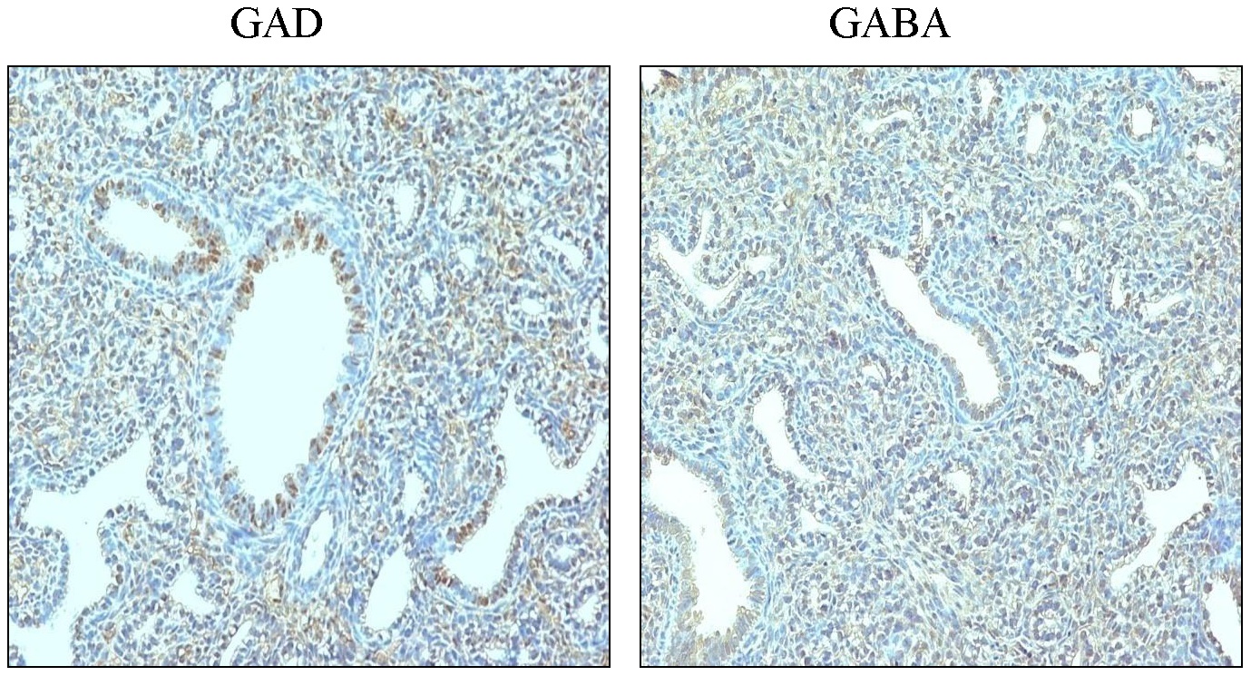
- ↑ Chintagari NR, Jin N, Gao L, Wang Y, Xi D, et al. (2010) Role of GABA Receptors in Fetal Lung Development in Rats. PLoS ONE 5(11): e14171. doi:10.1371/journal.pone.0014171
Lab 3 Assessment
<pubmed>22151899</pubmed> <pubmed>22214468</pubmed> <pubmed>12547712</pubmed>
Congenital Diaphragmatic Hernia
- Down-regulation of sonic hedgehog expression in pulmonary hypoplasia is associated with congenital diaphragmatic hernia.
- Computer simulation analysis of normal and abnormal development of the mammalian diaphragm.
- Outcomes of congenital diaphragmatic hernia: a population-based study in Western Australia.
- Congenital diaphragmatic hernia.
Laryngo-tracheo-oesophageal clefts
- Bronchopulmonary Dysplasia.
- Surfactant Metabolism Dysfunction and Childhood Interstitial Lung Disease (chILD).
- Evaluation of fetal vocal cords to select candidates for successful fetoscopic treatment of congenital high airway obstruction syndrome: preliminary case series.
- The epidemiology of meconium aspiration syndrome: incidence, risk factors, therapies, and outcome.
- Antenatal infection/inflammation and postnatal lung maturation and injury.
Lab 4 Assessment
Cord Stem Cell Therapeutics
Human umbilical cord blood-derived mesenchymal stem cell transplantation for the treatment of spinal cord injury[1]
Summary
The aim of this study was to find investigate if the transplantation of Human Umbilical Cord Blood Mesenchymal Stem Cells (HUCB-MSC’s) can offer an effective therapeutic treatment of Spinal Cord Injuries (SCI) and provide evidence for clinical applications. Recent studies have shown that there are possible treatments for SCI. These studies have shown that changing the local environment following after SCI (through transplantation of umbilical cord blood stem cells and other various cells and tissues) can aid in regenerating injured nerve axons and lead to functional restoration of SCI. In this study, HUCB-MSC’s were transplanted into rat models for the treatment of SCI and the therapeutic effects were evaluated through the observed behaviour and histological changes shown in the rats.
Method
- HUCB was retrieved from consenting donors from the Departments of Gynaecology and Obstetrics at the First and Third Affiliated Hospitals of Zhengzhou University and Zhengzhou People’s Hospital (Zhengzhou, China). The HUCB samples were screened against the hepatitis B virus.
- 46 adult female Wistar rats were used from the Experimental Animal Center of Henan (Zhengzhou, China). The rats were kept in a pathogen-free room at 25°C and humidity of 45% humidity and were 250-280g in weight.
- The Allen’s method (laminectomy of the spinous process and vertebral plates of T8-T10, exposing the dorsum of the spinal cord) was used to create the SCI rat models. After the exposing of the spinal cord at T-8-T10 a weight was dropped to simulate SCI and then the rats were separated into three groups: the injury group (received no treatment following injury), the control group (treated with saline) and the transplantation group (treated with HUCB-MSC suspension).
- The HUCB cells were isolated and screen for viability then cultured.
- The cultured HUCB-MSC’s were collected and diluted then the suspension was injected at the SCI site of the rat models. The same procedure was carried out on the control group using physiological saline.
- Following transplantation, locomotor ratings were obtained from the control and transplant groups at two and four weeks. Histological changes were observed via samples collected at week one and four. These samples were then underwent statistical analysis.
Findings
Following treatment, the transplantation group displayed recovery of spinal nerve function and immunohistochemistry identified that there was production of novel nerve cells at wee four. These findings suggest that transplanting HUCB-MSCs help the functional recovery of the damaged of spinal cord nerves in rats with SCI.
Reference
- ↑ <pubmed>24940417</pubmed>
Vascular Shunts
Foramen Ovale: located in the interatrial septum of the heart, allows blood to travel from the right atrium to the left atrium. Becomes fossa ovalis postnatally.
Ductus Venosus: located within the liver, becomes ligamentum venosum postnatally and allows blood from the umbilical vein to bypasses the liver and enter directly into the IVC.
Ductus Arteriosus: located within the aortich arch, allows blood to pass from the pulmonary artery into the descending aorta allowing blood from the right ventricle to bypass the non-functional lungs of the fetus. Postnatally it becomes the ligamentum arteriosum.