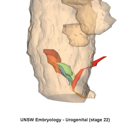
Urogenital System 3D stage 22 Movie
| Embryology - 23 Apr 2024 |
|---|
| Google Translate - select your language from the list shown below (this will open a new external page) |
|
العربية | català | 中文 | 中國傳統的 | français | Deutsche | עִברִית | हिंदी | bahasa Indonesia | italiano | 日本語 | 한국어 | မြန်မာ | Pilipino | Polskie | português | ਪੰਜਾਬੀ ਦੇ | Română | русский | Español | Swahili | Svensk | ไทย | Türkçe | اردو | ייִדיש | Tiếng Việt These external translations are automated and may not be accurate. (More? About Translations) |
| <mediaplayer width='503' height='480' image="http://embryology.med.unsw.edu.au/embryology/images/2/26/Stage22_URG3d.jpg">File:Stage22_URG3d.mp4</mediaplayer> |
|
Urinary System DevelopmentThe adult kidneys – the metanephroi – form from day 35, from a portion of the intermediate mesoderm called the metanephric blastema (or metanephric mesenchyme). They are induced to form by the ureteric buds, outgrowths from the end of the mesonephric ducts, which come into contact with the metanephric blastema. Upon contact, they begin to lengthen and bifurcate rapidly in the metanephric blastema – these branches differentiate into the collecting ducts. Both the ureteric buds and the metanephric blastema begin to differentiate; interestingly each induces differentiation in the other structure. The ureteric bud is induced by the metanephric blastema to form the collecting tubules, renal pelvis and ureters. The metanephric blastema is induced to form the nephrons. Development of the kidney is starts in week 5 and is completed by week 15. However, in week 6 the kidneys begin to ascend to their correct anatomical position. This movement is completed by week 9. During the ascent, the kidneys also become vascularised via the dorsal aorta. As this ascent occurs, the mesonephric ducts and the ureters enter the wall of the eventual bladder. Simultaneously, between weeks 4 and 6, the cloaca is partitioned into the urogenital sinus anteriorly, and the rectum posteriorly. This is achieved by downward growth of the urorectal septum, a portion of endoderm from the hindgut. The urogenital sinus has an area of enlargement – the bladder – and is superiorly continuous with the allantois. Further development of the urinary system varies depending on the sex of the embryo. In males, the pelvic urethra forms the membranous urethra, the prostatic urethra and penile urethra; in females it forms the membranous urethra and the vestibule of the vagina. The sex of the above model is male. Genital System DevelopmentTill the end of the 6th week the male and female genital systems are indistinguishable. Sex differentiation is based upon the presence of specific sex chromosomes. The female has two X chromosomes, while the male has an X and a Y chromosome. The specific gene involved in determining the male sex is the SRY transcription factor, which activates specific genes for male sex development. If SRY is damaged or absent, female development occurs. The development of the genital system occurs in 3 stages:
It should be noted that maturation of the genital system begins in the embryo but finishes in puberty.
|
|
|
|
|
|
|
These 3d movies were part of the UNSW Medical degree Independent Learning Project (ILP) prepared by Aashish Kumar (2006).
Glossary Links: A | B | C | D | E | F | G | H | I | J | K | L | M | N | O | P | Q | R | S | T | U | V | W | X | Y | Z | Numbers | Symbols | Movies
Cite this page: Hill, M.A. (2024, April 23) Embryology Urogenital System 3D stage 22 Movie. Retrieved from https://embryology.med.unsw.edu.au/embryology/index.php/Urogenital_System_3D_stage_22_Movie
- © Dr Mark Hill 2024, UNSW Embryology ISBN: 978 0 7334 2609 4 - UNSW CRICOS Provider Code No. 00098G