Ultrasound - Abnormal: Difference between revisions
mNo edit summary |
mNo edit summary |
||
| Line 5: | Line 5: | ||
The ultrasound technique can be used at any stage during pregnancy for embryo and placenta monitoring. Normal developmental ultrasound and features are listed on a separate page (More? [[Ultrasound]]) all content is for educational use only. | The ultrasound technique can be used at any stage during pregnancy for embryo and placenta monitoring. Normal developmental ultrasound and features are listed on a separate page (More? [[Ultrasound]]) all content is for educational use only. | ||
Some first trimester ({{GA}} 11-14 weeks) diagnostic measurements can include: | |||
* Trisomies - Nuchal translucency (NT), nasal bone length (NBL), prenasal thickness (PT) | |||
* Cardiovascular - tricuspid valve regurgitation (TR), "a"-wave pattern, DV PIV, S-wave (peak systolic velocity), D-wave (peak diastolic velocity), a-wave (atrial contraction in late diastole), time-averaged maximum velocity (TAMXV) | |||
{| | {| | ||
| [[File:Movie_help_01.jpg]] | | [[File:Movie_help_01.jpg]] | ||
| Line 22: | Line 27: | ||
{{Ultrasound Movies - abnormal}} | {{Ultrasound Movies - abnormal}} | ||
==Some Recent Findings== | ==Some Recent Findings== | ||
Latest revision as of 11:40, 15 April 2020
| Embryology - 24 Apr 2024 |
|---|
| Google Translate - select your language from the list shown below (this will open a new external page) |
|
العربية | català | 中文 | 中國傳統的 | français | Deutsche | עִברִית | हिंदी | bahasa Indonesia | italiano | 日本語 | 한국어 | မြန်မာ | Pilipino | Polskie | português | ਪੰਜਾਬੀ ਦੇ | Română | русский | Español | Swahili | Svensk | ไทย | Türkçe | اردو | ייִדיש | Tiếng Việt These external translations are automated and may not be accurate. (More? About Translations) |
Introduction
This page links to movies of abnormalities of human development as detected by ultrasound. Ultrasound movies or images are now commonly seen by parents in the first trimester as a clinical non-invasive early method of embryo staging (ageing) and early detection of abnormalities. Later in the second and third trimester ultrasound is used as a method for checking fetal growth and detection of developmental abnormalities. Ultrasound can also be used with other techniques to locate both embryo and placenta for other prenatal tests.
The ultrasound technique can be used at any stage during pregnancy for embryo and placenta monitoring. Normal developmental ultrasound and features are listed on a separate page (More? Ultrasound) all content is for educational use only.
Some first trimester (GA 11-14 weeks) diagnostic measurements can include:
- Trisomies - Nuchal translucency (NT), nasal bone length (NBL), prenasal thickness (PT)
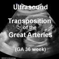
- Cardiovascular - tricuspid valve regurgitation (TR), "a"-wave pattern, DV PIV, S-wave (peak systolic velocity), D-wave (peak diastolic velocity), a-wave (atrial contraction in late diastole), time-averaged maximum velocity (TAMXV)

|
The ultrasound movies can be viewed in two ways:
|
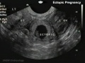
Ultrasound - Abnormalities
| Abnormal Ultrasound | |||||||||||||||
|---|---|---|---|---|---|---|---|---|---|---|---|---|---|---|---|
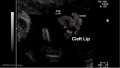
| Uterus | Cleft Lip | ||||||||||||||
|
|
|
| ||||||||||||
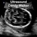
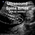
| Gastrointestinal | Neural | ||||||||||||||
|
|
|
| ||||||||||||
| Cardiac | |||||||||||||||
|
|
| |||||||||||||
| Placenta | |||||||||||||||
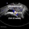
|
|
| |||||||||||||
| Ultrasound: normal movies | abnormal movies | all ultrasound movies | Movies | |||||||||||||||


Some Recent Findings
|
References
Glossary Links
- Glossary: A | B | C | D | E | F | G | H | I | J | K | L | M | N | O | P | Q | R | S | T | U | V | W | X | Y | Z | Numbers | Symbols | Term Link
Cite this page: Hill, M.A. (2024, April 24) Embryology Ultrasound - Abnormal. Retrieved from https://embryology.med.unsw.edu.au/embryology/index.php/Ultrasound_-_Abnormal
- © Dr Mark Hill 2024, UNSW Embryology ISBN: 978 0 7334 2609 4 - UNSW CRICOS Provider Code No. 00098G