Testis Development Movie: Difference between revisions
(Created page with "{{Movie header}} {| | width=380px|<mediaplayer width='350' height='650' image="http://embryology.med.unsw.edu.au/embryology/images/f/fe/Urogenital_sinus_001_icon.jpg">File:Testis 001...") |
No edit summary |
||
| Line 1: | Line 1: | ||
{{Movie header}} | {{Movie header}} | ||
{| | {| | ||
| width=380px|<mediaplayer width='350' height='650' image="http://embryology.med.unsw.edu.au/embryology/images/f/ | | width=380px|<mediaplayer width='350' height='650' image="http://embryology.med.unsw.edu.au/embryology/images/f/fa/Gonad-icon.jpg">File:Testis 001.mp4</mediaplayer> | ||
| valign="top" |[[File:Gonad-icon.jpg|100px|right]] | | valign="top" |[[File:Gonad-icon.jpg|100px|right]] | ||
Revision as of 15:23, 6 March 2013
| Embryology - 24 Apr 2024 |
|---|
| Google Translate - select your language from the list shown below (this will open a new external page) |
|
العربية | català | 中文 | 中國傳統的 | français | Deutsche | עִברִית | हिंदी | bahasa Indonesia | italiano | 日本語 | 한국어 | မြန်မာ | Pilipino | Polskie | português | ਪੰਜਾਬੀ ਦੇ | Română | русский | Español | Swahili | Svensk | ไทย | Türkçe | اردو | ייִדיש | Tiếng Việt These external translations are automated and may not be accurate. (More? About Translations) |
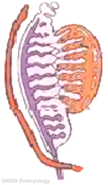
| <mediaplayer width='350' height='650' image="http://embryology.med.unsw.edu.au/embryology/images/f/fa/Gonad-icon.jpg">File:Testis 001.mp4</mediaplayer> | Testis and Ductus Deferens Development This animation shows the process of early testis and ductus deferens (vas deferens) development.
Embryonically, both gonads (testis and ovary) and internal genital tracts (ductus deferens and uterus) begin with the same basic elements that have divergent pathways under the initial influence of the sex chromosomes (Y and X).
|