Template:Third Trimester Timeline: Difference between revisions
From Embryology
mNo edit summary |
mNo edit summary |
||
| (8 intermediate revisions by the same user not shown) | |||
| Line 1: | Line 1: | ||
{| | {| | ||
|+ colspan=3|'''Third Trimester Timeline''' | |||
|- | |- | ||
| colspan=3 bgcolor="F5FFFA"|'''Links:''' {{human timeline}} | {{first trimester timeline}} | {{second trimester timeline}} | {{third trimester timeline}} | |||
| colspan= | |-bgcolor="CEDFF2" | ||
|-bgcolor=" | |||
| <center>'''Week'''</center> | | <center>'''Week'''</center> | ||
| <center>'''Stage'''</center> | | <center>'''Stage'''</center> | ||
| '''Event''' | | '''Event''' | ||
|- | |- | ||
| | | | ||
| Clinical third trimester | | Clinical third trimester | ||
| [[File:Fetal_size_change.jpg|90px|link=Fetal Development]] | | [[File:Fetal_size_change.jpg|90px|link=Fetal Development]] {{hearing}} 3rd Trimester - vibration acoustically of maternal abdominal wall induces startle respone in fetus. | ||
|-bgcolor=" | |-bgcolor="F5FAFF" | ||
| <center>27</center> | | <center>27</center> | ||
| | | | ||
| Line 22: | Line 20: | ||
| <center>28</center> | | <center>28</center> | ||
| | | | ||
| | | {{respiratory}} Month 7 - respiratory bronchioles proliferate and end in alveolar ducts and sacs | ||
|-bgcolor=" | |-bgcolor="F5FAFF" | ||
| <center>29</center> | | <center>29</center> | ||
| | | | ||
| | | | ||
{{tooth}} Week 29 - Permanent premolars (correspond to the milk molars) appear. | |||
|- | |- | ||
| <center>30</center> | | <center>30</center> | ||
| Line 36: | Line 34: | ||
[[Genital_System_Development|Genital]] male gonad (testes) descending | [[Genital_System_Development|Genital]] male gonad (testes) descending | ||
|-bgcolor=" | |-bgcolor="F5FAFF" | ||
| <center>31</center> | | <center>31</center> | ||
| | | | ||
| Line 44: | Line 42: | ||
| <center>32</center> | | <center>32</center> | ||
| | | | ||
| | | {{nail}} fingernails reach digit tip | ||
|-bgcolor=" | |-bgcolor="F5FAFF" | ||
| <center>33</center> | | <center>33</center> | ||
| | | | ||
| | | {{neural}} brain cortical sulcation - primary sulci present{{#pmid:11158907|PMID11158907}} | ||
|- | |- | ||
| <center>34</center> | | <center>34</center> | ||
| | | | ||
| | | {{neural}} brain cortical sulcation - insular, cingular, and occipital secondary sulci present{{#pmid:11158907|PMID11158907}} | ||
|-bgcolor=" | |-bgcolor="F5FAFF" | ||
| <center>35</center> | | <center>35</center> | ||
| | | | ||
| Line 66: | Line 64: | ||
| [[File:Frazer006_bw600.jpg|90px|link=Fetal Development]] [[Integumentary_System_-_Nail_Development|Nail Development]] toenails reach digit tip | | [[File:Frazer006_bw600.jpg|90px|link=Fetal Development]] [[Integumentary_System_-_Nail_Development|Nail Development]] toenails reach digit tip | ||
[[Vision_-_Lens_Development|Lens Development]] - lens growth and interocular distance plateaus after 36 weeks of gestation | [[Vision_-_Lens_Development|Lens Development]] - lens growth and interocular distance plateaus after 36 weeks of gestation{{#pmid:19541779|PMID19541779}} | ||
|-bgcolor=" | |-bgcolor="F5FAFF" | ||
| <center>37</center> | | <center>37</center> | ||
| | | | ||
| Line 80: | Line 78: | ||
[[Cardiovascular System Development|Heart]] pressure difference closes foramen ovale leaving a fossa ovalis | [[Cardiovascular System Development|Heart]] pressure difference closes foramen ovale leaving a fossa ovalis | ||
{{thyroid}} TSH levels increase, thyroxine (T3) and T4 levels increase to 24 h, then 5-7 days postnatal decline to normal levels | |||
{{adrenal}} - zona glomerulosa, zona fasiculata present | |||
|- | |- | ||
|}<noinclude>[[Category:Template]][[Category:Timeline]][[Category: | | | ||
| colspan=2| | |||
{| class="wikitable mw-collapsible mw-collapsed" | |||
! Systems | |||
|- | |||
| '''Systems:''' {{bone timeline}} | {{eye neural crest timeline}} | {{heart abnormality timeline}} | {{hearing EAM timeline}} | {{muscle timeline}} | {{ovary timeline}} | {{placental villi timeline}} | {{shoulder timeline}} | {{smell timeline}} | {{spleen timeline}} | {{ventricular timeline}} | |||
|} | |||
|}<noinclude>[[Category:Template]][[Category:Timeline]][[Category:Third Trimester]][[Category:Fetal]]</noinclude> | |||
Latest revision as of 12:37, 12 May 2018
| Links: human timeline | first trimester timeline | second trimester timeline | third trimester timeline | ||
| Event | ||
| Clinical third trimester | 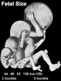 hearing 3rd Trimester - vibration acoustically of maternal abdominal wall induces startle respone in fetus. hearing 3rd Trimester - vibration acoustically of maternal abdominal wall induces startle respone in fetus.
| |
| respiratory Month 7 - respiratory bronchioles proliferate and end in alveolar ducts and sacs | ||
|
tooth Week 29 - Permanent premolars (correspond to the milk molars) appear. | ||
|
Genital male gonad (testes) descending | ||
| nail fingernails reach digit tip | ||
| neural brain cortical sulcation - primary sulci present[1] | ||
| neural brain cortical sulcation - insular, cingular, and occipital secondary sulci present[1] | ||
 Nail Development toenails reach digit tip Nail Development toenails reach digit tip
Lens Development - lens growth and interocular distance plateaus after 36 weeks of gestation[2] | ||
| Birth |  Clinical Week 40 Clinical Week 40
Heart pressure difference closes foramen ovale leaving a fossa ovalis thyroid TSH levels increase, thyroxine (T3) and T4 levels increase to 24 h, then 5-7 days postnatal decline to normal levels adrenal - zona glomerulosa, zona fasiculata present | |
- ↑ 1.0 1.1 Garel C, Chantrel E, Brisse H, Elmaleh M, Luton D, Oury JF, Sebag G & Hassan M. (2001). Fetal cerebral cortex: normal gestational landmarks identified using prenatal MR imaging. AJNR Am J Neuroradiol , 22, 184-9. PMID: 11158907
- ↑ Paquette LB, Jackson HA, Tavaré CJ, Miller DA & Panigrahy A. (2009). In utero eye development documented by fetal MR imaging. AJNR Am J Neuroradiol , 30, 1787-91. PMID: 19541779 DOI.