Template:Second Trimester table01
| Event | ||
| Fertilisation Age FA | Gestational Age GA | |
| 14 |  Week 12 - CRL 85 mm, femur length 15 mm, biparietal diameter 25 mm Week 12 - CRL 85 mm, femur length 15 mm, biparietal diameter 25 mm
Hearing Week 12-16 - Capsule adjacent to membranous labrynth undegoes vacuolization to form a cavity (perilymphatic space) around membranous labrynth and fills with perilymph Genital male and female external genital differences observable Respiratory Month 3-6 - lungs appear glandular, end month 6 alveolar cells type 2 appear and begin to secrete surfactant Tongue Week 12 - first differentiated epithelial cells (Type II and III) Genital female genital canal (80 days) formed with absorption of the median septum | |
| 15 | Tongue Week 12 to 13 - maximum synapses between cells and afferent nerve fibers
Hearing - Outer Ear Development Week 13 - Meatal plug disc-like, innermost surface in contact with the primordial malleus, contributes to the formation of the tympanic membrane. | |
| 16 | Tongue Week 14 to 15 - taste pores develop, mucous
Ovary Development 100 days - primary follicles present Nail Development toenails appear Head Development facial skeleton remodelling begins | |
| 17 | Pancreas glucagon detectable in fetal plasma.
Spleen Week 15 -alpha-smooth muscle actin (alpha-SMA)-positive reticulum cells scattered around the arterioles. [1] | |
| 18 | 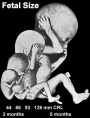 Hearing Week 16-24 - Centres of ossification appear in remaining cartilage of otic capsule form petrous portion of temporal bone. Continues to ossify to form mastoid process of temporal bone. Hearing Week 16-24 - Centres of ossification appear in remaining cartilage of otic capsule form petrous portion of temporal bone. Continues to ossify to form mastoid process of temporal bone.
Pituitary adenohypophysis fully differentiated Respiratory Week 16 to 25 lung histology - canalicular Hearing - Outer Ear Development Week 16.5 - External auditory meatus is fully patent throughout its length, lumen is still narrow and curved. Skin 4 months - basal cell- proliferation generates folds in basement membrane; neural crest cells- (melanocytes) migrate into epithelium; embryonic connective tissue- differentiates into dermis, a loose ct layer over a dense ct layer. Beneath the dense ct layer is another loose ct layer that will form the subcutaneous layer. Ectoderm contributes to nails, hair follictles and glands. Nails form as thickening of ectoderm epidermis at the tips of fingers and toes. These form germinative cells of nail field. Cords of these cells extend into mesoderm forming epithelial columns. These form hair follocles, sebaceous and sweat glands. primary follicles begin to form in the ovary and are characterized by an oocyte glandular urethra forms and skin folds present | |
| 19 |  Neural - Brain development histology week 17 Neural - Brain development histology week 17
| |
| 20 |  Tongue Week 18 - substance P detected in dermal papillae, not in taste bud primordia Tongue Week 18 - substance P detected in dermal papillae, not in taste bud primordia
Skin vernix caseosa covers skin Spleen Week 18 - alpha-SMA-positive reticulum cells increase in number and began to form a reticular framework. An accumulation of T and B lymphocytes occurred within the framework, and a primitive white pulp was observed around the arterioles. [1] Hearing - Outer Ear Development week 18 - External auditory meatus is already fully expanded to its complete form. | |
| 21 | ||
| 22 | Pituitary week 20 to 24 growth hormone levels peak, then decline
Skin lanugo, skin hair Skin 5 months - Hair growth initiated at base of cord, lateral outgrowths form associated sebaceous glands; Other cords elongate and coil to form sweat glands; Cords in mammary region branch as they elongate to form mammary glands. | |
| 23 | ||
| 24 |  Neural brain cortical sulcation - sylvian fissure, interhemispheric fissure, callosal sulcus, parietooccipital fissure, and hippocampic fissures present[2] Neural brain cortical sulcation - sylvian fissure, interhemispheric fissure, callosal sulcus, parietooccipital fissure, and hippocampic fissures present[2]
Spleen - Week 22 - antigenic diversity of the reticular framework was observed, and T and B lymphocytes were segregated in the framework. T lymphocytes were sorted into the alpha-smooth muscle actin-positive reticular framework, and the periarteriolar lymphoid sheath (PALS) was formed around the arteriole. B lymphocytes aggregated in eccentric portions to the PALS and formed the lymph follicle (LF). The reticular framework of the LF was alpha-SMA-negative. [1] | |
| 25 | ||
| 26 | Respiratory Week 24 to 40 lung histology - terminal sac
Spleen Week 24 - marginal zone appeared in the alpha-smooth muscle actin-positive reticular framework around the white pulp.[1] Earliest potential survival expected if born ovarian follicles can consist of growing oocytes surrounded by several layers of granulosa cells | |
| 27 | Respiratory end month 6 alveolar cells type 2 appear and begin to secrete surfactant | |