Template:Insert timeline: Difference between revisions
No edit summary |
No edit summary |
||
| Line 1: | Line 1: | ||
<gallery caption="Development of the Mouse Embryo" widths="175px" heights="175px" perrow="5"> | <gallery caption="Development of the Mouse Embryo" widths="175px" heights="175px" perrow="5"> | ||
File: Day 0 | File:Day 0 one cell stage.jpg|'''Day 0''' One Cell Egg | ||
File:Day 1 dividing egg.JPG|Day 1 Dividing Egg. Cleavage of cells has begun. The Day ends with 4 cells present. | File:Day 1 dividing egg.JPG|'''Day 1''' Dividing Egg. Cleavage of cells has begun. The Day ends with 4 cells present. | ||
File:Day 2 morula.JPG|Day 2 Morula | File:Day 2 morula.JPG|'''Day 2''' Morula | ||
File:Day 3 Advanced Segmentation.JPG|Day 3 Advanced segmentation of the Morula. 16-25 cells within same sized morula. | File:Day 3 Advanced Segmentation.JPG|'''Day 3''' Advanced segmentation of the Morula. 16-25 cells within same sized morula. | ||
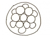
File:Day 4 Blastocyst.JPG|Day 4 Blastocyst formation | File:Day 4 Blastocyst.JPG|'''Day 4''' Blastocyst formation | ||
File:Day 4.5 Implantation.JPG|Day 4.5 Implantation and Invasion of the blastocyst into the uterine epithelium | File:Day 4.5 Implantation.JPG|'''Day 4.5''' Implantation and Invasion of the blastocyst into the uterine epithelium | ||
File:Day 5 Formation of Egg Cylinder.JPG|Day 5 Formation of the egg cylinder | File:Day 5 Formation of Egg Cylinder.JPG|'''Day 5''' Formation of the egg cylinder | ||
File:Day 6 Differentiation of Egg Cylinder.JPG|Day 6 Differentiation of the egg cylinder | File:Day 6 Differentiation of Egg Cylinder.JPG|'''Day 6''' Differentiation of the egg cylinder | ||
File:Day 6.5 Advanced Endometrial Reaction.JPG|Day 6.5 Gastrulation starts during the period of Advanced Endometrial Reaction | File:Day 6.5 Advanced Endometrial Reaction.JPG|'''Day 6.5''' Gastrulation starts during the period of Advanced Endometrial Reaction | ||
File:Day 7 Amnion.JPG|Day 7 Amnion formation | File:Day 7 Amnion.JPG|'''Day 7''' Amnion formation | ||
File:Day 7.5 Neural plate, presomite stage.JPG|Day 7.5 Neural plate and presomite stage | File:Day 7.5 Neural plate, presomite stage.JPG|'''Day 7.5''' Neural plate and presomite stage | ||
File:Day 8 First Somites.JPG|Day 8 Formation of the first seven somites. | File:Day 8 First Somites.JPG|'''Day 8''' Formation of the first seven somites. | ||
File:Day 8.5 Turning of embryo.JPG|Day 8.5 Rotation of the embryo. The embryo now has between 8 and 12 somites. | File:Day 8.5 Turning of embryo.JPG|'''Day 8.5''' Rotation of the embryo. The embryo now has between 8 and 12 somites. | ||
File:Day 9 Formation and closure of anterior neuropore.JPG|Day 9 Formation and closure of the anterior neuropore. There are 13 - 20 somites present in the embryo | File:Day 9 Formation and closure of anterior neuropore.JPG|'''Day 9''' Formation and closure of the anterior neuropore. There are 13 - 20 somites present in the embryo | ||
File:Day 9.5 Formation of posterior neuropore and forelimb bud.JPG|Day 9.5 Formation of the posterior neuropore and forelimb bud. There are 21 to 29 somites present and the embryo measures 1.8 - 3.3 mm in length. | File:Day 9.5 Formation of posterior neuropore and forelimb bud.JPG|'''Day 9.5''' Formation of the posterior neuropore and forelimb bud. There are 21 to 29 somites present and the embryo measures 1.8 - 3.3 mm in length. | ||
file:Day 10 Closure of posterior neuropore, hind limb bud and tail bud.JPG|Day 10 The posterior neuropore closes and formation of the hind limb bud and tail bud occurs. there are 30 - 34 somites present and the embryo is 3.1 - 3.9mm | file:Day 10 Closure of posterior neuropore, hind limb bud and tail bud.JPG|'''Day 10''' The posterior neuropore closes and formation of the hind limb bud and tail bud occurs. there are 30 - 34 somites present and the embryo is 3.1 - 3.9mm | ||
File:Day 10.5 Deep Lens Indentation.JPG|Day 10.5 Deep eye lens indentation occurs. There are 35-30 somites present and the embryo is now 3.5-4.9mm. | File:Day 10.5 Deep Lens Indentation.JPG|'''Day 10.5''' Deep eye lens indentation occurs. There are 35-30 somites present and the embryo is now 3.5-4.9mm. | ||
File:Day 11 Closure of lens vesicle.JPG|Day 11 Closure of the lens vesicle. There are 40-44 somites present and the embryo has reached 5-6mm in length. | File:Day 11 Closure of lens vesicle.JPG|'''Day 11''' Closure of the lens vesicle. There are 40-44 somites present and the embryo has reached 5-6mm in length. | ||
File:Day 11.5 Lens Vesicle completely separated from surface.JPG|Day 11.5 The lens vesicle is completely separated from the surface epithelium. There are currently 45 - 47 somites and the embryo reaches a length of 6 - 7mm | File:Day 11.5 Lens Vesicle completely separated from surface.JPG|'''Day 11.5''' The lens vesicle is completely separated from the surface epithelium. There are currently 45 - 47 somites and the embryo reaches a length of 6 - 7mm | ||
File:Day 12 Earliest signs of fingers.JPG|Day 12 First signs of fingers. The embryo is 7 - 9mm in length from crown to rump and there are 48 - 51 somites. | File:Day 12 Earliest signs of fingers.JPG|'''Day 12''' First signs of fingers. The embryo is 7 - 9mm in length from crown to rump and there are 48 - 51 somites. | ||
File:Day 13 Anterior Footplate Indented, marked pinna.JPG|Day 13 The anterior footplate becomes indented and the external ear structure is more defined. The embryo is now 9 - 11mm in length and has 52 - 55 somites. | File:Day 13 Anterior Footplate Indented, marked pinna.JPG|'''Day 13''' The anterior footplate becomes indented and the external ear structure is more defined. The embryo is now 9 - 11mm in length and has 52 - 55 somites. | ||
File:Day 14 Fingers Separate.JPG|Day 14 The fingers separate. The embryo has 56 - 66 somites and is 11-12mm in length. | File:Day 14 Fingers Separate.JPG|'''Day 14''' The fingers separate. The embryo has 56 - 66 somites and is 11-12mm in length. | ||
File:Day 15 toes separate.JPG|Day 15 The toes separate. At this stage the embryo is 12 - 14.4m from crown to rump | File:Day 15 toes separate.JPG|'''Day 15''' The toes separate. At this stage the embryo is 12 - 14.4m from crown to rump | ||
File:Day 16 Reposition of umbilical hernia.JPG|Day 16 The umbilical hernia is repositioned. The fetus now measures between 14 and 17 mm. | File:Day 16 Reposition of umbilical hernia.JPG|'''Day 16''' The umbilical hernia is repositioned. The fetus now measures between 14 and 17 mm. | ||
File:Day 17 fingers and toes joined together.JPG| | File:Day 17 fingers and toes joined together.JPG|'''Day 17''' The fingers and toes become joined together. The skin is wrinkled and thickened and the umbilical hernia has disappeared. The fetus measures 16.5 - 20mm depending on the degree of curvature. | ||
File:Day 18 long whiskers.JPG|Day 18 Whiskers elongate. The length of the embryo varies between 18 and 23 mm due to the degree of curvature of the fetus. | File:Day 18 long whiskers.JPG|'''Day 18''' Whiskers elongate. The length of the embryo varies between 18 and 23 mm due to the degree of curvature of the fetus. | ||
File:Day 19 Newborn Mouse.JPG|Day 19 The newborn mouse is between 23 and 27mm depending on the degree of flexure of the body axis | File:Day 19 Newborn Mouse.JPG|'''Day 19''' The newborn mouse is between 23 and 27mm depending on the degree of flexure of the body axis | ||
</gallery> | </gallery> | ||
Latest revision as of 18:32, 14 October 2009
- Development of the Mouse Embryo
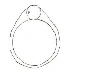
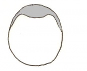
Day 0 One Cell Egg
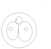
Day 1 Dividing Egg. Cleavage of cells has begun. The Day ends with 4 cells present.
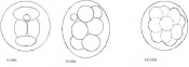
Day 2 Morula
Day 3 Advanced segmentation of the Morula. 16-25 cells within same sized morula.

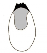
Day 4 Blastocyst formation
Day 4.5 Implantation and Invasion of the blastocyst into the uterine epithelium
Day 5 Formation of the egg cylinder
Day 6 Differentiation of the egg cylinder
Day 6.5 Gastrulation starts during the period of Advanced Endometrial Reaction
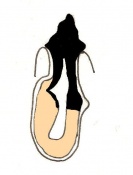
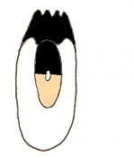
Day 7 Amnion formation
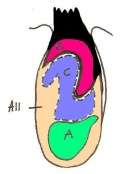
Day 7.5 Neural plate and presomite stage
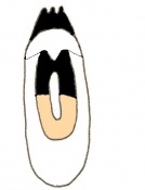
Day 8 Formation of the first seven somites.
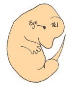
Day 8.5 Rotation of the embryo. The embryo now has between 8 and 12 somites.
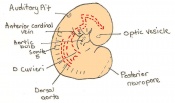
Day 9 Formation and closure of the anterior neuropore. There are 13 - 20 somites present in the embryo
Day 9.5 Formation of the posterior neuropore and forelimb bud. There are 21 to 29 somites present and the embryo measures 1.8 - 3.3 mm in length.
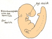
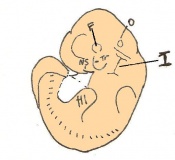
Day 10 The posterior neuropore closes and formation of the hind limb bud and tail bud occurs. there are 30 - 34 somites present and the embryo is 3.1 - 3.9mm
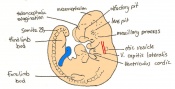
Day 10.5 Deep eye lens indentation occurs. There are 35-30 somites present and the embryo is now 3.5-4.9mm.
Day 11 Closure of the lens vesicle. There are 40-44 somites present and the embryo has reached 5-6mm in length.
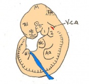
Day 11.5 The lens vesicle is completely separated from the surface epithelium. There are currently 45 - 47 somites and the embryo reaches a length of 6 - 7mm
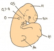
Day 12 First signs of fingers. The embryo is 7 - 9mm in length from crown to rump and there are 48 - 51 somites.
Day 13 The anterior footplate becomes indented and the external ear structure is more defined. The embryo is now 9 - 11mm in length and has 52 - 55 somites.
Day 14 The fingers separate. The embryo has 56 - 66 somites and is 11-12mm in length.
Day 15 The toes separate. At this stage the embryo is 12 - 14.4m from crown to rump
Day 16 The umbilical hernia is repositioned. The fetus now measures between 14 and 17 mm.
Day 17 The fingers and toes become joined together. The skin is wrinkled and thickened and the umbilical hernia has disappeared. The fetus measures 16.5 - 20mm depending on the degree of curvature.
Day 18 Whiskers elongate. The length of the embryo varies between 18 and 23 mm due to the degree of curvature of the fetus.
Day 19 The newborn mouse is between 23 and 27mm depending on the degree of flexure of the body axis


