Template:Insert timeline: Difference between revisions
From Embryology
No edit summary |
No edit summary |
||
| (6 intermediate revisions by the same user not shown) | |||
| Line 1: | Line 1: | ||
<gallery caption="Development of the Mouse Embryo" widths=" | <gallery caption="Development of the Mouse Embryo" widths="175px" heights="175px" perrow="5"> | ||
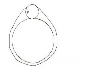
File:Day 0 one cell stage.jpg|Day 0 | File:Day 0 one cell stage.jpg|'''Day 0''' One Cell Egg | ||
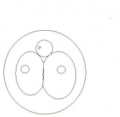
File:Day 1 dividing egg.JPG|Day 1 | File:Day 1 dividing egg.JPG|'''Day 1''' Dividing Egg. Cleavage of cells has begun. The Day ends with 4 cells present. | ||
File:Day 2 morula.JPG|Day 2 | File:Day 2 morula.JPG|'''Day 2''' Morula | ||
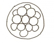
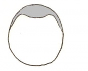
File:Day 3 Advanced Segmentation.JPG|Day 3 | File:Day 3 Advanced Segmentation.JPG|'''Day 3''' Advanced segmentation of the Morula. 16-25 cells within same sized morula. | ||
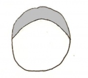
File:Day 4 Blastocyst.JPG|Day 4 | File:Day 4 Blastocyst.JPG|'''Day 4''' Blastocyst formation | ||
File:Day 4.5 Implantation.JPG|Day 4.5 | File:Day 4.5 Implantation.JPG|'''Day 4.5''' Implantation and Invasion of the blastocyst into the uterine epithelium | ||
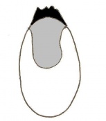
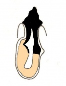
File:Day 5 Formation of Egg Cylinder.JPG|Day 5 | File:Day 5 Formation of Egg Cylinder.JPG|'''Day 5''' Formation of the egg cylinder | ||
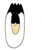
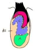
File:Day 6 Differentiation of Egg Cylinder.JPG|Day 6 | File:Day 6 Differentiation of Egg Cylinder.JPG|'''Day 6''' Differentiation of the egg cylinder | ||
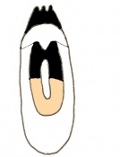
File:Day 6.5 Advanced Endometrial Reaction.JPG|Day 6.5 | File:Day 6.5 Advanced Endometrial Reaction.JPG|'''Day 6.5''' Gastrulation starts during the period of Advanced Endometrial Reaction | ||
File:Day 7 Amnion.JPG|Day 7 | File:Day 7 Amnion.JPG|'''Day 7''' Amnion formation | ||
File:Day 7.5 Neural plate, presomite stage.JPG|Day 7.5 | File:Day 7.5 Neural plate, presomite stage.JPG|'''Day 7.5''' Neural plate and presomite stage | ||
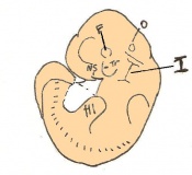
File:Day 8 First Somites.JPG|Day 8 | File:Day 8 First Somites.JPG|'''Day 8''' Formation of the first seven somites. | ||
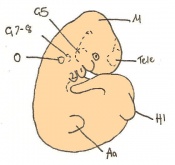
File:Day 8.5 Turning of embryo.JPG|Day 8.5 | File:Day 8.5 Turning of embryo.JPG|'''Day 8.5''' Rotation of the embryo. The embryo now has between 8 and 12 somites. | ||
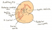
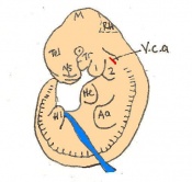
File:Day 9 Formation and closure of anterior neuropore.JPG|Day 9 | File:Day 9 Formation and closure of anterior neuropore.JPG|'''Day 9''' Formation and closure of the anterior neuropore. There are 13 - 20 somites present in the embryo | ||
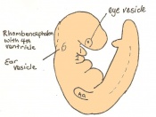
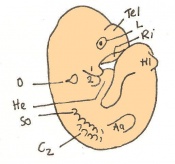
File:Day 9.5 Formation of posterior neuropore and forelimb bud.JPG|Day 9.5 | File:Day 9.5 Formation of posterior neuropore and forelimb bud.JPG|'''Day 9.5''' Formation of the posterior neuropore and forelimb bud. There are 21 to 29 somites present and the embryo measures 1.8 - 3.3 mm in length. | ||
File:Day 10.5 Deep Lens Indentation.JPG|Day 10.5 | file:Day 10 Closure of posterior neuropore, hind limb bud and tail bud.JPG|'''Day 10''' The posterior neuropore closes and formation of the hind limb bud and tail bud occurs. there are 30 - 34 somites present and the embryo is 3.1 - 3.9mm | ||
File:Day 11 Closure of lens vesicle.JPG|Day 11 | File:Day 10.5 Deep Lens Indentation.JPG|'''Day 10.5''' Deep eye lens indentation occurs. There are 35-30 somites present and the embryo is now 3.5-4.9mm. | ||
File:Day 11.5 Lens Vesicle completely separated from surface.JPG|Day 11.5 | File:Day 11 Closure of lens vesicle.JPG|'''Day 11''' Closure of the lens vesicle. There are 40-44 somites present and the embryo has reached 5-6mm in length. | ||
File:Day 12 Earliest signs of fingers.JPG|Day 12 | File:Day 11.5 Lens Vesicle completely separated from surface.JPG|'''Day 11.5''' The lens vesicle is completely separated from the surface epithelium. There are currently 45 - 47 somites and the embryo reaches a length of 6 - 7mm | ||
File:Day 13 Anterior Footplate Indented, marked pinna.JPG|Day 13 | File:Day 12 Earliest signs of fingers.JPG|'''Day 12''' First signs of fingers. The embryo is 7 - 9mm in length from crown to rump and there are 48 - 51 somites. | ||
File:Day 14 Fingers Separate.JPG|Day 14 | File:Day 13 Anterior Footplate Indented, marked pinna.JPG|'''Day 13''' The anterior footplate becomes indented and the external ear structure is more defined. The embryo is now 9 - 11mm in length and has 52 - 55 somites. | ||
File:Day 15 toes separate.JPG|Day 15 | File:Day 14 Fingers Separate.JPG|'''Day 14''' The fingers separate. The embryo has 56 - 66 somites and is 11-12mm in length. | ||
File:Day 16 Reposition of umbilical hernia.JPG|Day 16 | File:Day 15 toes separate.JPG|'''Day 15''' The toes separate. At this stage the embryo is 12 - 14.4m from crown to rump | ||
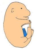
File:Day 17 fingers and toes joined together.JPG| | File:Day 16 Reposition of umbilical hernia.JPG|'''Day 16''' The umbilical hernia is repositioned. The fetus now measures between 14 and 17 mm. | ||
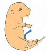
File:Day 18 long whiskers.JPG|Day 18 | File:Day 17 fingers and toes joined together.JPG|'''Day 17''' The fingers and toes become joined together. The skin is wrinkled and thickened and the umbilical hernia has disappeared. The fetus measures 16.5 - 20mm depending on the degree of curvature. | ||
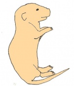
File:Day 19 Newborn Mouse.JPG|Day 19 | File:Day 18 long whiskers.JPG|'''Day 18''' Whiskers elongate. The length of the embryo varies between 18 and 23 mm due to the degree of curvature of the fetus. | ||
File:Day 19 Newborn Mouse.JPG|'''Day 19''' The newborn mouse is between 23 and 27mm depending on the degree of flexure of the body axis | |||
</gallery> | </gallery> | ||
Latest revision as of 18:32, 14 October 2009
- Development of the Mouse Embryo
