Talk:Sensory - Hearing Abnormalities
This current page introduces abnormalities associated with the development of hearing and associated ear structures. There is also some brief information about postnatal detection and treatment of abnormalities.

Inner Ear
File:Earlabyrinthsm.jpg[images/senses/earlabyrinth.jpg Inner ear labyrinth]
common cavity, severe cochlear hypoplasia
References
Mylanus EA, Rotteveel LJ, Leeuw RL. Congenital malformation of the inner ear and pediatric cochlear implantation. Otol Neurotol. 2004 May;25(3):308-17.
(See also [#Cochlear_Implant Cochlear Implant])
Blaser S, Propst EJ, Martin D, Feigenbaum A, James AL, Shannon P, Papsin BC. Inner ear dysplasia is common in children with Down syndrome (trisomy 21). Laryngoscope. 2006 Dec;116(12):2113-9.
Search PubMed: inner ear anomalies
Middle Ear
Middle ear abnormalities (ossicular anomalies) are rare and can be part of first arch syndrome.
Middle ear abnormalities include:
- familial expansile osteolysis
- malleus/incus fixation
- absence of the long process of the incus
- congenital fixation of stapes (stapes anchored to oval window)
- failure of annular ligament development
- cholesteatoma
Familial Expansile Osteolysis (FEO)
A rare congenital (autosomal dominant, 18q21.1-q22) disorder similar to Paget’s disease of bone. Osteolytic lesions occur in all bones (mainly long bones) causing medullar expansions and lead eventually to middle ear and jaw abnormalities.
Malleus/Incus Fixation
Congenital Fixation of Stapes
In this condition the stapes is anchored to oval window often by growth of bone around teh stapes (otosclerosis). Surgicallly treated by stapedectomy, where the bone and stapes is removed and replaced by a prosthesis.
Cholesteatoma
Squamous epithelium that has been trapped within the skull base during development (congenital) and also occurs in an acquired form. The presence of this abnormality leads to erosion of the bones (temporal bone, middle ear, or mastoid) in which the epithelium is embedded.
References
Daneshi A, Shafeghati Y, Karimi-Nejad MH, Khosravi A, Farhang F. Hereditary bilateral conductive hearing loss caused by total loss of ossicles: a report of familial expansile osteolysis. Otol Neurotol. 2005 Mar;26(2):237-40.
Seidman MD, Babu S. A new approach for malleus/incus fixation: no prosthesis necessary. Otol Neurotol. 2004 Sep;25(5):669-73.
Wehrs RE. Congenital absence of the long process of the incus. Laryngoscope. 1999 Feb;109(2 Pt 1):192-7.
Search PubMed: Middle ear ossicular anomalies | familial expansile osteolysis | cholesteatoma |
Outer Ear
Several genetic effects and syndromes can include impacts on developmental of the external ear either directly or by altering development of the skull or face. Several developmental environment effects can be indicated by changes in the relative position or appearance of the external ear at birth (More? [../Defect/maternal.htm Abnormal Development - Environment] | [../Defect/page5a.htm Abnormal Development - Fetal Alcohol Syndrome]).
External Ear: [#microtia Microtia] | [#Preauricular_sinus Preauricular Sinus] | [#Preauricular_tag Preauricular Tag] | [#Stenosis External Meatus Stenosis] |
Microtia
The condition in humans of an abnormally small external ear is called Microtia. This is generally surgically repaired by use of rib cartilage to reconstruct the external ear. A recent study has identified a mouse model for this condition with the knockout of the Pact gene.

OMIM Database Search: "Microtia" (2006 - 25 search results)
Search PubMed May 2006 "Microtia" 449 reference articles of which 37 were reviews.
Search PubMed: Microtia | external ear defects
References
Rowe TM, Rizzi M, Hirose K, Peters GA, Sen GC. A role of the double-stranded RNA-binding protein PACT in mouse ear development and hearing. Proc Natl Acad Sci U S A. 2006 Mar 29 ".. Pact(-/-) mouse were reduced size and severe microtia. As a result of the congenital abnormality of both outer and middle ears, these mice were hearing impaired. Our study demonstrated an essential role of PACT in mammalian ear development and produced the first animal model for studying human microtia."
Zim SA. Microtia reconstruction: an update. Curr Opin Otolaryngol Head Neck Surg. 2003 Aug;11(4):275-81. Review. "...autogenous rib cartilage continues to be the gold standard for microtia repair. Numerous refinements and modifications in the original technique described by Tanzer have paved the way for exceptional results in experienced hands. However, ideal results are not always achieved, and there continue to be drawbacks with the standard approach to reconstruction with autogenous rib cartilage. In an attempt to circumvent these shortcomings, surgeons have developed alternative or adjuvant techniques to repair the microtic ear, including the use of tissue expansion, alloplastic implants, and osseointegrated prostheses. Finally, greater emphasis is being placed on early atresia repair in appropriate candidates."
Preauricular Sinus
File:Preauricular sinus sm.jpg
Preauricular sinus in ascending limb of the helix
Preauricular sinus occurs in 0.25% births, is bilateral (hereditary) in 25-50% of cases and unilateral (mainly the left). They are developmental and generally occur on the surface in anterior margin of the ascending limb of the helix, and the duct runs inward to the perichondrium of the auricular cartilage and in some cases extend into the parotid gland. Postnatally they are a site for infection.
Search PubMed: Preauricular Sinus
Links: Medline Plus - Preauricular tag or pit
Preauricular Tag
File:Preauricular tag1.jpg File:Preauricular tag2.jpg
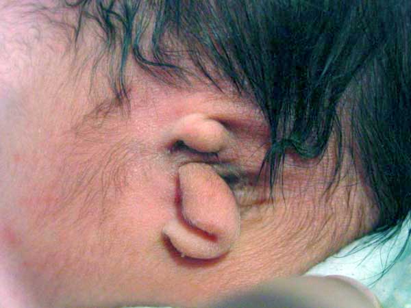
Skin tags in front of the external ear opening are common in neonates and in most cases are normal, though in some cases are indicative of other associated abnormalities.
Search PubMed: Preauricular Tag
Links: Medline Plus - Preauricular tag or pit
External Meatus Stenosis
Stenosis (narrowing) of the external auditory meatus is uncommon and can be due to chronic otitis externa or acquired atresia. The condition can be treated surgically by meatoplasty (reconstructive surgery of the canal) alone, though acquired atresia requires removal of the soft tissue plug and a split skin graft.
Search PubMed: external meatus stenosis
Congenital Deafness
The two main forms of congenital deafness are:
Conductive - disease of outer and middle ear
Sensorineural - cochlear or central auditory pathway
Outer ear Malformation
rare meatal atresia, canal narrow or not formed, part of first arch syndrome
Congenital malformations Statistics
Congenital sensorineural hereditary or acquired (see [#References recent reviews])
Hereditary
- recessive- severe
- dominant- mild
- can be associated with abnormal pigmentation
- hair and irises
Acquired
- rubella (German measles)
- maternal infection during 2nd month of pregnancy
- vaccination of young girls
- streptomycin
- antibiotic
- thalidomide
Conductive Hearing Loss
- produced by otitis media with effusion, is widespread in young children.
- temporary blockage of outer or middle ear
- See also: [#Conductive recent Ref] | [senseslink.htm#Hearing%20Links Senses WWW Link]
Conductive Hearing Loss
Moore DR, Hine JE, Jiang ZD, Matsuda H, Parsons CH, King AJ. Conductive hearing loss produces a reversible binaural hearing impairment. J Neurosci. 1999 Oct 1;19(19):8704-11. (J Neuroscience Link)
The authors of the above paper tested ferrets by long-term plugging of ear canal and found:
- Repeated testing during the 22 months after unplugging revealed a gradual return to normal levels of unmasking.
- Results show that a unilateral conductive hearing loss, in either infancy or adulthood, impairs binaural hearing both during and after the hearing loss.
- Show scant evidence for adaptation to the plug and demonstrate a recovery from the impairment that occurs over a period of several months after restoration of normal peripheral function.
Neonatal Hearing Screening
State Wide Infant Screening Hearing Program (SWISH) A newborn hearing testing program using an automated auditory response technology. Program was introduced in NSW Australia in 2002 across 17 area health service coordinators.
Automated Auditory Brainstem Response (AABR) - uses a stimulus which is delivered through earphones and detected by scalp electrodes. The test takes between 8 to 20 minutes and has a sensitivity 96-99%.
Puig T, Municio A, Meda C. Universal neonatal hearing screening versus selective screening as part of the management of childhood deafness. Cochrane Database Syst Rev. 2005 Apr 18;(2):CD003731.
(More? [../Child/neonatalscreening.htm#Hearing Child Notes - Neonatal Hearing Screening | ][../Child/neonatalscreening.htm Child Notes - Neonatal Screening])
Links: NSW Statewide Infant Screening - Hearing Program What is SWISH? | American Speech-Language-Hearing Association - Audiological Assessment of Children Birth to 5 Years of Age: 2004 (PDF Format)
Otitis Media
Otitis media (ear infection or "glue ear") is not an abnormality, but a very common developmental problem associated with fluid in the middle ear and can ooccur with or without accompanied signs and symptoms of ear infection. Prolonged or repeated occurance can lead to developmental delay in learning, speech and even damage to the middle ear structures.
Otitis media woth effusion (OME) is defined as middle ear effusion without signs or symptoms of an acute infection.
A recent [#CochraneOME Cochrane study] has shown that between the ages of one and three years it has a prevalence of 10% to 30% and a cumulative incidence of 80% at the age of four years.
Lous J, Burton MJ, Felding JU, Ovesen T, Rovers MM, Williamson I. Grommets (ventilation tubes) for hearing loss associated with otitis media with effusion in children. Cochrane Database Syst Rev. 2005 Jan 25;(1):CD001801.
Links: NIH National Institute on Deafness and Other Communication Disorders - Otitis Media | American Academy of Family Physicians - Otitis Media | Medline Plus - Otitis Media |
Cochlear Implant
Moller AR. Physiological basis for cochlear and auditory brainstem implants. Adv Otorhinolaryngol. 2006;64:206-23.
Kral A, Tillein J. Brain plasticity under cochlear implant stimulation. Adv Otorhinolaryngol. 2006;64:89-108.
Geers AE. Factors influencing spoken language outcomes in children following early cochlear implantation. Adv Otorhinolaryngol. 2006;64:50-65.
Das S, Buchman CA. Bilateral cochlear implantation: current concepts. Curr Opin Otolaryngol Head Neck Surg. 2005 Oct;13(5):290-3.
Search PubMed: Cochlear Implant
Human Genes
There are chromosomal abnormalities, such as trisomy 21 (Down Syndrome) that are commonly associated with hearing disorders associated with outer, middle and inner ear defects.
The following table lists only a few of the growing number of known genes associated with hearing loss.
| Symbol | Description | Position |
| DFN1 | deafness, X-linked 1, progressive | Xq22 |
| DFN2 | deafness, X-linked 2, perceptive, congenital | Xq22 |
| DFN4 | deafness, X-linked 4, congenital sensorineural | Xp21.2 |
| DFNB9 | deafness, autosomal recessive 9 | 2p23-p22 |
| DIAPH1 | diaphanous (Drosophila, homolog) 1 | 5q31 |
| GJB2 | gap junction protein, beta 2, 26kD (connexin 26) | 13q11-q12 |
| MYO7A | myosin VIIA (Usher syndrome 1B (autosomal recessive, severe)) | 11q13.5 |
| POU4F3 | POU domain, class 4, transcription factor 3 | 5q31 |
References
Recent Reviews Abnormal Development
- Zim SA. Microtia reconstruction: an update. Curr Opin Otolaryngol Head Neck Surg. 2003 Aug;11(4):275-81.
- Webster WS. [See Related Articles ] Teratogen update: congenital rubella. Teratology. 1998 Jul;58(1):13-23. Review.
- Yates JA, et al. [See Related Articles] Isolated congenital internal auditory canal atresia with normal facial nerve function. Int J Pediatr Otorhinolaryngol. 1997 Jul 18;41(1):1-8. Review.
- Lambert PR, et al. [See Related Articles] Congenital malformations of the external auditory canal. Otolaryngol Clin North Am. 1996 Oct;29(5):741-60. Review. PMID: 8893214; UI: 97048378.
- Lin AE, et al. [See Related Articles] Further delineation of the branchio-oculo-facial syndrome. Am J Med Genet. 1995 Mar 13;56(1):42-59. Review. MID: 7747785; UI: 95266633.
- Strasnick B, et al [See Related Articles] Teratogenic hearing loss. J Am Acad Audiol. 1995 Jan;6(1):28-38. Review. PMID: 7696676; UI: 95210704.
- Kossowska E, et al. [See Related Articles] Prenatal and neonatal prophylaxis in otorhinolaryngology. Int J Pediatr Otorhinolaryngol. 1980 Jun;2(2):85-98. Review. PMID: 6765128; UI: 84160924.
- Gottlieb G. [See Related Articles] Conceptions of prenatal development: behavioral embryology. Psychol Rev. 1976 May;83(3):215-34. Review. No abstract available.PMID: 188059; UI: 77079452.
- Holme RH, Steel KP Genes involved in deafness. Curr Opin Genet Dev 1999 Jun;9(3):309-314
- "Remarkable progress has been made over the past few years in the field of hereditary deafness. To date, mutations in at least 35 genes are known to cause hearing loss. We are now beginning to understand the function of many of these genes, which affect diverse aspects of ear development and function."
Articles
Rowe TM, Rizzi M, Hirose K, Peters GA, Sen GC. A role of the double-stranded RNA-binding protein PACT in mouse ear development and hearing. Proc Natl Acad Sci U S A. 2006 Mar 29; [Epub ahead of print]
List of references from a 1999 search [../Refer/senses/ear_rev.htm Ear Development Reviews] | [../Refer/senses/select.htm Selected Research Articles and Reviews]
WWW Links
Australian Newborn hearing screening program (about 1 in 500 babies are born with a hearing loss Factsheet)
Western Australian pamphlet PDF: Your Newborn Baby Hearing Test
Medline Plus Ear Disorders
New Zealand National Screening Unit - Newborn Hearing Screening