Talk:Paper - The development of the vena cava inferior in man
McClure CFW. and Butler EG. The development of the vena cava inferior in man. (1925) Amer. J Anat. 35(3): 331-383.
Numerous embryos have been examined, and those at important and critical stages were reconstructed in wax after the method of Born.
The following is the series of human embryos actually made use of in this investigation:
Carnegie Embryological Collection (Baltimore)
- No. 588, 4 mm embryo (reconstructed x 150)
- No. 800, 6.5 mm embryo
- No. 623, 10.1 mm embryo (reconstructed x 100)
- No. 841, 15 mm embryo (reconstructed x 100)
Embryological Collection of the College of Physicians and Surgeons (P And S) of Columbia University (New York)
- No. 1095, 11 mm embryo (reconstructed x 100)
- No. 1024, 16 mm embryo (reconstructed x 100)
- No. 1090, 22 mm embryo (reconstructed x 100)
Embryological Collection Of Cornell University Medical College (New York)
- 10.2 mm embryo
- 14 mm embryo
- 15 mm embryo
- 19 mm embryo
Harvard Embryological Collection (Boston)
- No. 2051, 15 mm embryo (reconstructed x 100).
- No. 1913, 18 mm embryo (reconstructed by Huntington and McClure in 1915).
- No. 2924, 25 mm embryo
- No. 2128, 45 mm embryo (reconstructed x 50).
Owing largely to the circumstance of our obtaining material from these several collections, we found that the recorded length of the various embryos cannot always be taken as indicating exactly their relative ages. Our studies have shown that the embryos examined by us can be arranged, at least so far as the development of the venous system is concerned, in the following series according to age: 4 mm (Carnegie, no. 588); 6.5 mm (Carnegie, no. 800); 10.2 mm (Cornell); 11 mm (P and S, no. 1095); 10.1 mm (Carnegie, no. 623); 15 mm (Cornell); 15 mm (Carnegie, no. 841); 15 mm (Harvard, no. 2051); 14 mm (Cornell); 16 mm (P and S, no. 1024) ; 18 mm (Harvard, no. 1913) ; 19 mm (Cornell) ; 22 mm (P and S, no. 1090) ; 25 mm (Harvard, no. 2924) and 45 mm (Harvard, no. 2128). We have also observed that all of the really significant transformations of the embryonic veins, leading up to the establishment of the inferior vena cava, take place largely in embryos measuring approximately between 10 and 18 mm in length.
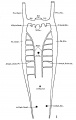
Fig. 1 human embryo 4 mm no. 588
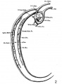
Fig. 2 mm human embryo 4 mm no. 588
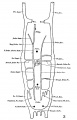
Fig. 3 human embryo 11 mm Columbia University Collection, no. 1095
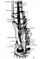
Fig. 4 human embryo 11 mm Columbia University Collection, no. 1095
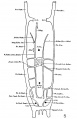
Fig. 5 human embryo 10.1 mm no. 588 (Carnegie stage 17)
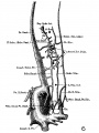
Fig. 6 human embryo 10.1 mm no. 588 (Carnegie stage 17)
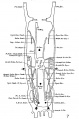
Fig. 7 human embryo 15 mm no. 841 (Carnegie stage 18)
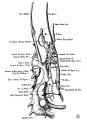
Fig. 8 human embryo 15 mm no. 841 (Carnegie stage 18)
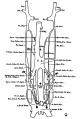
Fig. 9 human embryo 15 mm Harvard Collection, no. 2051
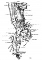
Fig. 10 human embryo 15 mm Harvard Collection, no. 2051 Ventrolateral aspect.
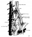
Fig. 11 human embryo 15 mm Harvard Collection, no. 2051 venous ring of right side

Fig. 12 human embryo 16 mm Columbia University Collection, no. 1024
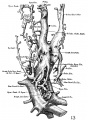
Fig. 13 human embryo 16 mm Columbia University Collection, no. 1024
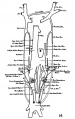
Fig. 14 human embryo 22 mm Columbia University Collection, no. 1090
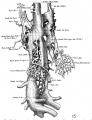
Fig. 15 human embryo 22 mm Columbia University Collection, no. 1090
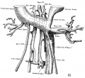
Fig. 16 human embryo 45 mm Harvard Collection, no. 2128
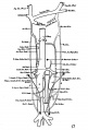
Fig. 17 venous system of adult man
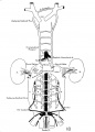
Fig. 18 embryonic veins of the domestic cat


