Talk:Lecture - Fertilization: Difference between revisions
From Embryology
No edit summary |
|||
| Line 26: | Line 26: | ||
* Process of oogonia mature into oocytes (ova, ovum, egg) | * Process of oogonia mature into oocytes (ova, ovum, egg) | ||
* all oogonia form primary oocytes before birth, therefore a maturation of preexisting cells in the female gonad, ovary | * all oogonia form primary oocytes before birth, therefore a maturation of preexisting cells in the female gonad, ovary | ||
[[ | [[File:Human_ovary_non-growing_follicle_model.jpg|300px]] | ||
* humans usually only 1 ovum released every menstrual cycle (IVF- superovulation) | * humans usually only 1 ovum released every menstrual cycle (IVF- superovulation) | ||
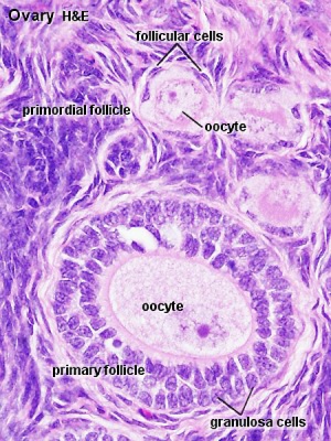
* oocyte and its surrounding cells = follicle | * oocyte and its surrounding cells = follicle | ||
Revision as of 12:06, 24 July 2012
2011 Audio Lecture 2 Audio
Fertilization
Fertilization Preparation
Prior to the fertilization process commencing both the gametes oocyte (egg) and spermatozoa (sperm) require completion of a number of biological processes.
- Oocyte Meiosis - completes Meiosis 1 and commences Meiosis 2 (arrests at Metaphase II).
- Spermatozoa Capacitation - following release (ejaculation) and mixing with other glandular secretions, activates motility and acrosome preparation.
- Migration - both Oocyte and Spermatozoa.
- oocyte ovulation and release with associated cells, from ovary into fimbria then into uterine tube (oviduct, uterine horn, fallopian tube) and epithelial cilia mediated movement.
- spermatozoa ejaculation, deposited in vagina, movement of tail to "swim" in uterine secretions through cervix, uterine body and into uterine tube, have approximately 24-48h to fertilize oocyte.
Endocrinology - Diagram of the comparative anatomy of the male and female reproductive tracts
Oogenesis
Oocyte Development (Oogenesis)
- Process of oogonia mature into oocytes (ova, ovum, egg)
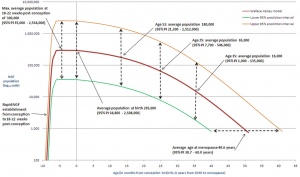
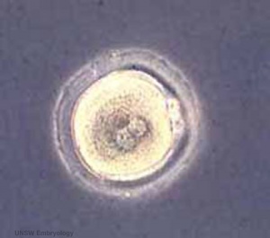
- all oogonia form primary oocytes before birth, therefore a maturation of preexisting cells in the female gonad, ovary
- humans usually only 1 ovum released every menstrual cycle (IVF- superovulation)
- oocyte and its surrounding cells = follicle
- primary -> secondary -> ovulation releases
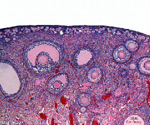
Ovary- Histology - whole transverse section (cortex, medulla)
Menstrual Cycle
- Primary Oocyte - arrested at early Meiosis 1
- diploid: 22 chromosome pairs + 1 pair X chromosomes (46, XX)
- autosomes and sex chromosome
- Oogenesis- pre-antral then antral follicle (Graafian follicle is mature antral follicle released)
- Secondary oocyte
- 1 Day before ovulation completes (stim by LH) Meiosis 1
- haploid: 22 chromosomes + 1 X chromosome (23, X)
- nondisjunction- abnormal chromosome segregation
- begins Meiosis 2 and arrests at metaphase
- note no interphase replication of DNA, only fertilization will complete Meiosis 2
Ovulation (HPG Axis)
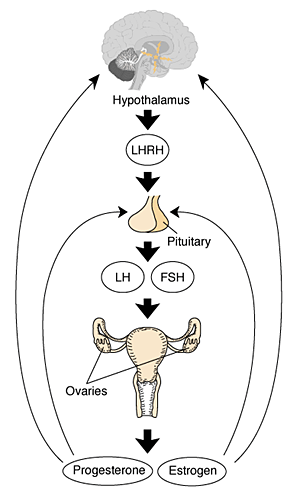
- Hypothalmus releases gonadotropin releasing hormone (GRH, luteinizing hormone–releasing hormone, LHRH) -> Pituitary releases follicle stimulating hormone (FSH) and lutenizing hormone (LH) -> ovary follicle development and ovulation.
- release of the secondary oocyte and formation of corpus luteum
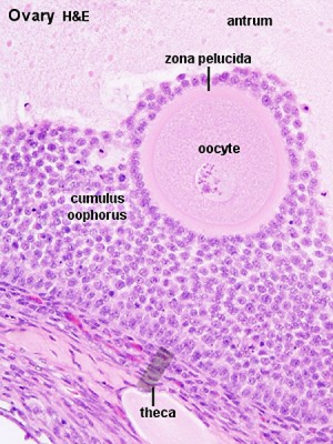
- secondary oocyte encased in zona pellucida and corona radiata
- Ovulation associated with follicle rupture and ampulla movement.
Zona Pellucida
MBoC - Figure 20-21. The zona pellucida
- glycoprotein shell ZP1, ZP2, ZP3
- mechanical protection of egg
- involved in the fertilization process
- sperm binding
- adhesion of sperm to egg
- acrosome reaction
- releases enzymes to locally breakdown
- block of polyspermy
- altered to prevent more than 1 sperm penetrating
- may also have a role in development of the blastocyst
Corona Radiata
- granulosa cells and extracellular matrix
- protective and nutritional role for cells during transport
- cells are also lost during transport along oviduct
Gamete formation- Spermatogenesis
Spermatozoa Development (Spermatogenesis)
- process of spermatagonia mature into spermatazoa (sperm)
- continuously throughout life occurs in the seminiferous tubules in the male gonad- testis (plural testes)
- at puberty spermatagonia activate and proliferate (mitosis)
- primary spermatocyte -> secondary spermatocyte-> spermatid->sperm
- Seminiferous Tubule is site of maturation involving meiosis and spermiogenesis
- Spermatogenesis- Meiosis
- meiosis is reductive cell division
- 1 spermatagonia (diploid) 46, XY (also written 44+XY) = 4 sperm (haploid); 23, X 23, X 23, Y 23, Y
Spermiogenesis
- morphological (shape) change from round spermatids to elongated sperm
- loose cytoplasm
- Transform golgi apparatus into acrosome (in head)
- Organize microtubules for motility (in tail, flagellum)
- Segregate mitochondria for energy (in tail)
Ejaculate
- By volume <10 % sperm and accessory glands contribute majority of volume (60 % seminal vesicle, 10 % bulbourethral, 30 % prostate)
- 3.5 ml, 200-600 million sperm
- Capacitation is the removal of glycoprotein coat and seminal proteins and alteration of sperm mitochondria
- Infertility can be due to Oligospermia, Azoospermia, Immotile Cilia Syndrome
- Oligospermia (Low Sperm Count) - less than 20 million sperm after 72 hour abstinence from sex
- Azoospermia (Absent Sperm) - blockage of duct network
- Immotile Cilia Syndrome - lack of sperm motility
Fertilization Site
- Fertilization usually occurs in first 1/3 of oviduct
- Fertilization can also occur outside oviduct, associated with In Vitro Fertilization (IVF, GIFT, ZIFT...) and ectopic pregnancy
- The majority of fertilized eggs do not go on to form an embryo
Fertilization - Spermatozoa
- Sperm Binding - zona pellucida protein ZP3 acts as receptor for sperm
- Acrosome Reaction - exyocytosis of acrosome contents (Calcium mediated) MBoC - Figure 20-31. The acrosome reaction that occurs when a mammalian sperm fertilizes an egg
- enzymes to digest the zona pellucida
- exposes sperm surface proteins to bind ZP2
- Membrane Fusion - between sperm and egg, allows sperm nuclei passage into egg cytoplasm
Fertilization- Oocyte
- Membrane Depolarization - caused by sperm membrane fusion, primary block to polyspermy
- Cortical Reaction - IP3 pathway elevates intracellular Calcium, exocytosis of cortical granules MBoC - Figure 20-32. How the cortical reaction in a mouse egg is thought to prevent additional sperm from entering the egg
- enzyme alters ZP3 so it will no longer bind sperm plasma membrane
- Meiosis 2 - completion of 2nd meiotic division
- forms second polar body (a third polar body may be formed by meiotic division of the first polar body)
Formation of the Zygote
- Pronuclei - Male and Female haploid nuclei approach each other and nuclear membranes break down
- chromosomal pairing, DNA replicates, first mitotic division
- Sperm contributes - centriole which organizes mitotic spindle
- Oocyte contributes - mitochondria (maternally inherited)
Sex Determination
- based upon whether an X or Y carrying sperm has fertilized the egg, should be 1.0 sex ratio.
- actually 1.05, 105 males for every 100 females, some studies show more males 2+ days after ovulation.
- cell totipotent (equivalent to a stem cell, can form any tissue of the body)
Men - Y Chromosome
- Y Chromosome carries Sry gene, protein product activates pathway for male gonad (covered in genital development)
Women - X Chromosome
- Gene dosage, one X chromosome in each female embryo cell has to be inactivated
- process is apparently random and therefore 50% of cells have father's X, 50% have mother's X
- Note that because men only have 1 X chromosome, if abnormal, this leads to X-linked diseases more common in male that female where bothe X's need to be abnormal.

