Talk:Carnegie stage 12
Template:Ref-MüllerO'Rahilly1987
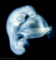
The development of the human brain, the closure of the caudal neuropore, and the beginning of secondary neurulation at stage 12
Anat Embryol (Berl). 1987;176(4):413-30.
Müller F1, O'Rahilly R. Author information
Abstract Twenty-four embryos of stage 12 (26 days) were studied in detail and graphic reconstructions of five of them were prepared. The characteristic features of this stage are 21-29 pairs of somites, incipient or complete closure of the caudal neuropore, and the appearance of upper limb buds. The caudal neuropore closes during stage 12, generally when 25 somitic pairs are present. The site of final closure is at the level of future somite 31, which corresponds to the second sacral vertebral level. Non-closure of the neuropore may be important in the genesis of spina bifida aperta at low levels. The primitive streak probably persists until the caudal neuropore closes, when it is replaced by the caudal eminence or end-bud (Endwulst oder Rumpfknospe). The caudal eminence, which appears at stage 9, gives rise inter alia to hindgut, notochord, caudal somites, and the neural cord. The material for somites 30-34 (which appear in stage 13) is laid down during stage 12, and its absence would be expected to result in sacral agenesis. Aplasia of the caudal eminence results in cloacal deficiency and various degrees of symmelia. The junction of primary and secondary development (primäre und sekundäre Körperentwicklung) is probably at the site of final closure of the caudal neuropore. Secondary neurulation begins during stage 12. The cavity of the already formed spinal cord extends into the neural cord, and isolated spaces are not found within the neural cord. Primary and secondary neurulation are probably coextensive with primary and secondary development of the body, respectively. The telencephalon medium has enlarged, two mesencephalic segments (M1 and M2) are distinguishable, and rhombomere 4 is reduced. The sulcus limitans is detectable in the spinal cord and hindbrain (RhD), and in the mesencephalon and diencephalon, where it extends as far rostrally as the optic sulcus in D1. A marginal layer is appearing in the rhombencephalon and mesencephalon. The first nerve fibres are differentiating, chiefly within the hindbrain (from the nucleus of the lateral longitudinal tract). Optic neural crest is at its maximum, and the otic vesicle is giving crest cells to ganglion 7/8. Neural crest continues to develop in the brain and contributes to cranial ganglia 5, 7/8, and 10/11. The spinal crest extends as far caudally as somites 18-19 but shows no subdivision into ganglia yet. Placodal contribution to the trigeminal ganglion is not certain at stage 12. Such a contribution to ganglion 7/8 is not unlikely.(ABSTRACT TRUNCATED AT 400 WORDS). PMID: 3688450
Carnegie Collection
209 | 24 somites 250 384 486 | 21 somites 1062 | 29 somites 2197 4245-7 4479 4736 | 26 somites 4759 4784 | 23 somites 5035 | 25-28 somites 5048 5056 5206 5300 5923 | 28 somites 6097 | 25 somites 6144 | 27 somites 6488 | 28 somites 6937 | 26 somites 7724 | 29 somites 7852 | 25 somites 7999 | 28 somites 8505a | 24 somites 8505b | 23 somites 8941 | 28 somites 8942 | 25 somites 8943 | 22 somites 8944 | 25 somites 8963 | 22 somites 8964 | 23 somites 9154 | 24 somites


