Talk:Carnegie stage 11
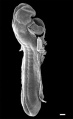
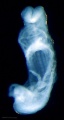
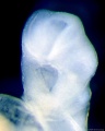
A fifteen-somite human embryo
Adv Anat Embryol Cell Biol. 1989;116:1-102.
Mizoguti H.
Abstract
The morphological features of a well-preserved human embryo having 15 pairs of somites are described and illustrated with a complete set of photomicrographs. This embryo was found during a forensic dissection of a Japanese woman, fixed in 10% formalin for 5 days, and embedded in an epoxy resin mixture according to routine procedures. Serial sections about 0.75 microns thick were made and stained with toluidine blue. The most important features were as follows: 1. The embryo measured 4.1 mm in greatest length and was quite symmetrical viewed dorsally. 2. Closure of the neural tube took place from the level of the first branchial groove, corresponding to the level of the anterior one-third of the rhombencephalon, to that of about 0.4 mm posterior to the 15th somite, coming across the anterior end of the cloacal membrane. The neural plate and the wall of the neural tube consisted exclusively of a pseudostratified columnar epithelium, and neither the mantle nor marginal layers were identified anywhere. 3. At the anterior end of the embryo there was a conspicuous optic evagination, and at the dorsal end of the second branchial groove a round otic placode. 4. Of the three segments of the entodermal tract the midgut was the largest, constituting about half the entire length, and opened ventrally into the large yolk sac, whose surface was covered by a meshwork of highly developed blood vessels. In the foregut two branchial pouches were seen, corresponding to the branchial grooves. Primordia of the thyroid gland and of the liver were also recognized. Among the entodermal epithelial cells lining the ventral half of the hindgut anterior to the origin of the allantois were found numerous primordial germ cells. 5. Among the 15 pairs of somites, the first two were small and consisted of dermomyotome of epithelial cell arrangement and of sclerotome mingled into mesenchyme. The next three were large, triangular on transverse section, and consisted of dorsolateral dermomyotome and ventromedial sclerotome representing a densely packed mesenchymal cell aggregation. The sixth to the tenth somites showed a well-defined triangular contour, each containing a lumen, the myocoele. The last five somites were progressively smaller and were quadrangular in shape. 6. The intermediate mesoderm was encountered at levels from the 7th to the 15th somite, but no indications of pronephric differentiation were detected. 7. The heart tube was relatively large, took an S-shaped tortuous course, and occupied almost the entire pericardiac cavity. It consisted of the bulbus cordis, the ventriculus, and the atrium, each of which consisted of a thick myocardial tube and a thin endocardial tube.(ABSTRACT TRUNCATED AT 400 WORDS)
PMID 2610025
Carnegie Collection
12 164 318 470 779 1182b 2053 4315 4529 4783 4877 5072 6050 6344 6784 7358 7611 7665 7702 7851 8005 8116 8962
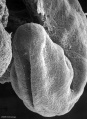
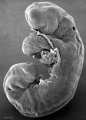
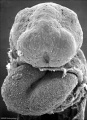
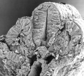
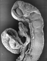
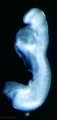
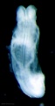
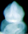
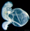
Carnegie Stage 11 Scanning EM Images
- Stage11 sem1.jpg
SEM1 -

SEM2 - Close up view of the degenerating Buccopharyngeal Membrane

SEM3 - Higher power ventrolateral view of the Buccopharyngeal Membrane

SEM4 - Low power ventral view of the Buccopharyngeal Membrane
SEM5 -
SEM6 -
SEM7 - Posterior Neuropore
SEM8 -
SEM9 - Anterior Neuropore
SEM10 -
SEM11 -
- Stage11 sem12.jpg
SEM12 -
SEM13 -
- Stage11 sem14.jpg
SEM14 -
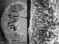
SEM21 - Scanning EM showing low power image of whole embryo and region shown in detail box right of neural crest cells


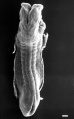
Carnegie Stage 11 Bright Field Images
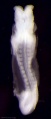
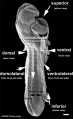
BF1 - dorsal view
BF2 - lateral view
BF3 - ventral with scale bar
BF4 - ventral view
BF5 - lateral view
BF6 - ventral view
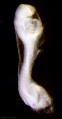
BF8 - ventral head
BF9 - ventral head
BF10 - dorsal neural
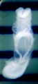
BF11 - ventral left
BF12 - ventral right


