Talk:Book - Contributions to Embryology Carnegie Institution No.39: Difference between revisions
No edit summary |
mNo edit summary |
||
| (3 intermediate revisions by the same user not shown) | |||
| Line 1: | Line 1: | ||
Carnegie Collection | |||
==Explanation of Figures== | |||
[[File:Lewis1920_abbreviations.jpg|thumb|Abbreviations]] | |||
:[[Book_-_Contributions_to_Embryology_Carnegie_Institution_No.39|'''Lewis Links''']]: [[:File:Lewis1920_Plate_1.jpg|Plate 1]] | [[:File:Lewis1920_Plate_2.jpg|Plate 2]] | [[:File:Lewis1920_Plate_3.jpg|Plate 3]] | [[:File:Lewis1920_Plate_4.jpg|Plate 4]] | [[:File:Lewis1920_Plate_5.jpg|Plate 5]] | [[Book_-_Contributions_to_Embryology_Carnegie_Institution_No.39|Contribution No.39]] | [[Book_-_Contributions_to_Embryology#Volume_IX|Volume IX]] | [[Book_-_Contributions_to_Embryology|Contributions to Embryology]] | |||
<gallery> | |||
File:Lewis1920_Plate_1.jpg|Plate 1 | |||
File:Lewis1920_Plate_2.jpg|Plate 2 | |||
File:Lewis1920_Plate_3.jpg|Plate 3 | |||
File:Lewis1920_Plate_4.jpg|Plate 4 | |||
File:Lewis1920_Plate_5.jpg|Plate 5 | |||
</gallery> | |||
<gallery> | |||
File:Lewis1920 fig01.jpg|Fig. 1. Dorsal aspect of base of the cartilaginous skull with the basioccipital in the horizontal plane. | |||
File:Lewis1920 fig02.jpg|Fig. 2. Right half of dorsal aspect of the base of the cartilaginous skull. | |||
File:Lewis1920 fig03.jpg|Fig. 3. Dorsal aspect of cartilaginous and membranous skull. | |||
File:Lewis1920 fig04.jpg|Fig. 4. Dorsal aspect of base of the adult skull. | |||
File:Lewis1920 fig05.jpg|Fig. 5. Median sagittal aspect of the cartilaginous skull. | |||
File:Lewis1920 fig06.jpg|Fig. 6. Ventral aspect of base of the cartilaginous skull. | |||
File:Lewis1920 fig07.jpg|Fig. 7. Lateral aspect of cartilaginous skull and cervical vertebrae, with the brain and cervical cord and hypophysis in position. | |||
File:Lewis1920 fig08.jpg|Fig. 8. Lateral aspect of cartilaginous skull and cervical vertebra; with the brain, cervical cord, and nerves. | |||
File:Lewis1920 fig09.jpg|Fig. 9. Lateral view of cartilaginous skull and cervical vertebrae with the overlying membranous skull and the dorsal membrane. | |||
File:Lewis1920 fig10.jpg|Fig. 10. Dorsal aspect of sphenoid cartilage, showing attachment of the orbital muscles to the basal part of the orbital wing. | |||
File:Lewis1920 fig11.jpg|Fig. 11. Dorsal aspect of sphenoid cartilage. | |||
File:Lewis1920 fig12.jpg|Fig. 12. Lateral view of the right otic region. Part of the malleus and incus cut away showing course of facial nerve and position of otic ganglion. | |||
File:Lewis1920 fig13.jpg|Fig. 13. Lateral view of right otic region showing relations of facial nerve. | |||
File:Lewis1920 fig14.jpg|Fig. 14. Lateral view of base of cartilaginous skull with deeper muscles of occipital region and of the mouth and pharynx. | |||
File:Lewis1920 fig15.jpg|Fig. 15. Lateral view of part of cartilaginous and membranous skull. | |||
File:Lewis1920 fig16.jpg|Fig. 16. Dorsal view of temporal and occipital cartilages, showing the relation of the inner ear to the otic capsule. | |||
</gallery> | |||
===Plate 1=== | |||
[[File:Lewis1920_Plate_1.jpg|600px]] | |||
[[:File:Lewis1920_Plate_1.jpg|Plate 1]]: [[:File:Lewis1920 fig01.jpg|Fig. 1]] | [[:File:Lewis1920 fig02.jpg|Fig. 2]] | [[:File:Lewis1920 fig03.jpg|Fig. 3]] | [[:File:Lewis1920 fig04.jpg|Fig. 4]] | |||
===Plate 2=== | |||
[[File:Lewis1920_Plate_2.jpg|600px]] | |||
[[:File:Lewis1920_Plate_2.jpg|Plate 2]]: [[:File:Lewis1920 fig05.jpg|Fig. 5]] | [[:File:Lewis1920 fig06.jpg|Fig. 6]] | [[:File:Lewis1920 fig07.jpg|Fig. 7]] | |||
===Plate 3=== | |||
[[File:Lewis1920_Plate_3.jpg|600px]] | |||
[[:File:Lewis1920_Plate_3.jpg|Plate 3]]: [[:File:Lewis1920 fig08.jpg|Fig. 8]] | [[:File:Lewis1920 fig09.jpg|Fig. 9]] | |||
===Plate 4=== | |||
[[File:Lewis1920_Plate_4.jpg|600px]] | |||
[[:File:Lewis1920_Plate_4.jpg|Plate 4]]: [[:File:Lewis1920 fig10.jpg|Fig. 10]] | [[:File:Lewis1920 fig11.jpg|Fig. 11]] | [[:File:Lewis1920 fig12.jpg|Fig. 12]] | [[:File:Lewis1920 fig13.jpg|Fig. 13]] | [[:File:Lewis1920 fig14.jpg|Fig. 14]] | |||
===Plate 5=== | |||
[[File:Lewis1920_Plate_5.jpg|600px]] | |||
[[:File:Lewis1920_Plate_5.jpg|Plate 5]]: [[:File:Lewis1920 fig15.jpg|Fig. 15]] | [[:File:Lewis1920 fig16.jpg|Fig. 16]] | |||
===Carnegie Collection=== | |||
* Serial No. - 460 | * Serial No. - 460 | ||
* Size ( mm) - | * Size ( mm) - greatest length 21 mm | ||
* Fixative - Bichlor. acetic | * Fixative - Bichlor. acetic | ||
* Embedding medium - | * Embedding medium - paraffin | ||
* Plane - transverse | * Plane - transverse | ||
* Thickness - 40 micron | * Thickness - 40 micron | ||
* Stain - H&E, coch, | * Stain - H&E, coch, | ||
* Sex - | * Sex - female | ||
1910 tojected | * 1910 tojected | ||
Latest revision as of 11:27, 5 July 2014
Explanation of Figures

- Lewis Links: Plate 1 | Plate 2 | Plate 3 | Plate 4 | Plate 5 | Contribution No.39 | Volume IX | Contributions to Embryology
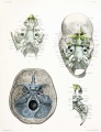
Plate 1
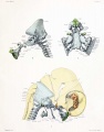
Plate 2
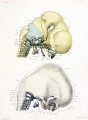
Plate 3
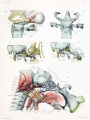
Plate 4
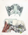
Plate 5
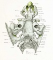
Fig. 1. Dorsal aspect of base of the cartilaginous skull with the basioccipital in the horizontal plane.
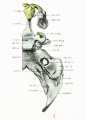
Fig. 2. Right half of dorsal aspect of the base of the cartilaginous skull.
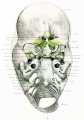
Fig. 3. Dorsal aspect of cartilaginous and membranous skull.
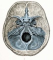
Fig. 4. Dorsal aspect of base of the adult skull.
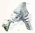
Fig. 5. Median sagittal aspect of the cartilaginous skull.
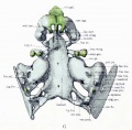
Fig. 6. Ventral aspect of base of the cartilaginous skull.
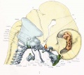
Fig. 7. Lateral aspect of cartilaginous skull and cervical vertebrae, with the brain and cervical cord and hypophysis in position.
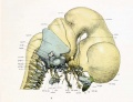
Fig. 8. Lateral aspect of cartilaginous skull and cervical vertebra; with the brain, cervical cord, and nerves.
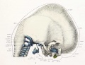
Fig. 9. Lateral view of cartilaginous skull and cervical vertebrae with the overlying membranous skull and the dorsal membrane.
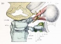
Fig. 10. Dorsal aspect of sphenoid cartilage, showing attachment of the orbital muscles to the basal part of the orbital wing.
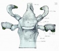
Fig. 11. Dorsal aspect of sphenoid cartilage.
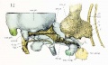
Fig. 12. Lateral view of the right otic region. Part of the malleus and incus cut away showing course of facial nerve and position of otic ganglion.
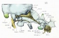
Fig. 13. Lateral view of right otic region showing relations of facial nerve.

Fig. 14. Lateral view of base of cartilaginous skull with deeper muscles of occipital region and of the mouth and pharynx.

Fig. 15. Lateral view of part of cartilaginous and membranous skull.
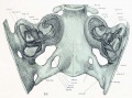
Fig. 16. Dorsal view of temporal and occipital cartilages, showing the relation of the inner ear to the otic capsule.


Plate 1

Plate 1: Fig. 1 | Fig. 2 | Fig. 3 | Fig. 4
Plate 2

Plate 2: Fig. 5 | Fig. 6 | Fig. 7
Plate 3

Plate 4

Plate 4: Fig. 10 | Fig. 11 | Fig. 12 | Fig. 13 | Fig. 14
Plate 5

Carnegie Collection
- Serial No. - 460
- Size ( mm) - greatest length 21 mm
- Fixative - Bichlor. acetic
- Embedding medium - paraffin
- Plane - transverse
- Thickness - 40 micron
- Stain - H&E, coch,
- Sex - female
- 1910 tojected
, ch. ty., cLl. g., coc. d., coc. pt., com. d., oris, gal., digas., dor. mem., dor. sel., ed. bl., end. d., end. sul., epiph., eth. for., fac. for., [ac. sul., fen. ves., for. rot., fos. inc., fos. sub., fron. bl., gang. I. C, gen. g., gen. glo., gen. hy., gr. sup. pet. n. hy. for., hyo. glo., hyp. c.,
hyp.
inf. alv., inf. col., inf. orb., in. sp. lig., int. ac. m., Jac. cart., jug. for., jug. pro., jug. v., lat. rec, lig. fla.. Ion. cjip., It. ma«., mand., mas. for., mas. pro..
- alar, lam. - alar laminsi
- alar, pro. - alar process.
- amp. lat. d. - ampulla lateral semicircular duct.
- amp. p. d. - ampulla posterior semicircular duct.
- amp. s. d. - ampulla superior semicircular duct.
- ansa, hy. - ansa hypoglossi.
- ant. arch. I. C - anterior arch atlas.
- ant. nar. - anterior nares.
- ant. rt. - anterior root occipital neural arch.
- aq. vcs. - aqueduct vcstibuli.
- atl. mas. - atlanto-mastoid muscle.
- atl. occ. art. - atlanto-occipital articulation.
- basiocc - basioccipital.
- bas. pt. - basal part orbital wing sphenoid.
- biventcr - biventer cervicis muscle.
- can. pt. - canalicular part otic capsule.
- chr. pl. - choroid plexus.
chordatympani.
ciliary ganglion.
cochlear duct.
cochlear part otic capsule.
common duct.
crista galli.
digastric muscle.
dorsal membrane.
dorsum sellae.
edge cranial blastema.
endolymphatic duct.
endolymphatic sulcus.
epiphysis.
ethmoid foramen.
facial foramen.
sulcus for facial nerve.
fenestra vestibuli.
foramen rotundum.
fossa incudis.
fossa subarcuata.
frontal bl;i.stema.
ganglion first cervical nerve.
geniculate ganglion.
genioglossus muscle.
geniohyoid muscle.
greater superficial petrosal nerve.
hypoglossal foramen.
hyoglossus muscle.
h>'pophyscal canal.
hypophysis.
inferior alveolar nerve.
inferior colliculus.
infraorbital nerve.
interspinous ligament.
internal acoustic meatus.
Jacobson's cartilage.
jugular foramen.
jugular process.
jugular vein.
lateral rectus muscle.
ligamenta subflava.
longLssimus capitis muscle.
lateral mass basioccipital.
mandible.
mastoid foramen.
mastoid process.
max..
maxillary blastema.
med. lac. for..
metlian lacerated foramen.
med. rt..
median root occipital neural arch.
Meek.,
Meckel's cartilage.
meseth..
mesethmoid.
nas. bl.,
iia-sal blastema.
nas. cap.,
nasal capsule.
neu. arch.
neural arch.
obi. cap. inf..
obliquus capitis inferior muscle.
obi. cap. sup..
obliquus capitis superior muscle.
occ. arch,
occipital neural arch.
occ. fis.,
occipital fissure.
occ. mas..
occipitomastoid muscle.
occ. con.,
occi|)ilal condyle.
olf. for..
olfactory foramen.
opt. for.,
optic foramen.
opt. n.,
optic nerve.
orb. wing,
orbital wing.
ot. cap.,
otic capsule.
otic gang.,
otic ganghon.
pal. n..
palatine nerve.
para, gl..
parathyroid gland.
post, rt..
posteiior root occipital neural arch.
rec. cap. ant..
rectus capitis anterior muscle.
rec. cap. lat.,
rectus capitis lateralis muscle.
rec. cap. post..
rectus capitis posterior muscle.
sel. tur..
sella turcica.
semispi. cap.,
semLspinalis capitis muscle.
sem. g..
semiluiKir ganglion.
squama,
squaiiKi occipital cartilage.
sph. pal. g.,
si>hcni)palatiiic ganglion.
sph. pal. n..
.sphcndpakitiiic nerve.
ster. thy..
sternothyroid muscle.
sty. glo..
styloglossus muscle.
sty. hy.,
stylohyoid muscle.
sty. ph.,
stylopharyngcus muscle.
submax. cap..
subina\illary gland capsule.
sup. can..
superior semicircular canal.
sup. ling.,
superior lingual muscle.
sup. orb. fis.,
suiierior orbital fissure.
sup. rec.,
superior rectus muscle.
temp, wing,
temporal wing.
thy. hy..
thjTohyoid muscle.
thym. gl.,
thymus gland.
thyr.,
thyroid cartilage.
thyr. gl..
thyroid gland.
tr. pro.,
transverse process atlas.
tr. sul.,
transverse sulcus.
zyg-.
zygoma blastema.
zyg. arc,
zygomatic arch.
zyg. pro.,
zygomatic process of frontal b'astema.
III.
III. cranial nerve.
IV.
IV. "
V.
V.
VI.
VI.
Vll.
VII. "
IX.
IX. "
x'S;}
X.I .. .. XL/
XII.
XII. "
I. C. N.
I. cervical "
II. C. N.
11.
line between sphenoid and mesethmoid