Talk:BGD Lecture - Sexual Differentiation
2017
Anomalies in human sex determination provide unique insights into the complex genetic interactions of early gonad development
Clin Genet. 2017 Feb;91(2):143-156. doi: 10.1111/cge.12932.
Bashamboo A1, Eozenou C1, Rojo S1, McElreavey K1.
Abstract Human sex determination (SD) involves complex mutually antagonistic genetic interactions of testis- and ovary-determining pathways. For many years, both male and female SD were considered to be regulated by a linear cascade of pro-male and pro-female genes, respectively; however, it has become clear that male and female development is achieved through the repression of the alternative state. A gene determining the formation of a testis may function by repressing the female state and vice versa. Uniquely in development, SD is achieved by suppression of the alternate fate and maintained in adulthood by a mutually antagonistic double-repressive pathway. Here, we review genetic data generated through large-scale sequencing approaches that are changing our view of how this system works, including the recently described recurrent NR5A1 p.R92W mutation associated with testis development in 46,XX children. We also review some of the unique challenges in the field to establish that mutations, such as this are pathogenic. The impending surge of new genetic data on human SD from sequencing projects will create opportunities for the development of mechanistic models that will clarify how the system operates and importantly provide data to understand how selection and developmental processes interact to direct the evolution of SD across species. © 2016 John Wiley & Sons A/S. Published by John Wiley & Sons Ltd.
KEYWORDS: NR5A1; cell-fate choice decisions; cellular models of human disease; disorders of sex development; gonadal dysgenesis; infertility; sex determination; testicular DSD PMID: 27893151 DOI: 10.1111/cge.12932
2016
- Hill, M.A. (2016) Embryology BGD Lecture - Sexual Differentiation. Retrieved March 16, 2016, from https://embryology.med.unsw.edu.au/embryology/index.php/BGD_Lecture_-_Sexual_Differentiation
- Hill, M.A. (2016) Embryology BGDB Practical - Sexual Differentiation. Retrieved March 16, 2016, from https://embryology.med.unsw.edu.au/embryology/index.php/BGDB_Practical_-_Sexual_Differentiation
- Moore, K.L., Persaud, T.V.N. & Torchia, M.G. (2015). The developing human: clinically oriented embryology (10th ed.). Philadelphia: Saunders. Chapter 12 http://www.unsw.eblib.com.wwwproxy0.library.unsw.edu.au/patron/Read.aspx?p=2074364&pg=329 Urogenital System
- Schoenwolf, G.C., Bleyl, S.B., Brauer, P.R., Francis-West, P.H. & Philippa H. (2015). Larsen's human embryology (5th ed.). New York; Edinburgh: Churchill Livingstone. Chapter 15 http://www.unsw.eblib.com.wwwproxy0.library.unsw.edu.au/patron/Read.aspx?p=2074524&pg=393 Development of the Urinary System Chapter 16 http://www.unsw.eblib.com.wwwproxy0.library.unsw.edu.au/patron/Read.aspx?p=2074524&pg=412 Development of the Reproductive System
- Hill, M.A. (2016) Embryology Genital System Development. Retrieved March 16, 2016, from https://embryology.med.unsw.edu.au/embryology/index.php/Genital_System_Development
- Nussey, S and Saffron Whitehead, S. Endocrinology: An Integrated Approach. Oxford: BIOS Scientific Publishers; 2001. Chapter 6 http://www.ncbi.nlm.nih.gov/books/NBK29 The gonad
- 1 June 2013, at 12:53. - page has been accessed 12,947 times.
- 29 May 2011 at 17:18 - page has been accessed 375 times.
--Mark Hill 13:38, 28 May 2011 (EST) If you have an early print version or 2009 lecture slides, the final 2011 updated presentation may differ, as I probably won't be finished updating here until before lecture. I also remove the draft banner when I am satisfied with the content. If you cannot wait, here is a similar 2010 Science Lecture. Some useful images used in the lecture from a recent review[1] are shown as external links and are not reproduced here, due to copyright restrictions. As always the animations will help you understand dynamic processes and will be covered again during the associated practical class.
2009 Lecture | 2009 Lecture slides PDF |
References
<pubmed>17245850</pubmed>| PDF
--Mark Hill 11:26, 28 May 2011 (EST) This is a good recent review paper from one of the original researchers who discovered SRY. Listed below the reference are links to the review figures in various formats.
<pubmed>17237341</pubmed>| Physiol. Rev.
Figure Pages
These are links to the images with descriptive legends.
- Signaling in genital development
- Sex reversal in humans caused by abnormal X-Y exchange
- Mesonephric tubules in the 11.5 dpc mouse urogenital ridge
- Development and differentiation of the genital duct system
- The migratory pathway of primordial germ cells
- Structure of the early fetal testis
- Differentiation of pre-Sertoli cells into Sertoli cells
- Model for cell-autonomous and prostaglandin-mediated upregulation of Sox9 in pre-Sertoli cells
- Visualization of testicular cell types
- Ovary and follicle development and differentiation
- Postulated molecular pathway leading to the formation of the bipotential genital ridge
- Structure of mouse and human SRY protein
- Postulated interaction of molecular players involved in early testicular development
Table 1. Genes implicated in sexual development in mammals
Figures
These are links to just the images.
- Fig. 1 - Signaling in genital development
- Fig. 2 - Sex reversal in humans caused by abnormal X-Y exchange
- Fig. 3 - Mesonephric tubules in the 11.5 dpc mouse urogenital ridge
- Fig. 4 - Development and differentiation of the genital duct system
- Fig. 5 - The migratory pathway of primordial germ cells
- Fig. 6 - Structure of the early fetal testis
- Fig. 7 - Differentiation of pre-Sertoli cells into Sertoli cells
- Fig. 8 - Model for cell-autonomous and prostaglandin-mediated upregulation of Sox9 in pre-Sertoli cells
- Fig. 9 - Visualization of testicular cell types
- Fig. 10 - Ovary and follicle development and differentiation
- Fig. 11 - Postulated molecular pathway leading to the formation of the bipotential genital ridge
- Fig. 12 - Structure of mouse and human SRY protein
- Fig. 13 - Postulated interaction of molecular players involved in early testicular development
Table 1. Genes implicated in sexual development in mammals
Figure Pages and Figures
These are links to the figure pages with descriptive legends and the images alone.
- Signaling in genital development | Fig. 1 - Signaling in genital development
- Sex reversal in humans caused by abnormal X-Y exchange | Fig. 2 - Sex reversal in humans caused by abnormal X-Y exchange
- Mesonephric tubules in the 11.5 dpc mouse urogenital ridge | Fig. 3 - Mesonephric tubules in the 11.5 dpc mouse urogenital ridge
- Development and differentiation of the genital duct system | Fig. 4 - Development and differentiation of the genital duct system
- The migratory pathway of primordial germ cells | Fig. 5 - The migratory pathway of primordial germ cells
- Structure of the early fetal testis | Fig. 6 - Structure of the early fetal testis
- Differentiation of pre-Sertoli cells into Sertoli cells | Fig. 7 - Differentiation of pre-Sertoli cells into Sertoli cells
- Model for cell-autonomous and prostaglandin-mediated upregulation of Sox9 in pre-Sertoli cells | Fig. 8 - Model for cell-autonomous and prostaglandin-mediated upregulation of Sox9 in pre-Sertoli cells
- Visualization of testicular cell types | Fig. 9 - Visualization of testicular cell types
- Ovary and follicle development and differentiation | Fig. 10 - Ovary and follicle development and differentiation
- Postulated molecular pathway leading to the formation of the bipotential genital ridge | Fig. 11 - Postulated molecular pathway leading to the formation of the bipotential genital ridge
- Structure of mouse and human SRY protein | Fig. 12 - Structure of mouse and human SRY protein
- Postulated interaction of molecular players involved in early testicular development | Fig. 13 - Postulated interaction of molecular players involved in early testicular development
Genes implicated in sexual development in mammals
Table below modified from Table 1. Genes implicated in sexual development in mammals in recent review article.<pubmed>17237341</pubmed>| Physiol. Rev.
| Gene | Protein Function | Gonad Phenotype of Null Mice | Human Syndrome | |
| Bipotential gonad | ||||
| Wt1 | Transcription factor | Blockage in genital ridge development | Denys-Drash, WAGR, Frasier syndrome | |
| Sf1 | Nuclear receptor | Blockage in genital ridge development | Embryonic testicular regression syndrome | |
| Lhx9 | Transcription factor | Blockage in genital ridge development | a | |
| Emx2 | Transcription factor | Blockage in genital ridge development | a | |
| M33 | Transcription factor | Gonadal dysgenesis | a | |
| Testis-determining pathway | ||||
| Gata4/Fog2 | Transcription/cofactor | Reduced Sry levels, XY sex reversal | a | |
| Sry | Transcription factor | XY sex reversal | XY sex reversal (LOF); XX sex reversal (GOF) | |
| Sox9 | Transcription factor | XY sex reversal | Campomelic dysplasia, XX sex reversal (GOF) | |
| Sox8 | Transcription factor | XY sex reversal in combination with partial loss of Sox9 function | a | |
| Fgf9 | Signaling molecule | XY sex reversal | a | |
| Dax1 | Nuclear receptor | Impaired testis cord formation and spermatogenesis | Hypogonadism | |
| Pod1 | Transcription factor | XY sex reversal | a | |
| Dhh | Signaling molecule | Impaired differentiation of Leydig and PM cells | XY gonadal dysgenesis | |
| Pgdra | Receptor | Reduction in mesonephric cell migration | a | |
| Pgds | Enzyme | No phenotype | a | |
| Arx | Transcription factor | Abnormal testicular differentiation | X-linked lissencephaly with abnormal genitalia | |
| Atrx | Helicase | ND | ATRX syndrome | |
| Insl3 | Signaling factor | Blockage of testicular descent | Cryptorchidism | |
| Lgr8 | Receptor | Blockage of testicular descent | Cryptorchidism | |
| Hoxa10 | Transcription factor | Blockage of testicular descent | Cryptorchidism | |
| Hoxal1 | Transcription factor | Blockage of testicular descent | Cryptorchidism | |
| Amh | Hormone | No Müllerian duct degeneration | Persistent Müllerian duct syndrome | |
| Misrl1 | Receptor | No Müllerian duct degeneration | Persistent Müllerian duct syndrome | |
| Pax2 | Transcription factor | Dysgenesis of mesonephric tubules | a | |
| Lim1 | Transcription factor | Agenesis of Wolffian and Müllerian ducts | a | |
| Dmrt1 | Transcription factor | Loss of Sertoli and germ cells | XY femaleb | |
| Ovary-determining pathway | ||||
| Wnt4 | Signaling molecule | Müllerian duct agenesis, testosterone synthesis, and coelomic vessel formation | XY female (GOF) | |
| FoxL2 | Transcription factor | Premature ovarian failure | BPES | |
| Dax1 | Nuclear receptor | XY sex reversal (GOF) | XY sex reversal (GOF) | |
|
a No mutations in human sexual disorders identified to date. b Candidate gene for 9p deletion, XY sex reversal. |
2009 BGD Lecture Slide Text
Note that not all slide text shown and text out of context may not make sense to you in this bullet list form.
Internal Genitalia
- Reproductive Tract
External Genitalia
- Development
- Function
- Abnormalities
Background Notes
- Fertilization
- Week 1
- Kidney
- Endocrine
Online References
UNSW Embryology
- Gonad Development http://embryology.med.unsw.edu.au/Notes/urogen.htm
NIH Bookshelf
- Developmental Biology (Gilbert) - Chapter 17 http://www.ncbi.nlm.nih.gov/books/bv.fcgi?rid=dbio.chapter.4101
- Endocrinology (Nussey and Whitehead) - The Gonad http://www.ncbi.nlm.nih.gov/books/bv.fcgi?rid=endocrin.chapter.972
Textbook References
- Human Embryology (3rd ed.) Larson Chapter 10 pp266-313
- The Developing Human (6th ed.) Moore & Persaud Chapter 13 p303-346
- Before We Are Born (5th ed.) Moore & Persaud Chapter 14 p289-326
- Essentials of Human Embryology, Larson Chapter 10 p173-205
- Human Embryology, Fitzgerald and Fitzgerald Chapter 21-22 p134-152
Genital System Development
3 stages
- Differentiation of gonad (Sex determination)
- Differentiation of internal genital organs
- Differentiation of external genital organs
- 2nd and 3rd stages dependent on endocrine gonad
Long Maturation Timecourse
- Begins in embryo
- Finishes in puberty
Sex Chromosomes
X Chromosome
- 1400+ genes
- 150 million base pairs
- 95% determined
Chromosome Y
- 200+ genes
- 50 million base pairs
- 50% determined
Human Female Differentiation
- Genes such as Wnt-4 and DAX-1
- necessary for initiation of female pathway ovary development
- female not considered a default process
Sry on Gonad
- Sry gene on Y chromosome
- responsible for testis-determining function
- TDF
- expressed in a subset of somatic cells in developing gonad
- induces cells to differentiate into Sertoli cells
Sertoli cells
- produce signals that promote development of male characteristics
- suppress development of female characteristics
- induce primordial germ cells to commit to sperm development
DNA with SRY Protein - SRY protein binds DNA
- Testes determining factor (TDF)
- Transcription factor
Bends DNA 70–80 degrees
- Black
- SRY protein HMG box
- Red coil - DNA double helix (Image after Haqq et al. 1994 and Werner et al. 1995)
SRY on Gonad Development
Germ Cells Development
- Early germ line separation from somatic cells
- germ cells do not arise within gonad
Precursors primordial germ cells (PGCs)
- arise elsewhere and migrate into developing gonads
Gametogenesis
- forming PGCs and getting them into genital ridge as gonad forms
- formation of germ plasm and determination of PGCs
- migration of PGCs into developing gonads
- process of meiosis and modifications of meiosis for forming sperm and eggs
- differentiation of sperm and egg
- hormonal control of gamete maturation and ovulation
Movie: Germ Cell Migration
Sex Determination
Humans (week 5-6)
- Germ cells migrate into gonadal ridge
- Gonads (male/female) identical at this stage
- Indifferent
- Gonad development dependent on sex chromosome
- Y testes
- No Y ovary
Movie: Germ Cell Migration
Primary Sex Cords Carnegie stage 13/14
Internal Genital Organs
All embryos form paired
- Nephric duct
- Mesonephric duct (See kidney development)
- Paramesonephric duct
- Humans 7th week
- Invagination of coelomic epithelium
- Cord grows and terminates on urogenital sinus
Male Gonad (testes) secretes
- Mullerian duct inhibitory factor (MDIF) - Causes regression of paramesonephric duct
- Testosterone - Retains mesonephric duct
Female - opposite
Carnegie stage 13/14 Movie: Mesonephros and Gonad Movie: Urogenital Sinus Male Mesonephric Duct (st22) Movie: Gonad Development Gonad Differentiation Gonadal Cell Types Differentiation of Human Gonads Germ Cells and Ovary Folliculogenesis After colonization of gonad
female germ cells enter prophase of first meiotic division as a mid-gestational hallmark of gender
Perinatally oocytes interact with granulosa cells to form primordial follicles
- cyclic periodicity enter a 3 week growth phase that culminates in meiotic maturation and ovulation
- (study in mouse)
Folliculogenesis Spermatogenesis Inactive until puberty
Seminiferous tubules
- Initially solid (cellular)
- Hollow and active at Puberty
- Cyclic sperm production along length of tubule
Movie: Internal Female Genitalia
Genital Ligaments
Movie: Gonad Descent External Genitalia
Human Sex Hormones
- Hormonal production of differentiated gonads
- required for differentiation of internal and external genitalia during fetal life
- development of secondary sex characteristics at puberty
- Antimullerian hormone (AMH) secreted by Sertoli cells
- Testosterone secreted by Leydig cells
- External male genitalia
- requires transformation of testosterone to dihydrotestosterone
- 5alpha reductase type 2 expressed in genital skin and urogenital sinus
- effects of androgens occur in presence of functional androgen receptor protein
- Mutations of genes coding for steroidogenic enzymes, AMH, AMH receptor, AR and 5alpha reductase are all associated with impairment of sex differentiation and result in genital ambiguity
Hormone Axis
Hormone Dependent Genitalia
External Genital Organs
All embryos initially same (indifferent)
Testosterone
- Differentiates male
Fetal Development
External Genitalia Genital Abnormalities Chromosomal
Hermaphroditism
Gonadal Dysfunction
Tract Abnormalities
External Genitalia
Gonadal Descent
Gonadal Descent Cryptorchadism
- One or both testes fail to descend into scrotum
- 1:30 live male births
- May be associated with other abnormalities
Chromosomal Turner’s Syndrome
- Monosomy XO
- 99% non-viable embryos
- Fail to sexually mature at puberty
Klinefelter's Syndrome
- 47, XXY
- Begin normal male, become infertile
- Tall, mental dullness, behaviour problems
Males
- 46, XX
- Develop as male, infertile adults
- Portion of SRY gene located on one X
Sex Reversal in Humans Hermaphroditism True
- 46,XX
- Gonads both ovary and teste tissues
- Ovotestes or ovary and testes
Male Pseudohermaphrodites
- 46,XY
- Gonads of one sex, external genitalia of opposite
- Various causes
Female Intersex (old term Pseudohermaphrodites)
- 46,XX
- Gonads are ovaries, external genitalia ambiguous
- Hyperplastic adrenals secrete androgens
Gonadal Dysfunction
- Gonads fail to develop properly
Gonadal Dysgenesis
- Swyer’s syndrome
- 46,XX
Mixed Gonadal Dysgenesis
- 45,X/46,XY
Primary Hypogonadism
- Affected females 46,XX
Primary Hypogonadism
- Defective anterior pituitary production of gonadotropin
- Lack of gonadotropin-releasing hormone
Tract Abnormalities
- Many different forms
- Uterine - Associated with other anomolies
- Vagina - Agenesis, atresia (See endocrine lecture DES)
- Ductus Deferens - Uni- or bilateral absence - Failure of mesonephric duct to differentiate
External Genitalia
Multi-factoral
- Chromosomal, single gene, environmental
- Developmental arrest gives ambiguous
Androgen Insensitivity Syndrome
XY karyotype and presence of testes
Externally - develop female secondary sex characteristics
Internally
- women lack Müllerian duct derivatives
- have undescended testes
Summary of Gonad Development
Sex Differences in Adult and Developing Brains
- not known significance of brain sex differences
- transient sex differences in gene expression in developing brains may cause permanent differences in brain structure
- may prevent as well, by compensating for potentially differentiating effects of sex differences in gonadal hormone levels and sex chromosomal gene expression
Sex Chromosomes and Brain Sexual Differentiation
Brains of males and females differ
- in regions specialized for reproduction
- in other regions (controlling cognition, etc) where sex differences are not necessarily expected
Differentially susceptible to neurological and psychiatric disease
2 sources of sexually dimorphic information
- complement of sex chromosome genes
- mix of gonadal hormones
- sex differences in the brain have been attributed to the differential action of gonadal hormones
- evidence for sex chromosome effects on both neural and non-neural systems
- XX and XY cells differentiate even before they are influenced by gonadal hormones
- even if exposed to similar levels of gonadal steroids
Molecular Signaling Cascades
Hormone Axis - Puberty LH & FSH
Factors that increase gonadotrophin secretion and stimulate gonadal maturation
Bars show proportional rise in testosterone and estradiol secretions in males and females respectively
Activation of positive feedback mechanism of estradiol in females also occurs during puberty
Hormone Axis - Reproduction
Human Sexual Differentiation
gonads, internal genital ducts, and external genital structures develop from bipotential embryologic tissues
Male or female phenotype develops through a cascade of processes which initiate with sex determination and follow with sex differentiation
Karyotype (46, XY or 46, XX)
- of embryo (genetic sex) determines whether primordial gonad differentiates into a testis or an ovary respectively (gonadal differentiation)
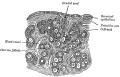
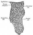
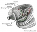
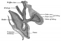
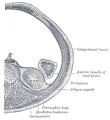
Historic Genital Images
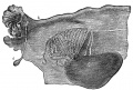
Broad ligament of adult showing Epoöphoron
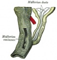
Urogenital Sinus of Female Human Embryo of 8.5 to 9 weeks old
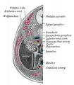
Transverse section of Human Embryo 8.5 to 9 Weeks Old
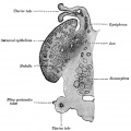
Longitudinal Section of Ovary of Cat Embryo of 9.4 cm long
Section of the Ovary of a Newly Born Child
Human Embryo (3.5 cm long) Testis Section of a Genital Cord
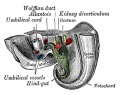
Tail end of Human Embryo 25 to 29 Days Old
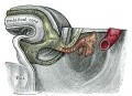
Tail end of human embryo eight and a half to nine weeks old
Primitive Kidney and Bladder
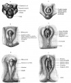
Stages in the development of the external sexual organs in the male and female
Retroperitoneal structures


- ↑ <pubmed>17237341</pubmed>| Physiol. Rev. | Figure Links