Talk:ANAT2341 Lab 7: Difference between revisions
mNo edit summary |
mNo edit summary |
||
| Line 1: | Line 1: | ||
==2015== | |||
==Group Project== | ==Group Project== | ||
* Peer Assessment of your project should now appear on your group discussion page. | * Peer Assessment of your project should now appear on your group discussion page. | ||
Revision as of 17:13, 13 September 2016
2015
Group Project
- Peer Assessment of your project should now appear on your group discussion page.
- Now you can make changes to your project based upon these reviews and your own concepts of how the project can be improved.
- At this time it is important that you interact with other group members to ensure they agree on the changes you may make.
- You may add additional content to your project.
- Look through the comments made by your peers and assemble a summary of good (positive) and bad (negative) feedback on your project.
- What are the common features of peer comments?
- Fix those minor problems (formatting, image size, image labelling, text, referencing)
- Are there parts of your project where group members have not worked appropriately or contributed their work?
- I will initially be discussing this with the groups individually. You will also have the opportunity to "feed back" contribution issues.
Plagiarism
Currently all students originally assigned to each group are listed as equal authors/contributors to their project. If you have not contributed the content you had originally agreed to, nor participated in the group work process, then you should contact the course coordinator immediately and either discuss your contribution or request removal from the group author list. Remember that all student online contributions are recorded by date, time and the actual contributed content. A similar email reminder will be sent to all current students.
Please note the Universities Policy regarding Plagiarism
In particular this example:
- "Claiming credit for a proportion of work contributed to a group assessment item that is greater than that actually contributed;"
Academic Misconduct carries penalties. If a student is found guilty of academic misconduct, the penalties include warnings, remedial educative action, being failed in an assignment or excluded from the University for two years.
- 2014 this was presented as Lab 9 without Steve Palmer exercise.
Muscle Development
Introduction
This laboratory concerns the development and differentiation of skeletal muscle, muscle stem cells, hypertrophy, fibre type differentiation and plasticity. After an introductory tutorial on muscle development, students do exercises looking at experimental images of muscle undergoing hypertrophy and fibre type conversion. Students do some simple analysis of the images in their work groups and we discuss the results. Then we look at a series of images from a mouse that expresses a transgene that influences fibre type choice and we discuss what has happened using the same structure.
Objectives
- Understand the origin, differentiation and development of skeletal muscle tissue.
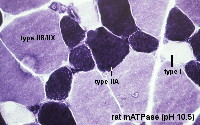
- Know what is meant by patterning, conversion and adult plasticity of muscle fibre type.
- Develop an understanding of research methods for studying skeletal muscle development and function.
Lab Tutorial
Today's laboratory will begin with a tutorial presentation on muscle development and research into muscle disorders. With any new concepts or terms, firstly use the terms list below or the glossary and feel free to ask questions about any concepts you may not still understand during or after the presentation.
Slides for tutorial: Muscle Development - Part 1
Lab Exercises
The following exercises need to be completed in your group in today's lab and are not part of your assessment questions.
There are 3 exercises:
1. Maturation Hypertrophy - In your groups, design a method to estimate the degree of hypertrophy that occurs during the transition from birth to adulthood. Use the rulers to make rough measurements on the screen and then estimate the degree of hypertrophy as a mean increase in fibre size. Discuss the result as a class.
2. The role of satellite cells in muscle hypertrophy - class discussion of data presented in a recent paper - McCarthy et al. 2011
- Synergist Ablation (SA) surgery - removal of the muscles surrounding the plantaris in the lower hind limb to increase the work load.
- BRDU - Bromodeoxyuridine; can be used as a marker to identify cells that have recently undergone DNA replication for cell division.
3. Fibre type shift - Discuss an experiment on fibre type shift that has been induced by chronic low frequency stimulation - Martins et al. 2006
Lab 7 Assessment Questions
Answer the questions shown below on your own student page.
1. (a) Provide a one sentence definition of a muscle satellite cell (b) In one paragraph, briefly discuss two examples of when satellite cells are activated ?
2. In one brief paragraph, describe what happens to skeletal muscle fibre type and size when the innervating motor nerve sustains long term damage such as in spinal cord injury?
Questions are also shown listed on the Students Page Lab 7 Assessment
Terms
anterior tibialis - (tibialis anterior) skeletal muscle situated on the lateral side of the tibia and is a direct flexor of the foot at the ankle-joint.
cis-acting elements - DNA sequences that through transcription factors or other trans-acting elements or factors, regulate the expression of genes on the same chromosome.
enhancer - A cis-regulatory sequence that can regulate levels of transcription from an adjacent promoter. Many tissue-specific enhancers can determine spatial patterns of gene expression in higher eukaryotes. Enhancers can act on promoters over many tens of kilobases of DNA and can be 5' or 3' to the promoter they regulate.
extensor digitorum longus - (EDL) skeletal muscle situated at the lateral part of the front of the leg and extend the phalanges of the toes, and, continuing their action, flex the foot upon the leg.
Gtf2ird1 - General Transcription Factor 2 -I Repeat domain-containing protein 1. OMIM: Gtf2ird1
MusTRD1 - muscle TFII-I repeat domain-containing protein 1.
MyHC - acronym for myosin heavy chain.
myoblast - the undifferentiated mononucleated muscle cell progenitor, which in skeletal muscle fuses to form a myotube, that in turn expresses contractile proteins to form a muscle fibre.
myosin heavy chain - protein forming the thick filament of the sarcomere and the motor for actin-myosin contraction. There are 17 different myosin classes.
myotube - the initial multinucleated cell formed by fusion of myoblasts during skeletal muscle development.
promoter - A regulatory region a short distance upstream from the 5' end of a transcription start site that acts as the binding site for RNA polymerase II. A region of DNA to which RNA polymerase IIbinds in order to initiate transcription.
regulatory sequence - (regulatory region, regulatory area) is a segment of DNA where regulatory proteins such as transcription factors bind preferentially.
soleus - skeletal muscle situated immediately in front of the gastrocnemius and when standing taking its fixed point from below, steadies the leg upon the foot and prevents the body from falling forward.
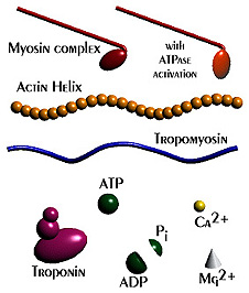
Troponin - striated muscle contraction is regulated by the calcium-ion-sensitive, multiprotein complex troponin and the fibrous protein tropomyosin. Troponin has 3 subunits (TnC, TnI, TnT) and is located on the actin filament. OMIM: Troponin I slow
visuospatial deficiency - performing the Delis hierarchical processing task, subjects are asked to copy a large global figure made of smaller local forms. Both Down syndrome (DS) and William-Beuren syndrome (WBS) groups fail but in different ways. WBS individuals produce the local elements and DS individuals produce only the global forms. PMID: 12952863 Hum Mol Genet.
William-Beuren syndrome - (WBS) rare developmental disorder (1/20,000–1/50,000 live births). A contiguous gene deletion syndrome resulting from the hemizygous deletion of several genes on chromosome 7q11.23. The syndrome has associated craniofacial abnormalities, hypersociability and visuospatial defects. OMIM: William-Beuren syndrome
References - Dr Palmer
- Mutation of Gtf2ird1 from the Williams-Beuren syndrome critical region results in facial dysplasia, motor dysfunction, and altered vocalisations. Howard ML, Palmer SJ, Taylor KM, Arthurson GJ, Spitzer MW, Du X, Pang TY, Renoir T, Hardeman EC, Hannan AJ. Neurobiol Dis. 2012 Mar;45(3):913-22. PMID: 22198572
- Negative autoregulation of GTF2IRD1 in Williams-Beuren syndrome via a novel DNA binding mechanism. Palmer SJ, Santucci N, Widagdo J, Bontempo SJ, Taylor KM, Tay ES, Hook J, Lemckert F, Gunning PW, Hardeman EC. J Biol Chem. 2010 Feb12;285(7):4715-24. PMID: 20007321
- Expression of Gtf2ird1, the Williams syndrome-associated gene, during mouse development. Palmer SJ, Tay ES, Santucci N, Cuc Bach TT, Hook J, Lemckert FA, Jamieson RV, Gunnning PW, Hardeman EC. Gene Expr Patterns. 2007 Feb;7(4):396-404. PMID: 17239664
- Sex determination and gonadal development in mammals. Wilhelm D, Palmer S, Koopman P. Physiol Rev. 2007 Jan;87(1):1-28. Review. PMID: 17237341
- MusTRD can regulate postnatal fiber-spec fic expression. Issa LL, Palmer SJ, Guven KL, Santucci N, Hodgson VR, Popovic K, Joya JE, Hardeman EC. Dev Biol. 2006 May 1;293(1):104-15. PMID: 16494860
- hMusTRD1alpha1 represses MEF2 activation of the troponin I slow enhancer. Polly P, Haddadi LM, Issa LL, Subramaniam N, Palmer SJ, Tay ES, Hardeman EC. J Biol Chem. 2003 Sep 19;278(38):36603-10. PMID: 12857748
Search
- Bookshelf muscle development | muscle fiber type | troponin
- Pubmed muscle fiber type | troponin | Gtf2ird1 |
UNSW Embryology Links
- File:Muscle development 1-Talk1 muscle development.pdf
- File:Talk1 muscle development 4 per page.pdf
- File:Part 2 Muscle Development.pdf
- File:Part 2 Muscle Development 4 per page.pdf
External Links
External Links Notice - The dynamic nature of the internet may mean that some of these listed links may no longer function. If the link no longer works search the web with the link text or name. Links to any external commercial sites are provided for information purposes only and should never be considered an endorsement. UNSW Embryology is provided as an educational resource with no clinical information or commercial affiliation.
- Blue Histology Skeletal Muscle histology
- Primer Skeletal Muscle Fiber Type: Influence on Contractile and Metabolic Properties
- Gray's Anatomy of the Human Body - The Muscles and Fasciæ of the Leg | Cross-section through middle of leg
- Wiki Skeletal muscle | Troponin
- OMIM: Gtf2ird1
- OMIM: William-Beuren syndrome
Glossary Links
- Glossary: A | B | C | D | E | F | G | H | I | J | K | L | M | N | O | P | Q | R | S | T | U | V | W | X | Y | Z | Numbers | Symbols | Term Link
Cite this page: Hill, M.A. (2024, April 20) Embryology ANAT2341 Lab 7. Retrieved from https://embryology.med.unsw.edu.au/embryology/index.php/Talk:ANAT2341_Lab_7
- © Dr Mark Hill 2024, UNSW Embryology ISBN: 978 0 7334 2609 4 - UNSW CRICOS Provider Code No. 00098G


