Talk:2014 Group Project 4
This is the discussion page for your project.
- Use this page to discuss online the project with your group members.
- Paste useful resources here.
- Remember to use your signature button to identify who you are when adding content here.
- The following collapsed tables provide starting points for students during project work, you also have tutorials built into practical classes and practice exercises for individual assessmet items.
|
|
|
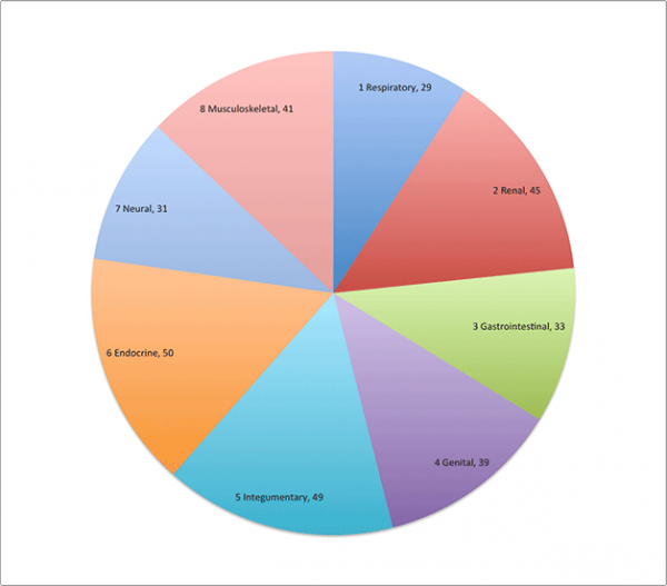
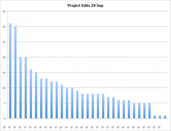
| Project Analysis 24 Sep |
|---|

--Mark Hill (talk) 09:57, 24 September 2014 (EST) Individual student data for each group has also been analysed. |

--Mark Hill (talk) 09:57, 24 September 2014 (EST) I have masked student ID.
|
2014 Student Projects: Group 1 | Group 2 | Group 3 | Group 4 | Group 5 | Group 6 | Group 7 | Group 8
--Mark Hill (talk) 17:54, 31 October 2014 (EST) These student projects have now been finalised and undergoing final assessment.
| Group Assessment Criteria |
|---|
|
Peer Reviews
The Introduction, Current Models and Current Research section all in dot-point form, which obviously allowed you to more easily, put information on the page. These need to be converted into paragraph form to give the content greater readability and flow.
Presuming the system development is supposed to be the introduction, there should be inclusion of current research, historical research and abnormalities. Without these the reader will not know all the sections of the page after reading the introductory section, which is the intros purpose. The use of bold and capital letters is unneeded. The existence of a table is good though has a bunch of formatting and text problems (capitals, bold, captions, lack of lines). “(around week 4-6) that sexual differentiation occurs in the fetus ” this statement is incorrect since it is an embryo during the week4-6, it becomes a later around week 10. “450px” has not been inserted properly, the sexual differentiation image requires caption and references.
Current Research and Models has in-depth content for undifferentiated and male, though limited information on current female genital research. The headings are repetitive also many without any content, similar content needs to be merged under single headings. In Historic findings the content and wording is good but same trend continues significantly more text on Male development compared to female.
Abnormalities section is great with even attention given to female, male and both. Information is appropriately in-depth and referenced, addressing causes, process and treatment. Addition of 1-2 images in the “both” section is advised, to allow readers to identify clinical features of the diseases. Like the use of drawings especially “Abnormalities of the Uterus and Vagina” and “Anat of Testes”, you should change the caption of the testes drawing from “alt text”.
For improvement; covert of dot-points into paragraphs, expand on female sections of “current research” and “historical research”, fix a few image problems and remove unnecessary bold/capitals/captions.
A great start on tabulating the information about the development of this system. There are references but I don’t see any in-text citations. The image used in this section is really good and relevant. It clearly shows the major processes in the development of the genital system. However, it is a bit pixelated so maybe try resizing the image to a smaller size. Maybe try uploading the image again with a different filename, change it to something more appropriate rather than “Image.jpg”. And also, if possible, try to include it in the table. Good job on embedding a video! I think this is the only group so far that has included a video. It’s a good video about the development, I just wish it had a voice-over explaining what is happening but that’s not really the group’s fault. Nonetheless, great job on the development section.
With the current research section, great use of dot points but a bit excessive. Maybe try to make paragraphs where it is appropriate. It is well-researched, very detailed and very informative. It’s good to see student drawings. Great job on that. I see that an image was not properly uploaded into the page, so just fix that. Good job on referencing. All research articles seem to be relevant to this section but try to incorporate some of the in-text citations of the remaining articles, not just the first three. Overall, really great job on the content of this section. It is evident that the person responsible for this section put a lot of effort in research.
As for historic findings, great job! I know this is probably the hardest of all the four sections in terms of finding information and this section is well-researched, very detailed, and very informative much like the current research section. Maybe try to use some dot points to lessen the bulk of this section. Great drawing included in this section. Try to add more, especially for the males since that is the bulk of this section.
Lastly, for abnormalities, great job on finding lots of abnormalities! Lots of references and each area of this section seems to be well-cited. The content of this section is very concise. All the important information about the disease is included, from the cause to the treatment. Good work! Try to find more images for the other abnormalities. It may be tedious but it will help in visualising the clinical manifestations of each disease. Overall, this group has done their research and did it well. Great job on the table for development and images. Their page is very clean and very organised, particularly the references. Don’t forget to write an introduction for your project’s page.
Just looking at the contents, if feels a little intimidating both in that it is so long as well as the use of caps. You should try and limit both; the use of all caps can be quite annoying in text and the extensive contents list can make people dread reading through your page if it looks like it’s quite long.
An introduction is recommended as it is usually a good starting point to provide the reader as sense of everything the page will cover. The system development is a little messy, but I will heed your note and pay attention to only the table. The table itself is a great idea to lay out all the events happening in the corresponding weeks, making it look neat and concise. However, the use of all caps, bold text, and two different fonts still makes this section look messy. Having both male and female events on the same table makes it look as if there is a chunk of info missing for the female side as well. I would suggest having them in separate tables next to each other, which would eliminate the empty rows in both areas. Both the image and the video (congrats on finding a video! Really good addition to the page) should be captioned.
The current research, models and findings seems well researched as there are a lot of points made. However, it is all presented in bullet points which can be visually unappealing. Some sections look incomplete as well, so an effort needs to be made to finish these areas as well as present them in an appealing manner e.g. in paragraph form with a picture next to it to both describe the text visually and offset the amount of text. The drawing of the testes should be captioned appropriately instead of the ‘alt text’ provided. It should also be enlarged, as its current size isn’t large enough to view any of the labels properly.
Historic findings looks well researched on first glance, but then I saw that only 4 sources were used to reference the section. It looks really bad when only one source is used to reference a large slab of text, which you have done twice. I suggest finding articles that state similar information and using them as references as well, to back up your current information found. Other than that, I suggest possibly formatting your section in a more appealing way; either summarize some areas in dot points, and add a picture.
The abnormalities section is nice and concise, without going into too much detail which is good. Just make sure you explain what it is, how it is formed/how you get it, some statistics and possibly an image to show what it looks like, and that’s all I believe you really need for this section.
Overall, your page is well researched with lots of info. Just make sure it looks visually appealing, is consistent in terms of font and presentation, images are used and captioned correctly, and all references are placed at the bottom of the page.
Firstly, great job on all the contents you guys managed to present, it’s quite detailed. There seems to be no introduction though, and the page jumps straight into explaining genital development. I think if an introduction were added, it would give the whole page better structure and formatting so the reader knows what to expect when they decide if they want to read on. The dot points used for the developmental section allows for easy readability of the contents, however, the use of caps lock and arrows takes away from the overall presentation of the page. I would suggest any text you want to emphasize to make bold or underline the word. I also noticed that there was a note stating the attempt to put all the developmental information into a table, but had issues. I suggest you look at the editing basic page you can search for in the top right hand corner as it outlines a step-by-step guide into making tables etc.
In regards to referencing, there are no in-text citations for the first two subheadings. The sections were they do have citations also have a list of references at the bottom of each section. I would recommend just adding a final list of references at the bottom of the page, as it looks much neater.
I’m impressed with the level of hand-drawn diagrams uploaded. I would also recommend adding captions to the image. For example:
. The “alt text” should be edited to describe the caption of the drawing. This particular image seems to have a broken link though; the “alt text” also appeared in the labeled diagram of the testes. Otherwise, good job on the other images.
The current findings section seems to be untouched, with the exception of some pubmed journal article links, I’m assuming you are still in the process of adding content. The historic findings, however, is extensive and well researched. Good job.
The abnormalities section is done well. There is more than enough abnormalities listed, and they are researched well, I would just suggest adding a few more images for better visualization. Overall, great page, just needs better formatting for the mentioned sections.
This project would benefit from having an introduction to prepare the reader for what is to come and summarise everything briefly. The system development part is interesting and clearly there has been a lot of research put into finding the information. I suggest adding pictures or student-drawn diagrams, particularly of the chromosome and the SRY gene location to make it more comprehendible. If you’re not a geneticist, it can be difficult to picture that much detail at an embryonic level.
It is clear you have considered inserting images so it would be important to follow through with that before the final stages of marking. I’m not really sure why you’ve inserted a table here as well since a lot of the information was already covered previously. Maybe use less information in the table. The references at the end of this section should appear at the very end of the wiki page. A lot of other groups have already done that so if you need to copy the formatting, it’s definitely possible. The use of a video on your page is commendable and sets this project above others in that sense. It’s a great idea to have a youtube clip. However, it is 9 minutes long which is a bit long for a student page that is designed to inform students on the genital system on a wholistic scale rather than tackling complicated ideas. Maybe try editing the youtube video so you only use a 30 second or 1minute clip.
The male and female genital development section is clearly presented and the use of bullet points make it easy to follow. However reading the information, it appears that a lot of it I recognised from the lectures. This doesn’t suggest the student explored external embryology sources. On another note, perhaps the lecture on the genital system was very indepth and this student did do research but found all the relevant information had already been covered. None the less, I think it would be advantageous to add a subheading in the section that looks at recent findings. This would broaden the understanding an embryology student can achieve by reading this wiki-page. Also there has been an error uploading an image so that should be fixed.
Although the information is presented well, the bulk of references should be included at the very end of the page. This project is very good but there is still some further research needed, particularly under the current findings subheading. The information presented under the historical findings subheading is quite dense and would benefit from being broken up into a table or simple bullet points. The abnormalities part is excellent and there has clearly been broad research into different embryological resources.
Group Topic
--Z3417458 (talk) 14:08, 18 August 2014 (EST) Hi everyone :), We all need to decide on a system for our group asap, does anyone have any suggestions ? I was thinking we could do the Genital or Musculoskeletal ?
--Z3415716 (talk) 17:45, 19 August 2014 (EST) Hello, I was thinking of covering the genital system development as well.
--Z3417753 (talk) 20:39, 19 August 2014 (EST) Genital it is :)
--Z3416697 (talk) 11:07, 20 August 2014 (EST) Great can't wait! there seems to be a lot of info about genital embryogenesis
--Z3417458 (talk) 21:07, 26 August 2014 (EST) Hey everyone, just wanted to make a note of what each of us was going to research. So as we all discussed last week, I am happy to do part 5. Abnormalities :)
--Z3417753 (talk) 23:18, 26 August 2014 (EST) Hey ! Yes im doing current research models and findings :)
--Z3415716 (talk) 01:05, 27 August 2014 (EST) Thank you all for referencing your articles. I am having some difficulty with referencing 1 of my 3 articles mainly because they are not from Pubmed. I will consult with Mark tomorrow and have my part completely uploaded during the lab. Thanks for your understanding.
--Z3417458 (talk) 14:57, 1 September 2014 (EST) Hey All, just wanted to let you know that there are some really good pictures showing the differentiation between the male and female genital development in the textbooks. So maybe this week we could decide which ones we like and then I can try to draw them. :)
--Z3415716 (talk) 17:27, 2 September 2014 (EST) That sounds really good. If we are not given some time tomorrow during the lab to meet with our group and if you all don't mind we can stay back for 10 minutes or so to have a look at the images you found and if anyone has found any interesting material. See you all tomorrow in the lab.
--Z3417458 (talk) 18:06, 9 September 2014 (EST) Hi, I know we can only use one image from wikipedia so maybe we could use this one ? Or has anyone found any others ? Heres the link -- > http://en.wikipedia.org/wiki/Sexual_differentiation#mediaviewer/File:2915_Sexual_Differentation-02.jpg
--Z3417458 (talk) 18:14, 16 September 2014 (EST) Hi everyone, I am going to post 2 images on here tonight, please let me know which you prefer :)
--Z3417458 (talk) 21:04, 16 September 2014 (EST) Or this one ->
2. File:Sexual Differentiation.jpg
--Z3415716 (talk) 14:42, 21 September 2014 (EST) Since my part is historical findings, I have found a few old articles around 50-100+ years old. Below I'm going to past a paragraph about the female genital system development I have composed from information of two articles, one is from the 1950s and the other is 1890s. My only concern is what I have written doubles up with the system development part of this assignment so I have not uploaded onto the page but if you guys think it's fine for historical finding then I will, if not we can add that into system development and the timeline. I am still searching for historical teachings and images that can be used in this assignment.
The mullerian (paramesonephric) ducts, found laterally to the wolffian ducts, are the original structures of the female reproductive system. Female sexual organs (the fallopian tubes, uterus and vagina) originate from the mullerian ducts, which differentiates within the foetal developmental phase. Initially the foetus contains two mullerian ducts, however by the ninth week fusion of the lower portion of the ducts is complete, creating the fundamental structure of the uterus and the vagina, however the these two organs are not continuous with vagina being solid. The non-fused upper part of the ducts emerge into the fallopian tubes. It is not until the fourth and fifth month of development that the uterus becomes continuous with the vagina, with both organs developing a hollow lumen. The muscular layers of the uterus is also present by this stage. The cervix begins to form within the fifth month, between the continuous vagina and uterus. Also within the same month, the formation of the hymen occurs. The hymen is described as a pouting vertical slit and represents the remains of the mullerian eminence
--Z3417458 (talk) 21:05, 22 September 2014 (EST) I think it can be added under your heading of historical findings :)
--Z3417753 (talk) 12:26, 1 October 2014 (EST) hey guys hope you are all enjoying your break :) Hope your assignments are all going well :)
Also, I found this article that might be useful if you havent already found it - it goes under historic findings - it is from 1942!!
Schonfeld WABeebe GW Normal growth and variation in the male genitalia from birth to maturity. J Urol 1942;8759- 777
--Z3417458 (talk) 21:59, 2 October 2014 (EST) Hey, hope your enjoying your break too. Thats great :). If you any of you guys come across an image that we could use for the first page, post in on here so we can decide if we want to use it. :)
--Z3415716 (talk) 16:16, 5 October 2014 (EST) Thank you, I'm doing the historical findings and I will have a look into that article. Thanks again. I have just redrawn an image from one of my articles about the Mullerian ducts and forming the female genital system. I will try and upload it following the steps Mark gave to us in the first lab so once it is up please let me know if you guys like it or not. Thanks
--Z3415716 (talk) 16:57, 5 October 2014 (EST) Also another thing, please let me know if I am being too specific in my part (Historical findings). I still have more to add on other areas of genital development, so if what I am doing is fine then I will continue this way, if not please let me know so I can change what I have. Thanks again.
--Z3417458 (talk) 16:15, 6 October 2014 (EST) Hey Everyone, I've found a video we could use on our page, the background music is a bit annoying but the drawings are really good, detailed and clear heres a link. Let me know if any of you have found some too. :) https://www.youtube.com/watch?v=MureNA-RSZM
- Great progress on the table. Maybe once you've managed to format everything you need into it, don't forget to reference bits you need to
- I liked the diagram you used to show the different pathways of genital development for the different genders. It's just a bit blurry so maybe think of re-uploading a clearer image or of making the image on your page a little smaller
- Good use of dot points under the "current research" section but maybe think of connecting the separate points a bit more as it seems a bit disjointed and difficult to follow. Maybe think of having your write-up as normal and using points in particular parts that show a sequence of events, or separate components of something
- Look to getting more references for the current research and models section because you're just using 1 at the moment
- Proofread. I know maybe you guys are still at the collation of information stage, but I find it's easier to get it right as you go along rather than coming back to it later
- Re-phrase some bits like: female and male fetuses’ external genitalia --> The external genitalia of the female and male fetus
- Great drawn images! They're all so clear, well thought out and identify all relevant components of what you're trying to show all throughout your page
- I liked the detail of your "historical findings" section
