Talk:2010 BGD Practical 6 - Week 3
Week 3

Week 3
Mesoderm means the "middle layer" and it is from this layer that nearly all the bodies connective tissues are derived. In early mesoderm development a number of transient structures will form and then be lost as tissue structure is patterned and organised. Humans are vertebrates, with a "backbone", and the first mesoderm structure we will see form after the notochord will be somites.
Facts: Week 4, 22 - 23 days, 2 - 3.5 mm, Somite Number 4 - 12
View: This is a dorsal view of the human embryo, the amniotic membrane has been removed. Top embryo is an early stage 10, bottom is late stage 10.
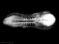
Early stage 10
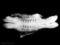
Late stage 10
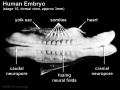
Labeled stage 10


trilaminar embryo
mesoderm regions
somite coelom

neural tube and neural crest


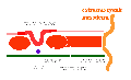
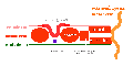
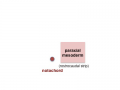
Mesoderm organization: lateral plate - intermediate mesoderm - paraxial mesoderm - axial mesoderm - paraxial mesoderm - intermediate mesoderm - lateral plate

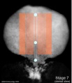
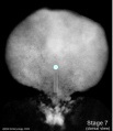
Stage 7 paraxial mesoderm
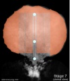
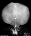
Stage 7 intermediate mesoderm
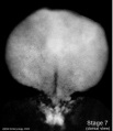
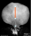
Stage 7 lateral plate


Axial Mesoderm
- notochord
- mechanical role in embryonic disc folding
- molecular role in patterning surrounding tissues
Stage 7 embryonic disc
Stage 7 primitive-streak-node
Stage 7 cloacal-oral-membranes
Stage 7 notochord


Adult - contributes to the nucleus pulposis of the intervertebral disc
Paraxial Mesoderm
- differentiates rostro-caudally (head to tail)
- remains unsegmented in the head region.
- segments in the body region to form pairs of somites along the length of the embryo.
Adult - contributes vertebral column (vertebra and IVD), dermis of the skin, skeletal muscle of body and limbs
Intermediate Mesoderm
- named by position (between paraxial and lateral plate)
- differentiates rostro-caudally (head to tail)
- forms 3 sets of "kidneys" in sequence
- pronephros
- mesonephros
- metanephros
Adult - metanephros forms the kidney
Lateral Plate Mesoderm
- a "horseshoe shaped" space forms in the middle
- somatic mesoderm - closest to ectoderm
- space - forms the 3 body cavities (pericardial, pleural, peritoneal)
- splanchnic mesoderm - closest to endoderm
Adult - body connective tissues, gastrointestinal tract (connective tissues, muscle, organs), heart
Somite Development

Somite initially forms 2 main components
- ventromedial- sclerotome forms vertebral body and intervertebral disc
- dorsolateral - dermomyotome forms dermis and skeletal muscle
paraxial mesoderm
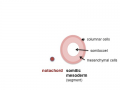
early somite
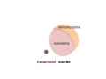
sclerotome and dermomyotome
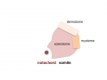
dermatome and myotome
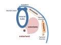
epaxial and hypaxial muscles


Sclerotome
- sclerotome later becomes subdivided
- rostral and caudal halves separated laterally by von Ebner's fissure
- half somites contribute to a single vertebral level body
- other half intervertebral disc
- therefore final vertebral segmentation “shifts”
Myotome
- Body - epaxial and hypaxial muscles
- Limbs - flexor and extensor muscles
Dermatome
- connective tissue underlying epidermis
- begins as a dorsal thickening
- spreads throughout the body
![]()
![]()
![]()