Unused files
From Embryology
The following files exist but are not embedded in any page. Please note that other web sites may link to a file with a direct URL, and so may still be listed here despite being in active use.
Showing below up to 500 results in range #1 to #500.
 Chick15h.jpg 143 × 140; 3 KB
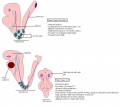
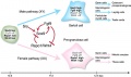
 Gray1110 common male female genital.gif 276 × 800; 55 KB
 Gray1116 hindgut 32-33days.gif 485 × 350; 52 KB
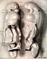
 Braune2 B1.jpg 1,200 × 3,263; 413 KB
 Gray0005.gif 460 × 538; 104 KB
 Icon-Quiz.jpg 199 × 151; 5 KB
 Stage7 SEM1.jpg 450 × 333; 40 KB
 Stage7 SEM2.jpg 450 × 300; 27 KB
 Gray0064.gif 469 × 515; 30 KB
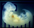 Stage14 human scale.jpg 646 × 530; 40 KB
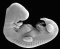 Stage14 SEM.jpg 646 × 530; 36 KB
 Mark Hill 08.jpg 180 × 240; 20 KB
 Stumbleupon 16x16.png 16 × 16; 901 bytes
 Citeulike 16x16.png 16 × 16; 413 bytes
 Icon citeulike 16x16.gif 16 × 16; 79 bytes
 Connotea 32x32.png 32 × 32; 1 KB
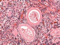
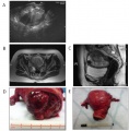 Uterine teratoma.jpg 600 × 609; 73 KB
 Neural-crest-icon.png 120 × 101; 7 KB
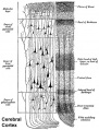 Historic-Cerebral-cortex.jpg 483 × 634; 65 KB
 Csf cartoon1.jpg 600 × 307; 35 KB
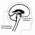 Csf cartoon3.jpg 600 × 588; 37 KB
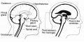
 CNS later development.jpg 961 × 462; 59 KB
 VIT Gene Evolution.jpg 668 × 405; 76 KB
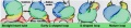
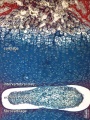
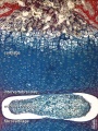
 Preimplantation Development in rabbits.jpg 860 × 445; 51 KB
 Stage14 planes.gif 305 × 436; 8 KB
 Stages 1-5 mouse.pdf ; 29 KB
 Stage10 bf2a.jpg 800 × 647; 21 KB
 Stage10 bf2b.jpg 600 × 485; 14 KB
 Language perisylvian connections.jpg 600 × 396; 50 KB
 Chicken-gastrulation.jpg 800 × 773; 128 KB
 Ovary-human-follicle.jpg 492 × 1,000; 103 KB
 1755-8166-1-21-3-l.jpg 1,200 × 321; 28 KB
 Mouse cerebellum.jpg 600 × 591; 200 KB
- Early Growth of Trophoblast.pdf ; 234 KB
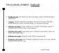 FROG DEVELOPMENT.jpg 672 × 608; 53 KB
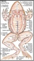 ANATOMY OF FROG.jpg 252 × 445; 37 KB
 Branchial arch muscle cartoon.jpg 454 × 381; 40 KB
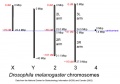 Drosophila-chromosome-diagram.jpg 800 × 557; 45 KB
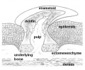 Odontodes.jpg 455 × 376; 93 KB
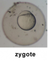 ZYgote image 1.png 142 × 181; 37 KB
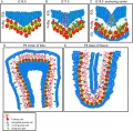
 Palatal shelves animation.gif 550 × 400; 134 KB
 Palate.gif 550 × 400; 135 KB
 Theiler stages 12-14 mouse.JPG 1,025 × 933; 94 KB
 Theiler stages 13-14 mouse.JPG 1,013 × 792; 93 KB
 Tongue cartoon1.gif 373 × 277; 6 KB
 Tongue cartoon2.gif 327 × 318; 6 KB
 Tongue cartoon3.gif 249 × 269; 5 KB
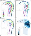
 Neural-crest-migration1.jpg 430 × 377; 17 KB
 Neural-crest-migration2.jpg 430 × 415; 20 KB
 Neural-crest-migration3.jpg 500 × 465; 34 KB
 Neural-crest-migration4.jpg 488 × 481; 20 KB
 Neural-crest-migration5.jpg 434 × 422; 19 KB
 Stage14 GIT3d sm.mov ; 67 KB
- Stage14 GIT3d med.mov ; 166 KB
 Carnegie stages species.gif 556 × 357; 8 KB
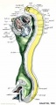
 Stage 11 historic-Atwell1930-1b.jpg 323 × 600; 26 KB
 Stage 11 historic-Atwell1930-1c.jpg 215 × 400; 14 KB
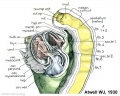
 Stage 11 historic-Atwell1930-2.jpg 800 × 639; 87 KB
 Stage 11 historic-Atwell1930-2b.jpg 600 × 479; 50 KB
 Stage 11 historic-Atwell1930-2c.jpg 400 × 319; 23 KB
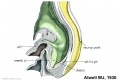 Stage 11 historic-Atwell1930-3.jpg 1,000 × 679; 87 KB
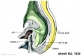 Stage 11 historic-Atwell1930-3c.jpg 400 × 271; 15 KB
 Stage 11 historic-Heuser1930-1a.jpg 417 × 800; 57 KB
 Stage 11 historic-Heuser1930-1b.jpg 313 × 600; 30 KB
 Stage 1.JPG 640 × 512; 18 KB
 1.doc ; 130 KB
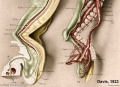
 Stage 11 historic-Davis1923-1a.jpg 643 × 800; 61 KB
 Stage 11 historic-Davis1923-1b.jpg 482 × 600; 41 KB
 Stage 11 historic-Davis1923-1c.jpg 321 × 400; 20 KB
 Stage 11 historic-Davis1923-2a.jpg 614 × 800; 83 KB
 Stage 11 historic-Davis1923-2b.jpg 461 × 600; 48 KB
 Stage 11 historic-Davis1923-2c.jpg 307 × 400; 23 KB
 Stage 11 historic-Davis1923-3a.jpg 800 × 492; 59 KB
 Stage 11 historic-Davis1923-3b.jpg 600 × 369; 41 KB
 Stage 11 historic-Davis1923-3c.jpg 400 × 246; 22 KB
 Stage 11 historic-Davis1923-4a.jpg 800 × 578; 63 KB
 Stage 11 historic-Davis1923-4b.jpg 600 × 434; 39 KB
 Stage 11 historic-Davis1923-4c.jpg 400 × 289; 21 KB
 Stage13.jpg 490 × 337; 6 KB
 Stage23 bf1a.jpg 800 × 616; 19 KB
 Stage23 bf1b.jpg 600 × 462; 12 KB
 Stage22 bf1b.jpg 600 × 450; 11 KB
 Stage21 bf1b.jpg 600 × 450; 14 KB
 Stage19 bf1a.jpg 800 × 600; 17 KB
 Stage19 bf1b.jpg 600 × 450; 11 KB
 Stage20 bf1a.jpg 800 × 600; 19 KB
 Stage20 bf1b.jpg 600 × 450; 11 KB
 Stage18 bf1a.jpg 800 × 600; 15 KB
 Stage18 bf1b.jpg 600 × 450; 10 KB
 Stage17 bf1a.jpg 800 × 600; 21 KB
 Stage17 bf1b.jpg 600 × 450; 13 KB
 Stage16 bf1a.jpg 800 × 600; 17 KB
 Stage16 bf1b.jpg 600 × 450; 11 KB
 Stage15 bf1a.jpg 800 × 600; 17 KB
 Stage15 bf1b.jpg 600 × 450; 11 KB
 Stage14 bf2a.jpg 800 × 600; 45 KB
 Stage14 bf2b.jpg 600 × 450; 26 KB
 Stage13 bf2a.jpg 800 × 600; 40 KB
 Stage13 bf2b.jpg 600 × 450; 24 KB
 HeartILP002.jpg 514 × 944; 60 KB
 HeartILP-draft004.jpg 1,508 × 329; 70 KB
 Cover of magazine.jpg 600 × 530; 91 KB
 Lightbulb.jpg 318 × 359; 13 KB
 Neural crest-derived cartilage.jpg 478 × 356; 40 KB
 Somite patterning.jpg 600 × 366; 58 KB
 Muscle elongation.png 518 × 600; 194 KB
 Picture 1.png 693 × 386; 158 KB
 Picture 2.png 1,028 × 640; 368 KB
 Mousevasc.gif 478 × 300; 29 KB
 Ossification endochondral 1a.jpg 600 × 800; 103 KB
 Ossification endochondral 1b.jpg 450 × 600; 64 KB
 Picture 3.png 861 × 636; 316 KB
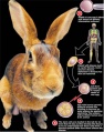
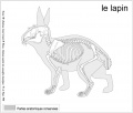 Le lapin.JPG 995 × 843; 78 KB
 Mouse forelimb.jpg 600 × 812; 71 KB
 Cartilage oligomeric protein.jpg 600 × 681; 258 KB
 Stage14-sem2a-limb.jpg 504 × 800; 40 KB
 Rabbithydrocephalus.jpg 1,518 × 549; 46 KB
 Rabbitspinabifida.jpg 1,707 × 573; 41 KB
 Rabbitmalformation2.jpg 354 × 497; 49 KB
 Cleftim1.jpg 754 × 370; 48 KB
- Walter Heape.pdf ; 1.32 MB
 Lab7 muscle-1.jpg 1,000 × 844; 92 KB
 Lab7 muscle-2.jpg 1,000 × 1,007; 164 KB
 Spina BifidaMyelomeningocele.jpg 886 × 299; 39 KB
 SpinabifidaMyeloschisis1.jpg 576 × 251; 30 KB
 Rabbitcleft1.jpg 737 × 437; 40 KB
 Bugs1.jpg 723 × 1,223; 78 KB
 Bugs5.jpg 727 × 1,224; 84 KB
 Day 4.5 Implantation 2.JPG 425 × 345; 9 KB
 Zygote Period.png 547 × 260; 31 KB
 Cleavage.png 956 × 185; 240 KB
 Blastula.png 677 × 184; 174 KB
 Larvae.png 610 × 317; 191 KB
 Cleaveage Period.png 563 × 260; 48 KB
 Last time Cleavage.png 747 × 169; 179 KB
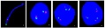
 Mouse fragilis2 expression.jpg 600 × 675; 60 KB
 Mouse genital ridge fragilis expression.jpg 600 × 457; 33 KB
 NIDCR.jpg 300 × 302; 143 KB
 Day 10.5.JPG 129 × 132; 8 KB
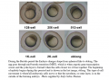
 Sizes.JPG 549 × 379; 18 KB
 Rabbit hybrid.jpg 372 × 472; 63 KB
 Bone-femur-a.jpg 638 × 800; 100 KB
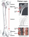 Bone-femur-b.jpg 479 × 600; 61 KB
 Stage31lat.gif 108 × 36; 3 KB
 File-Figure3-3.jpg 650 × 375; 123 KB
 Figure3-3.jpg 650 × 375; 123 KB
 Genetics of laboratory rodents.jpg 416 × 480; 19 KB
 Mice expressing GFP.jpg 400 × 300; 20 KB
 Morula.JPG 436 × 278; 10 KB
 Gastrula Period Drawing.png 970 × 408; 76 KB
 Pill.jpg 800 × 493; 115 KB
 Genetics.jpg 262 × 300; 11 KB
 Transgenicmouse.jpg 262 × 300; 11 KB
 Sperm-egg.jpg 400 × 274; 13 KB
 Mouse inner ear.jpg 478 × 508; 47 KB
 Adult hearing embryonic origins c.jpg 400 × 270; 20 KB
 External ear stages-14-23-adult a.jpg 800 × 524; 30 KB
 External ear stages-14-23-adult b.jpg 600 × 393; 20 KB
 External ear stages-14-23-adult c.jpg 400 × 262; 11 KB
 Mouse tooth stem cell.png 600 × 161; 70 KB
 Permanentteeth.jpg 400 × 280; 23 KB
- Looping animation 002.mov ; 212 KB
- Folding animation 001.mov ; 1.32 MB
- Folding animation 002.mov ; 503 KB
 Logonew.png 135 × 135; 8 KB
 Stage22 pancreas c.jpg 400 × 320; 38 KB
- Septation A 02 draft1.ppt ; 1.83 MB
- Septation A 02 draft2.ppt ; 2.41 MB
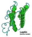 Leptin.jpg 317 × 363; 17 KB
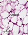 White adipose histology.jpg 400 × 500; 57 KB
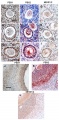
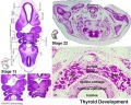
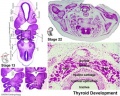
 Stage13 and 22 thyroid development.jpg 1,000 × 800; 283 KB
 Stage13 and 22 thyroid development b.jpg 600 × 480; 61 KB
 Stage13 and 22 thyroid development c.jpg 400 × 320; 32 KB
 Mouse seminiferous tubule EM.jpg 1,152 × 862; 352 KB
- Transrabbit.mov ; 142 KB
 FlashVideo.png 256 × 256; 18 KB
 Stage01animal.jpg 443 × 407; 102 KB
 Stage03animal.jpg 431 × 412; 111 KB
 XMRV-infected prostate cells.png 485 × 599; 528 KB
 Stage07dorsal.jpg 417 × 383; 73 KB
 Stage08dorsal.jpg 417 × 387; 84 KB
 Stage105vega.jpg 425 × 420; 80 KB
 Crabbit 3.jpg 700 × 700; 85 KB
 Cephalic plexus.png 600 × 557; 559 KB
 Stage19ant.jpg 358 × 398; 25 KB
 Rabbit1.jpg 700 × 700; 76 KB
 Crabbit 2.jpg 700 × 700; 73 KB
 5chris6m.jpg 200 × 141; 5 KB
 HeartILP draft heartwatermark.jpg 984 × 1,163; 78 KB
 HeartILP draft navigation.jpg 1,620 × 501; 69 KB
 Heart historic 002icon.jpg 320 × 240; 4 KB
 HeartILP draft Navigation2.jpg 1,278 × 323; 52 KB
 Blastomere mitosis 01 icon.jpg 150 × 102; 3 KB
- Newborn n 01.flv ; 2.41 MB
 Newborn-cranial-nerves.jpg 320 × 240; 9 KB
 HeartILP draft loopingseries.jpg 1,548 × 577; 85 KB
 Col00he.jpg 1,280 × 1,024; 117 KB
 HeartILP draft septationbloodflow.jpg 1,003 × 974; 101 KB
 ARecVG02.jpg 300 × 400; 113 KB
 Stage11 bf7a.jpg 800 × 600; 23 KB
 Stage11 bf7c.jpg 400 × 300; 4 KB
 Stage12 bf5a.jpg 800 × 600; 39 KB
 Stage12 bf5c.jpg 400 × 300; 6 KB
 Stage10 bf5a.jpg 800 × 600; 37 KB
 Stage10 bf5c.jpg 400 × 300; 7 KB
 Stage10 bf4a.jpg 800 × 600; 33 KB
 Stage10 bf4c.jpg 400 × 300; 11 KB
 HeartILP draft atimeline.jpg 1,772 × 769; 158 KB
 HeartILP draft itimeline.jpg 1,772 × 687; 133 KB
 HeartILP draft btimeline.jpg 1,658 × 556; 73 KB
 Foundmedium.jpg 180 × 135; 27 KB
 Aortic Arches (Drawing).jpg 499 × 418; 29 KB
 Caesarean delivery.jpg 450 × 310; 49 KB
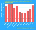 Caesarean rate 1999.jpg 440 × 365; 40 KB
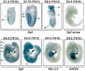 Mouse-Sp2 mRNA expression.png 600 × 503; 877 KB
 Mouse-E11.5.png 505 × 600; 337 KB
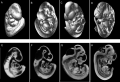 Mouse-E9.5 E10.5 E11.5 E12.5.png 599 × 407; 170 KB
 Mouse-E12.5.png 496 × 600; 278 KB
 Mouse-spermatozoa SLY protein.jpg 637 × 767; 352 KB
- Error creating thumbnail: File missingUSA birth location 1990–2006.jpg 600 × 415; 40 KB
 Uterus anatomical position.jpg 500 × 373; 62 KB
 Mouse primitive node cilia.jpg 592 × 981; 130 KB
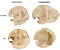
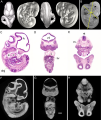
 Mouse CT E10.5 head 01.jpg 1,000 × 636; 154 KB
 Mark Hill 1989.jpg 1,420 × 2,089; 249 KB
 Bacterial vaginosis.png 1,000 × 755; 134 KB
 Mouse-model ovarian cord formation.jpg 600 × 368; 48 KB
 Human- adult ovary epithelial cords and primary follicles.jpg 600 × 384; 51 KB
 Menstrual cycle 02.jpg 467 × 515; 70 KB
 Ovary Histology - tunica albuginea.jpg 1,280 × 1,024; 336 KB
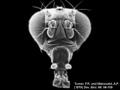 Fly-wild type head.jpg 320 × 240; 10 KB
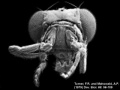 Fly-antennapedia head.jpg 320 × 240; 12 KB
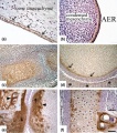
 Human Carnegie stage 15 HOXC5 expression.jpg 670 × 504; 90 KB
 Human Carnegie stage 13 GJA1 expression.jpg 706 × 470; 96 KB
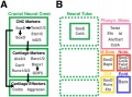
 Human Carnegie stage 13 stem cell markers.jpg 657 × 585; 78 KB
 Huntingtin structure.jpg 595 × 266; 39 KB
 Uterine tube histology 04.jpg 1,280 × 1,024; 253 KB
 Causes of neonatal death globally 2000.jpg 600 × 427; 135 KB
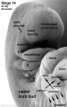
 Posterior forelimb bud identity.png 600 × 406; 895 KB
 Posterior forelimb bud identity.jpg 600 × 406; 46 KB
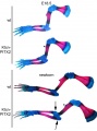
 Mouse- Hand2 forelimb buds.jpg 458 × 600; 48 KB
 Mouse-kidney model GDNF-FGF10.jpg 405 × 928; 59 KB
 Human- fetal week 10 sagittal planes.jpg 600 × 250; 29 KB
 Human- fetal week 10 upper body A.jpg 600 × 450; 104 KB
 Human- fetal week 10 upper body B.jpg 600 × 450; 105 KB
 Human- fetal week 10 upper body C.jpg 600 × 450; 109 KB
 Human- fetal week 10 upper body D.jpg 600 × 450; 106 KB
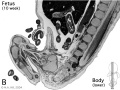
 Human- fetal week 10 lower body A.jpg 600 × 450; 96 KB
 Human- fetal week 10 lower body B.jpg 600 × 450; 93 KB
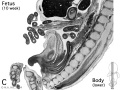 Human- fetal week 10 lower body C.jpg 600 × 450; 94 KB
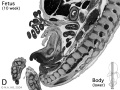 Human- fetal week 10 lower body D.jpg 600 × 450; 91 KB
 File-Human- fetal week 10 cerebellum A.jpg 347 × 284; 24 KB
 10wkcerebellumB.jpg 347 × 284; 21 KB
 Human- fetal week 10 heart ABCD.jpg 600 × 450; 133 KB
 Mouse- pancreas differentiation model.jpg 1,200 × 435; 96 KB
 Mouse- axial skeleton intervertebral disc.jpg 600 × 300; 40 KB
 Mouse- postnatal muscle-extensor digitorum longus.jpg 600 × 800; 44 KB
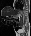
 Human- neural Chiari malformation.jpg 1,200 × 1,344; 212 KB
 Human- ventricular system cartoon 02.jpg 1,179 × 1,254; 115 KB
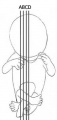 Human- fetal week 10 planes.jpg 174 × 365; 7 KB
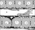 Liver-sinusiod cartoon.jpg 600 × 523; 51 KB
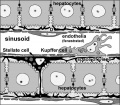 Liver-sinusoid-label cartoon.jpg 600 × 523; 58 KB
 Muscle-reorganization microtububules and centrosome protein.jpg 1,000 × 1,125; 140 KB
 Muscle- centrosome protein localizes cytoplasmic site nuclear envelope.jpg 1,000 × 1,840; 293 KB
 Mouse- germ cell development.jpg 1,000 × 588; 74 KB
 Stage10 bf6a.jpg 800 × 600; 63 KB
 Stage10 bf6c.jpg 400 × 300; 22 KB
 Stage11day25somite19-dorsal-sem2.jpg 668 × 1,000; 132 KB
 Blastocyst- TPR53 expression.jpg 1,000 × 1,089; 68 KB
 Ovary- atretic follicle 03.jpg 790 × 593; 202 KB
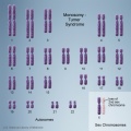 Chromosome- monosomy.jpg 504 × 504; 164 KB
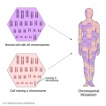 Chromosome- mosaicism.jpg 504 × 504; 136 KB
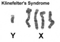 Klinefelter syndrome YXXXX.jpg 244 × 172; 5 KB
 Endoderm 001 icon.jpg 556 × 600; 28 KB
- 2010-Embryo Lab 170510-603.mp3 ; 2.02 MB
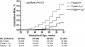 Preconceptional folate and risk preterm birth.png 600 × 329; 92 KB
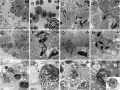
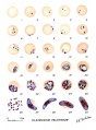 Stages Plasmodium falciparum.jpg 674 × 914; 65 KB
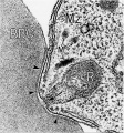 Malaria and red blood cell em.jpg 500 × 536; 82 KB
 Placental cord ultrasound 01.jpg 585 × 1,309; 192 KB
 Placenta- first trimester histology x40.jpg 1,000 × 800; 124 KB
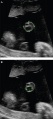
 Ultrasound 12 week icon.jpg 500 × 374; 46 KB
- PediNeuroLogic Larsen welcome.flv ; 5.3 MB
- PediNeuroLogic Intro 01.flv ; 848 KB
- PediNeuroLogic Intro 02.flv ; 147 KB
- PediNeuroLogic Intro 03.flv ; 468 KB
- PediNeuroLogic Intro 04.flv ; 407 KB
- PediNeuroLogic Intro 05.flv ; 923 KB
- PediNeuroLogic Intro 06.flv ; 872 KB
- PediNeuroLogic Intro 07.flv ; 294 KB
- PediNeuroLogic Intro 08.flv ; 781 KB
- Newborn n 03.flv ; 410 KB
- Newborn n 04.flv ; 1.06 MB
- Newborn n 05.flv ; 1.11 MB
- Newborn n 06.flv ; 1.03 MB
- Newborn n 07.flv ; 1.49 MB
- Newborn n 08.flv ; 465 KB
- Newborn n 09.flv ; 2.4 MB
- Newborn n 10.flv ; 349 KB
- Newborn n 11.flv ; 542 KB
- Newborn n 12.flv ; 2.4 MB
- Newborn n 13.flv ; 1.24 MB
- Newborn n 14.flv ; 487 KB
- Newborn n 16.flv ; 543 KB
- Newborn n 17.flv ; 1.26 MB
- Newborn n 18.flv ; 732 KB
- Newborn n 19.flv ; 436 KB
- Newborn n 20.flv ; 2.14 MB
- Newborn n 21.flv ; 1.14 MB
- Newborn n 22.flv ; 1.52 MB
- Newborn n 23.flv ; 815 KB
- Newborn n 24.flv ; 744 KB
- Newborn n 25.flv ; 710 KB
- Newborn n 26.flv ; 2.12 MB
- Newborn ab 27.flv ; 1.39 MB
- Newborn n 28.flv ; 899 KB
- Newborn ab 01.flv ; 2.61 MB
- Newborn ab 02.flv ; 1.14 MB
- Newborn ab 03.flv ; 235 KB
- Newborn ab 04.flv ; 1.41 MB
- Newborn ab 05.flv ; 907 KB
- Newborn ab 06.flv ; 1.01 MB
- Newborn ab 07.flv ; 854 KB
- Newborn ab 08.flv ; 894 KB
- Newborn ab 09.flv ; 1.76 MB
- Newborn ab 10.flv ; 501 KB
- Newborn ab 11.flv ; 702 KB
- Newborn ab 12.flv ; 741 KB
- Newborn ab 13.flv ; 779 KB
- Newborn ab 14.flv ; 416 KB
- Newborn ab 15.flv ; 934 KB
- Newborn ab 16.flv ; 922 KB
- Newborn ab 17.flv ; 1.57 MB
- Newborn ab 18.flv ; 1.36 MB
- Newborn ab 19.flv ; 878 KB
- Newborn ab 20.flv ; 2.56 MB
- Newborn ab 21.flv ; 1.54 MB
- Newborn ab 23.flv ; 1.18 MB
- Newborn ab 24.flv ; 1,008 KB
- Newborn ab 25.flv ; 518 KB
- Newborn ab 26.flv ; 1.45 MB
- Newborn ab 28.flv ; 920 KB
- Newborn n 27.flv ; 1.44 MB
- Dev anat 01.flv ; 288 KB
- Dev anat 02.flv ; 100 KB
- Dev anat 03.flv ; 325 KB
- Dev anat 04.flv ; 532 KB
- Dev anat 05.flv ; 168 KB
 Newborn n 01.jpg 320 × 240; 11 KB
 PediNeuroLogic Intro 08.jpg 320 × 240; 13 KB
 PediNeuroLogic Intro 07.jpg 320 × 240; 11 KB
 PediNeuroLogic Intro 06.jpg 320 × 240; 16 KB
 PediNeuroLogic Intro 05.jpg 320 × 240; 6 KB
 PediNeuroLogic Intro 04.jpg 320 × 240; 21 KB
 PediNeuroLogic Intro 03.jpg 320 × 240; 11 KB
 PediNeuroLogic Intro 02.jpg 320 × 240; 14 KB
 PediNeuroLogic Intro 01.jpg 320 × 240; 9 KB
 Dev anat 05.jpg 320 × 240; 16 KB
 Dev anat 03.jpg 320 × 240; 10 KB
 Model for multiple XIST RNA anchor points.jpg 1,000 × 622; 138 KB
 Non-canonical Wnt signaling.jpeg 1,280 × 980; 215 KB
 Pig sperm capacitation 01.jpg 1,000 × 840; 204 KB
 Mean maternal age term or preterm birth.jpg 600 × 393; 22 KB
 Spontaneous births.jpg 1,000 × 431; 63 KB
 Multibrowser logo circle.jpg 265 × 203; 37 KB
 XIST human embryonic stem cells 01.jpg 1,000 × 388; 50 KB
 Stage19 bf2a.jpg 683 × 1,024; 242 KB
 Stage15 bf2a.jpg 959 × 1,024; 344 KB
 Stage7 bf5a.jpg 1,024 × 721; 690 KB
 Stage23 bf2a.jpg 683 × 1,024; 349 KB
 Stage23 bf2c.jpg 160 × 240; 32 KB
 Stage19 bf3a.jpg 1,024 × 685; 231 KB
 Stage19 bf3b.jpg 500 × 334; 83 KB
 Figure 1 Adult Embryo Ear - final.psd ; 5.87 MB
- Figure 2 Stage 13 - final.psd ; 6.4 MB
- Figure 3 Stage 19 - final.psd ; 7.82 MB
- Figure 4 stage22 - final400dpi.psd ; 5.6 MB
- Figure 5 Fetal - final400dpi.psd ; 5.47 MB
 Limb bud growth model 01.jpg 600 × 261; 30 KB
 Limb bud growth model 02.jpg 600 × 637; 80 KB
- 2001winston.doc ; 68 KB
 Notochordal interaction with nucleus pulposus.jpg 478 × 360; 92 KB
 Cervical vertebra.jpg 767 × 514; 71 KB
 Gastrointestinal tract growth 01.jpg 100 × 176; 5 KB
- Fetal heartbeat 01.mp3 ; 225 KB
 Stage 13 image 100-icon.jpg 140 × 200; 17 KB
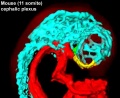 Mouse-Cephalic-plexus-11somite.jpg 600 × 491; 59 KB
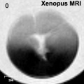
 Xenopus MRI 01.jpg 512 × 512; 28 KB
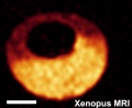 Xenopus MRI 02.jpg 544 × 445; 23 KB
 Xenopus MRI 03.jpg 320 × 320; 10 KB
- Human development 001.mov ; 2 MB
- Ovulation 001.mov ; 376 KB
- Ovulation 002.mov ; 422 KB
- Somitogenesis 001.mov ; 359 KB
- Primordial germ cell 001.mov ; 680 KB
- Primordial germ cell 002.mov ; 3.7 MB
- Primordial germ cell 003.mov ; 695 KB
 Spermatozoa animation.gif 300 × 200; 123 KB
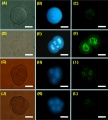
 Oct4 staining on USSC-derived spheres-2.jpg 1,200 × 1,504; 355 KB
 Mouse - stomach 01.png 599 × 600; 1.45 MB
 Omphalocoele MRI.jpg 553 × 631; 52 KB
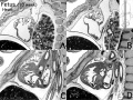
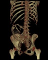 Fetus 35 week CT.jpg 700 × 874; 62 KB
 Liver hepatocyte from stem cell.png 600 × 444; 96 KB
- Neural - Sylvian fissure.mov ; 476 KB
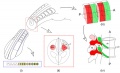
 Mouse head-neural crest 01.jpg 900 × 339; 48 KB
 Mouse-E10.5-Sox10.jpg 400 × 463; 24 KB
 Mouse-E8.5-Sox10.jpg 400 × 463; 20 KB
 Nejm199208273270903 t4.gif 111 × 103; 5 KB
 CVS table.gif 111 × 103; 5 KB
 Spina bifida occulta.jpg 2,480 × 3,507; 1.4 MB
 Muscle tutorial front.jpg 850 × 632; 130 KB
 In vitro kidney development.png 600 × 412; 1.08 MB
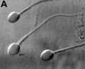 Human-spermatozoa 01.jpg 1,000 × 805; 90 KB
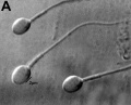 Human-spermatozoa 01a.jpg 800 × 644; 66 KB
 Human-spermatozoa 01c.jpg 400 × 322; 22 KB
 Chicken - somite.jpg 1,200 × 375; 269 KB
 Muscle- C2C12 differentiation.jpg 600 × 889; 101 KB
 Mouse-neural crest Sox10 E10.5.jpg 1,000 × 420; 55 KB
 Mouse-heart E17.5.jpg 353 × 1,000; 145 KB
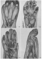
 Mouse-E17.5 normal and abnormal limb.jpg 661 × 848; 105 KB
 Mouse-E17.5 foot.jpg 385 × 500; 29 KB
 Mouse-E17.5-foot-large.jpg 600 × 779; 53 KB
 Mouse- facial branchiomotor neuron migration.jpg 600 × 841; 104 KB
 Pineal histology 002.jpg 1,000 × 800; 241 KB
 Pineal histology 003.jpg 800 × 640; 166 KB
 Mouse-pancreas islets-sweet taste receptor.jpg 1,050 × 261; 38 KB
 Salamander- early development.jpg 1,200 × 887; 85 KB
 Human- Stage 22 integument 01.jpg 1,000 × 750; 205 KB
 Human- Stage 22 integument 03.jpg 600 × 450; 95 KB
 Human- Stage 22 integument 04.jpg 400 × 300; 48 KB
 Protein synthesis - translation.gif 250 × 250; 5.54 MB
- Protein synthesis - translation.mov ; 4.54 MB
- Protein synthesis - translation.mp4 ; 2.7 MB
 Chromoabno.JPG 672 × 633; 33 KB
- Timeline Embryology.doc ; 44 KB
 Human bilateral renal agenesis-hypoplasia-dysplasia.png 1,613 × 906; 2.75 MB
- Timeline Embryology.pdf ; 196 KB
 Human- fetal week 10 head A1.jpg 1,200 × 1,088; 159 KB
 Australian nursing and midwifery labour force 2008.jpg 800 × 657; 110 KB
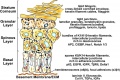 Epidermis cartoon.jpg 800 × 536; 126 KB
 Mouse-embryo granzyme G.jpg 899 × 1,000; 90 KB
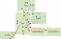 Y chromosome haplogroup distribution.jpg 800 × 522; 42 KB
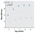 Anogenital distance from birth to 2 years.jpg 565 × 545; 34 KB
 Mouse- early gene expression 01.jpg 800 × 660; 49 KB
 Disomic XY spermatozoa.jpg 393 × 440; 7 KB
 Corpora amylacea in prostatic stromal smooth muscle.jpg 800 × 728; 167 KB
- Error creating thumbnail: File with dimensions greater than 12.5 MPWorm - embryonic cell lineage 01.jpg 10,389 × 1,336; 598 KB
 Mouse- intervertebral disc development.jpg 1,454 × 728; 95 KB
 Pre-auricular appendages locations.jpg 306 × 400; 17 KB
 Pre-auricular fistulae locations.jpg 296 × 398; 16 KB
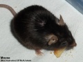 Mouse- C57BL6J strain.jpg 1,000 × 750; 125 KB
 Human- spermatozoa NANOG expression.jpg 1,000 × 333; 77 KB
 Mouse- E7.5 early late.jpg 490 × 1,108; 68 KB
 Mouse- E7.5 late bud 02.jpg 442 × 1,000; 77 KB
 Mouse- E7.5 late bud 03.jpg 442 × 1,000; 50 KB
 Mouse-mammary-E10-E11.jpg 600 × 406; 40 KB
 Mouse-mammary-E11.5.jpg 600 × 406; 78 KB
 Mouse-mammary-E12.0.jpg 600 × 398; 89 KB
 Mouse-mammary-E13.jpg 600 × 415; 57 KB
 Beckwith-Wiedemann syndrome ear lobe creases.jpg 257 × 414; 12 KB
 Mouse- postnatal osteoblasts.jpg 400 × 537; 59 KB
 Mouse- preimplantation gene expression 01.jpg 726 × 1,959; 226 KB
- Human blastocyst day 5-6 small.mov ; 509 KB
 Feverfew 01.jpg 600 × 900; 76 KB
 Human ovulation 04.jpg 734 × 583; 64 KB
 Human ovulation 02.jpg 734 × 583; 68 KB
 Human ovulation 03.jpg 734 × 583; 76 KB
 Human ovulation 05.jpg 734 × 583; 71 KB
 Telencephalon development signals.jpg 1,000 × 711; 62 KB
 SHmedium.jpg 180 × 135; 30 KB
- 2010 Wiki Stats.xls ; 9 KB
 Forster034.jpg 1,003 × 717; 158 KB
 Baileytable03.jpg 821 × 667; 73 KB
 226.jpg 565 × 705; 71 KB
 231.jpg 747 × 493; 69 KB
 Bailey239+240.jpg 944 × 496; 128 KB
 Bailey239 240.jpg 944 × 496; 128 KB
 Bailey324 325.jpg 706 × 888; 114 KB
 Baileytable08.jpg 968 × 570; 76 KB
 Appendicular skeleton small.jpg 220 × 407; 12 KB
 Primordial follicle activation signaling model.jpg 403 × 440; 32 KB
 Clinical Embryology M. Brookes and A. Zietman.jpg 371 × 498; 43 KB
 Clinical Maternal-Fetal Medicine H.N. Winn and J.C. Hobbins.jpg 345 × 475; 28 KB
 Conjoined Twins R.S. Spencer.jpg 301 × 475; 21 KB
 Human Fertilisation & Embryology R.G. Lee and D. Morgan.jpg 327 × 500; 43 KB
 Molecular Embryology J.M. Barry.jpg 320 × 500; 25 KB
 Borwn014.jpg 800 × 770; 91 KB
 Carnegie Institute of Washington small logo.jpg 300 × 297; 30 KB
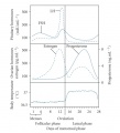
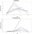 Menstrual cycle- estradiol and progesterone graph.jpg 558 × 593; 95 KB
 Model - TGF regulation of SHH.jpg 600 × 372; 45 KB
 Chicken neural Plxdc2 expression.jpg 1,000 × 850; 121 KB
 Chicken neural tube thickening.jpg 529 × 600; 69 KB
 Lymph node 05.jpg 1,000 × 800; 180 KB
- Mouse adult lymph node 01.mov ; 1.58 MB
- Mouse adult lymph node 02.mov ; 2.3 MB
- Mouse adult lymph node 03.mov ; 1.11 MB
- Mouse adult lymph node 04.mov ; 568 KB
- Mouse adult lymph node 05.mov ; 1.41 MB
- Mouse adult lymph node 06.mov ; 2.17 MB


.jpg)


