Search results
From Embryology
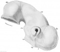
File:Keibel Mall 2 165.jpg ==Fig. 165. Mesencephalon and telencephalon of a human embryo from the end of the fourth week== he, cerebral vesicle; in, infundibulum; ma, mammillary process; mi, mesencephalon; op, torus opticus; t, lamina ferminalis; zw, diencephalon.(1,037 × 900 (166 KB)) - 10:32, 21 February 2014
File:Primary Brain Vesciles.pdf ...3 primary brain vesicles are formed; forebrain (prosencephalon) midbrain (mesencephalon), and hindbrain (rhombencephalon). These structures then differentiate into(453 KB) - 09:52, 25 October 2017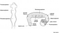
File:CNS primary vesicles.jpg # midbrain (mesencephalon) ...ondary vesicles.jpg|5 secondary vesicles]] (telencephalon - diencephalon - mesencephalon - metencephalon - myelencephalon)(987 × 562 (49 KB)) - 16:48, 18 May 2017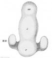
File:Keibel Mall 2 161.jpg .... II2, p. 183, Fig. 187.) Xca. 50. au, opiio vesicle; e, telencephalon; m, mesencephalon; z, diencephalon.(621 × 700 (46 KB)) - 10:34, 21 February 2014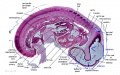
File:Embryo stage13- brain flexures.jpg # cranial (cephalic) flexure - pushes mesencephalon upwards(1,000 × 623 (82 KB)) - 11:43, 17 May 2010
File:Week4.jpg ...arising from the neural crest cells contributed by the prosencephalon. The mesencephalon is also believed to contribute neural crest cells for migration.(521 × 712 (75 KB)) - 19:12, 3 October 2012
File:Primary Brain.png ...3 primary brain vesicles are formed; forebrain (prosencephalon) midbrain (mesencephalon), and hindbrain (rhombencephalon). These structures then differentiate into(1,005 × 655 (681 KB)) - 12:39, 26 October 2017
File:Johnson1917 plate02fig01.jpg ...numbers are indicated brain and spinal-cord neuromeres, numbered from the mesencephalon backward. The black lines and numbers indicate the position of the body-seg(800 × 460 (46 KB)) - 10:29, 27 April 2012
File:Bailey363.jpg P.. prosencephalon; M., mesencephalon; R., rhombencephalon ; Ms., spinal cord; cw., chiasma eminence; J., infundi(876 × 373 (46 KB)) - 16:35, 29 January 2011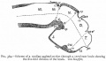
File:Bailey364.jpg T., Telencephalon; D., diencephalon; M., mesencephalon; Mt., metencephalon; Ml., myelencephalon; c., cerebellum; cc., cerebellar c(809 × 465 (53 KB)) - 15:13, 19 June 2011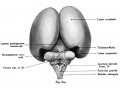
File:Kollmann614.jpg ...nvexity of the thalamus. The occipital lobes diverge, making the midbrain (mesencephalon) namely the quadrigeminal plate in large part is visible. occipitales weichen auseinander, wodurch das Mittelhirn (Mesencephalon) und zwar(679 × 502 (33 KB)) - 18:11, 18 October 2011
File:Johnson1917 plate02.jpg ...numbers are indicated brain and spinal-cord neuromeres, numbered from the mesencephalon backward. The black lines and numbers indicate the position of the body-seg(827 × 1,000 (114 KB)) - 17:17, 24 April 2012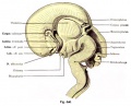
File:Kollmann608.jpg ...Monro), the roof of the diencephalon, the investment of the corpus of the mesencephalon, aqueduct, and the Isthmus bridge curvature highlighted by labels. The corp pineale, des Mesencephalon, Aquaeductus, Isthmus und die Brückenkrümmung(697 × 563 (77 KB)) - 19:50, 17 October 2011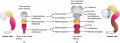
File:Secondary brain vesicle.jpeg - Mesencephalon does not further develop into a secondary brain vesicle(1,204 × 431 (209 KB)) - 16:23, 24 October 2017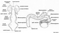
File:CNS secondary vesicles.jpg # [[Neural_-_Mesencephalon_Development|Mesencephalon]](987 × 562 (81 KB)) - 11:04, 15 May 2019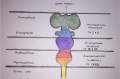
File:Brain Development 2.png ...e secondary subdivisions: Prosencephalon (Telencephalon and Diencephalon), Mesencephalon, and Rhombencephalon (Metencephalon, Myelencephalon). Also, shows the crani(1,490 × 986 (2.12 MB)) - 16:25, 25 October 2017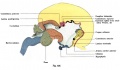
File:Kollmann626.jpg des Dieocephaloo (Zwischenhims), des Mesencephalon (Mittelhirns), des Klein-(971 × 569 (76 KB)) - 17:19, 17 October 2011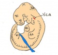
File:Day 11.5 Lens Vesicle completely separated from surface.JPG V.ca = vena cardinalis anterior, Tel= telenchephalon, M= mesencephalon, Rh= rhombencephalon, He= heart, Hl= hindlimb bud, Aa= forelimb bud, 1= 1st(511 × 484 (29 KB)) - 13:01, 25 June 2014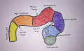
File:Brain Development.png ...e secondary subdivisions: Prosencephalon (Telencephalon and Diencephalon), Mesencephalon, and Rhombencephalon (Metencephalon, Myelencephalon)(1,390 × 842 (1.92 MB)) - 16:22, 25 October 2017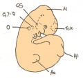
File:Day 11 Closure of lens vesicle.JPG M= Mesencephalon, Tel = telencephalon, Hl = hindlimb bud, Aa= Forelimb bud, O= otic vesicle,(560 × 528 (27 KB)) - 13:19, 18 August 2014


