Search results
From Embryology
Page title matches
- ...egory includes pages and images that relate to the [[Carnegie Collection]] Embryo No. {{CE190}}. [[Carnegie Embryos]]2 members (0 subcategories, 0 files) - 17:48, 8 June 2020
Page text matches
- ...e>[[Category:Template]][[Category:Carnegie Collection]][[Category:Carnegie Embryo]][[Category:Fetal]][[Category:Human]]</noinclude>183 bytes (19 words) - 17:41, 8 June 2020
- ===All Carnegie Embryos listed=== [[Category:Carnegie Embryo 6]]5 KB (496 words) - 11:36, 29 July 2018
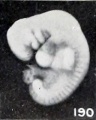
File:Mall Meyer1921 fig190.jpg ==Fig. 190. A well-preserved cyema 5.5 mm== [[Category:Carnegie Embryo 1380]](256 × 320 (14 KB)) - 07:28, 22 January 2020- '''Modern Notes:''' [[Carnegie stage 2]] | [[Week 2]] [[:Category:Carnegie Embryo 8190|'''Carnegie, No. 8190''']]3 KB (382 words) - 11:34, 26 July 2020
- HPL (human placental lactogen) - consists of 190 amino acids. It modifies metabolic state of mother during the pregnancy to 1. What Carnegie stages occur during week 3 and week 4?7 KB (1,020 words) - 08:50, 25 October 2010
- [[Carnegie stage 3]] ...eeding stage in development. It is to be remembered that at all stages the embryo is a living organism, that is, it is a going concern with adequate mechanis13 KB (1,877 words) - 15:40, 26 June 2019
- [[Carnegie stage 1]] ...a. 0.1 mm) and weight (ca. 0.004 mg) of the organism at fertilization, the embryo is "''schon ein individual-spezifischer Mensch''" (Blechschmidt, 1972). The11 KB (1,686 words) - 23:53, 6 June 2018
- ...10) the right side of the neck and thorax was cut in sagittal sections. In embryo (9) and the six foetuses the neck and upper part of the thorax were cut in (10) Embryo, 9th-10th week, no measurement recorded, sagittal sections at 15 9, of righ47 KB (7,825 words) - 22:31, 6 March 2017
- ...istoric human study. This embryo has been classified as [[Carnegie stage 7|Carnegie Stage 7]]. [[Carnegie stage 8|Carnegie Stage 7]]32 KB (5,184 words) - 13:46, 31 July 2017
- ....jpg|90px|left]] This historic 1941 paper by Gilmour describes early human embryo blood formation. ....065 x 0.045 mm. Age about 16 days, probably slightly younger than Peters’ embryo (1899).92 KB (14,488 words) - 11:45, 28 July 2020
- ...ibes gastrointestinal tract smooth muscle development using a number of [[Carnegie Collection]] embryos. {{Carnegie Collection fetal table}}28 KB (4,448 words) - 11:30, 28 May 2018
- ...y of fertilization and early cleavage in the human. In vitro fertilization embryo. London: Churchill Livingstone. ...e human. In Trounson, A.O. and Wood, C. (eds.). In Vitro Fertilization and Embryo Transfer. Churchill Livingstone, London.46 KB (6,369 words) - 07:52, 30 December 2018
- 190 Old Derby Street Bilaminar embryo (days 6— 14)17 KB (2,253 words) - 13:01, 31 July 2019
- ...|90px|left]] This historic 1929 paper by Ingalls (1880-1949) describes a [[Carnegie Collection]] human embryos segmental thickenings in the dorsal ectoderm of ...(726), appear as shown in figure A. This represents a left lateral view of embryo no. 155, C.R. 11.8 mm. The thickenings or dises are indicated by the row of25 KB (4,158 words) - 21:41, 11 May 2019
- resulting brain in the human embryo. Cells Tissues Organs. 2013;197(3):178-95. of the human brain. Ann Anat. 2008;190(2):105-18. doi:29 KB (3,670 words) - 11:12, 23 July 2015
- ...the ossicles in the middle ear were independent in different locations. At Carnegie Stage 17 a homogeneous interzone clearly defined the incus and malleus anla Cross-section of human embryo [[Carnegie stage 22]] during [[Week 8]].32 KB (4,766 words) - 04:18, 5 July 2022
- ...in the embryo as at birth, but the deformities of the head and neck of the embryo are of such a nature that it can not survive long enough to admit of compar ...xomphaly. Other anomalies, however, are more difficult to recognize in the embryo as sharply defined malformations.32 KB (4,425 words) - 11:17, 22 November 2012
- ...bryo microfiche images were developed by '''Dr M. Smith '''and the 6mm Pig Embryo microfiche images by '''Dr M. Smith''' and '''Prof. I. Tork'''. Original website undergraduate Notes (Anat 2006) included with embryo serial images are edited with permission of '''Dr B. Freeman''', from his '34 KB (4,907 words) - 12:42, 19 May 2020
- ...onare Geschlechtzellen’ in the intestinal epithelium of a four~weeks human embryo. The supposed sex cells were disposed in such a way as to suggest an active ...considered to be germ cells in the lateral plates of mesoderm of a 2.3—mm. embryo, and as these plates were folded under the gut in 2.8-mm. embryos, the germ56 KB (9,121 words) - 18:37, 25 May 2019
- ...Mall describes the human embryos in the collection that would become the [[Carnegie Collection]]. There is also a [[:File:1904 - Catalogue of the collection of [[Carnegie Collection]] | [[Carnegie Embryos]]21 KB (2,470 words) - 23:39, 9 August 2018
