Search results
From Embryology
- ===All Carnegie Embryos listed=== [[Category:Carnegie Embryo 6]]5 KB (496 words) - 11:36, 29 July 2018

File:Hertig1946b fig09b.jpg ==Fig. 9.B. A 3.2 mm embryo with amnion removed== ...lom (body cavity) within the embryo. Carnegie {{CE6488}}, sequence 2, X15. 104(800 × 586 (26 KB)) - 17:35, 7 August 2017- ==Human Embryo (17.8mm) == ...nd dimensions suggest that it is a [[Carnegie_stage_19|Carnegie stage 19]] embryo (Week 7, 48 - 51 days, 16 - 18 mm).856 bytes (121 words) - 10:05, 9 April 2014
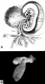
File:Hertig1946b fig09.jpg ==Fig. 9. A 2.5 mm and 3.2 mm embryo== ...telline duct as the digestive tract becomes more mature. The growth of the embryo and its surrounding amnion have resulted in the partial approximation of th(800 × 1,481 (169 KB)) - 17:34, 7 August 2017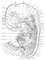
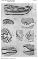
File:Human Embryo 17.8mmCNS GIT.jpg ==Human Embryo (17.8mm) Gastrointestinal Tract== ...icle of the heart, and in part the urogenital system of a 17.8 mm. hurrian embryo (H.E.C.839).(500 × 678 (105 KB)) - 17:27, 6 November 2015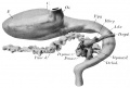
File:Thyng1914 fig02.jpg ==Fig. 2. Gastrointestinal Tract Embryo HEC839== ...nd dimensions suggest that it is a [[Carnegie_stage_19|Carnegie stage 19]] embryo (Week 7, 48 - 51 days, 16 - 18 mm).(1,189 × 800 (112 KB)) - 13:22, 18 May 2014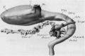
File:Human Embryo 17.8mm GIT.jpg ==Human Embryo (17.8mm) Gastrointestinal Tract== ...nd dimensions suggest that it is a [[Carnegie_stage_19|Carnegie stage 19]] embryo (Week 7, 48 - 51 days, 16 - 18 mm).(600 × 401 (38 KB)) - 17:55, 23 May 2016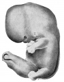
File:Thyng1914 fig01.jpg ==Fig. 1. External features of Embryo HEC839== ...embryo are seen in profile view in text figure 1, a reproduction of figure 104 in Minot's (1910) "Laboratory text-book of embryology," also in part in pla(786 × 1,000 (140 KB)) - 17:32, 6 November 2015- ! Length of Embryo (mm) ! Embryo No.,3 KB (282 words) - 12:25, 6 February 2018
File:Gage1905-plate05.jpg Fig. 14. From a photograph (X 47%) of Sec. 25 of the human embryo 148 (cf. Figs. 1, 2, 4). It shows: The neural tube just at the ventral bord Fig. 18. From a photograph (X 160) of Sec. 104, Homo 148, showing the pronephric tubule of the right side (Fig. 17) with i(979 × 1,500 (271 KB)) - 14:34, 18 August 2016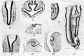
File:Gage1905-plate5.jpg Fig. 14. From a photograph (X 47%) of Sec. 25 of the human embryo 148 (cf. Figs. 1, 2, 4). It shows: The neural tube just at the ventral bord Fig. 18. From a photograph (X 160) of Sec. 104, Homo 148, showing the pronephric tubule of the right side (Fig. 17) with i(2,244 × 1,500 (522 KB)) - 14:34, 18 August 2016- ...hilly 1987|link=Embryology History - Ronan O'Rahilly|Ronan O'Rahilly (1987 Carnegie Labs)]] ...t study,<ref name=Weller1933>{{Ref-Weller1933}}</ref> used the following [[Carnegie Collection]] embryos: stage {{CS9}} (No. {{CE1878}}), {{CS10}} ({{CE391}};8 KB (1,113 words) - 18:19, 16 March 2020
- This {{Embryology}} category shows pages and media related to [[Carnegie stage 8]] of embryonic development that occurs during [[Week 3]] (post-fert {{Carnegie stage 8 links}}169 members (30 subcategories, 81 files) - 11:07, 8 August 2017
- ...the earlier months are even more rare. Streeter (’19) reported that in the Carnegie collection there were only forty—three specimens, of which all but two we ...hments of the two yolk stalks lay at different regions of the chorion. An embryo was present in each amniotic sac (fig. 1).8 KB (1,338 words) - 16:32, 27 November 2017
- The embryo is now 1.0 - 1.5 mm in size. {{Carnegie stage 8 links}}21 KB (2,879 words) - 00:32, 13 April 2018
- ...ng the ""Biggart" [[Carnegie stage 7]] and the younger "Macafee" embryo [[Carnegie stage 5]]. {{Carnegie stage 5 links}}20 KB (3,236 words) - 08:38, 5 September 2017
- == Carnegie Stage Table 1== <center> [[Carnegie_stage_13_-_serial_sections|Stage 13/14 shown in serial embryo sections]] series of Embryology Program</center>39 KB (4,806 words) - 23:27, 21 November 2013
- ...er birth. This development generates the most complex structure within the embryo and the long time period of development means in utero insult during pregna Neuralation begins at the trilaminar embryo with formation of the notochord and somites, both of which underly the ecto9 KB (1,372 words) - 09:17, 14 May 2020
- [[File:Human Carnegie stage 10-23.jpg|thumb|300px|Carnegie Embryos]] ...collection numbering also incorporated the Blechschmidt embryo collection (Carnegie Nos. 10315-10434 ) in 1972, the collection embryos have now been returned t43 KB (5,162 words) - 16:44, 28 April 2018
- ...his historic 1945 paper by Shaner describes a Carnegie Stage {{CS9}} human embryo of two to three pairs of somites. {{Carnegie stage 9 links}}20 KB (3,219 words) - 08:51, 13 October 2020


