Search results
From Embryology
Page title matches
- [[Penis Development|penis]]<noinclude>[[Category:Template]][[Category:Term Link]][[Category:Male]][[C130 bytes (14 words) - 08:17, 14 April 2018
- ...are: (1) The corpus cavernosum in males and females extends into the glans penis and clitoris, respectively, during the ambisexual stage (9 weeks) and thus External genitalia; Genital tubercle; Hypospadias; Penis; Prepuce; Urethra14 KB (2,019 words) - 14:05, 29 January 2019
- The external male genitalia consists of the {{penis}} and scrotum containing the {{testis}}. ...bryology_19-4#Development_of_the_Penis_and_Scrotum|1912 Development of the Penis and Scrotum]] | [[Paper - Notes on the development of the prepuce (1935)|1922 KB (3,254 words) - 13:19, 7 February 2020
- ...x|left]] This historic 1914 paper by deLima is an early description of a {{penis}} abnormality. '''Modern Notes:''' {{Hypospadia}} | {{penis}}9 KB (1,548 words) - 23:22, 3 March 2020
- NOTE ON A CASE OF BIFID PENIS, WITH PENIAL HYPOSPADIA. By J. A. Pires ve Lima, Professor of Topographical On the mere inspection of the penis, this organ is found to be short and9 KB (1,531 words) - 23:19, 3 March 2020
- ...24 paper by Lipschutz describes rare male human genital abnormality of the penis. '''Modern Notes:''' {{genital abnormalities}} | {{penis}}2 KB (386 words) - 08:33, 6 February 2020
- A NOTE ON A CASE OF BIFID PENIS A cass of bifid penis with penile hypospadias is described by Pires de Lima (1),2 KB (336 words) - 08:32, 6 February 2020
Page text matches
- [[Penis Development|penis]]<noinclude>[[Category:Template]][[Category:Term Link]][[Category:Male]][[C130 bytes (14 words) - 08:17, 14 April 2018
- ...Intestine]][[Category:Genital]][[Category:Abnormal Development]][[Category:Penis]][[Category:Historic Embryology]][[Category:1910's]]</noinclude>454 bytes (57 words) - 23:17, 3 March 2020
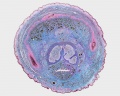
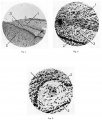
File:Male histology 002.jpg ==Male Histology - Penis== * cross-section of developing penis (Trichrome stain).(1,280 × 1,024 (490 KB)) - 00:21, 23 May 2012
File:Prepuce.jpeg ...1935. The drawings resemble and illustrate the observations of the foetal penis at different gestational age, depicting that the prepuce is a gradually gro The three images (A, B, C) show the overall continuous change of the penis in terms of the prepuce.(2,293 × 767 (382 KB)) - 17:27, 24 October 2014- ...aper - A note on a case of bifid penis (1924)|'''A note on a case of bifid penis''']]. (1924) {{J Anat.}} 58: 254-255. [https://www.ncbi.nlm.nih.gov/m/pubm385 bytes (50 words) - 23:32, 5 February 2020
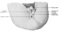
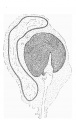
File:Keibel Mall 2 642.jpg The phallus has become the glans penis, the scrotal swellings are fully developed and the unpaired scrotal area is ...egory:Genital]][[Category:Male]][[Category:Historic Embryology]][[Category:Penis]](1,200 × 644 (95 KB)) - 12:57, 5 May 2019- | on inferior surface of glans penis ...'''Links:''' [[Genital_System_-_Abnormalities|Genital Abnormalities]] | [[Penis Development]]689 bytes (86 words) - 14:11, 6 June 2017
- ...nile] - abnormally placed urinary meatus that opens along the shaft of the penis.<br> ...otal] - abnormally placed urinary meatus that opens where the shaft of the penis meets the scrotum.<br>1 KB (175 words) - 11:59, 2 May 2019
- A NOTE ON A CASE OF BIFID PENIS A cass of bifid penis with penile hypospadias is described by Pires de Lima (1),2 KB (336 words) - 08:32, 6 February 2020
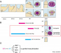
File:Mouse external genital development.jpg ...b, baculum (os penis); EB, estradiol benzoate; Flut, flutamide; gp, glans penis; p, prepuce; u, urethra.(800 × 701 (75 KB)) - 15:46, 8 June 2017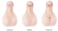
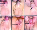
File:Hypospadias.jpg ...l''': The opening of the urethra is located somewhere near the head of the penis. ...'Midshaft''': The opening of the urethra is located along the shaft of the penis.(600 × 325 (53 KB)) - 22:57, 8 November 2014- ...24 paper by Lipschutz describes rare male human genital abnormality of the penis. '''Modern Notes:''' {{genital abnormalities}} | {{penis}}2 KB (386 words) - 08:33, 6 February 2020
File:Keibel Mall 2 639.jpg [[Category:Penis]](700 × 367 (25 KB)) - 12:57, 5 May 2019- ...ude>[[Category:Template]][[Category:Term Link]][[Category:Male]][[Category:Penis]][[Category:Abnormal Development]][[Category:Genital]][[Category:Urethra]]<217 bytes (22 words) - 23:17, 3 June 2019
- | urinary meatus that opens along the shaft of the penis | urinary meatus opens where the shaft of the penis meets the scrotum1 KB (142 words) - 13:28, 25 March 2019
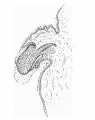
File:Hunter1935 text-fig02.jpg ==Text-fig. 2. L.S. Penis of human foetus 70 mm C.R. length==(483 × 612 (41 KB)) - 12:32, 4 January 2017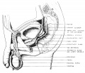
File:Nelsen1953 fig002.jpg [[Category:Male]][[Category:Genital]][[Category:Testis]][[Category:Penis]](1,200 × 1,037 (249 KB)) - 12:05, 7 September 2018
File:Fetal corpus cavernosum and corpus spongiosum 01.jpg ==Fetal Penis Development== ...lagen in the corpus cavernosum and corpus spongiosum (blue color) of fetal penis at different ages in weeks post-conception (WPC).(1,795 × 2,082 (919 KB)) - 11:42, 7 September 2014- | valign=top|prepubertal (testis volume < 1.5 ml<br>small penis (3 cm or less) <br>(age 9 and younger) ...estis volume 1.6 to 6 ml<br>skin on scrotum thins, reddens and enlarges<br>penis length unchanged<br>(age 9–11)3 KB (403 words) - 11:46, 3 June 2019
File:Hunter1935 plate01.jpg ...of the layer between the prepuee and glans. Pointers: 1, prepuce; 2, glans penis; 3, cell nest.(1,790 × 2,107 (711 KB)) - 16:27, 4 January 2017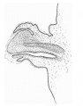
File:Hunter1935 text-fig03.jpg ==Text-fig. 3. L.S. Penis of human foetus 100 mm C.R. length==(666 × 845 (77 KB)) - 12:32, 4 January 2017- ...][[Category:Genital]][[Category:Testis]][[Category:Spermatozoa]][[Category:Penis]]</noinclude>199 bytes (22 words) - 10:09, 11 June 2018
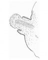
File:Hunter1935 text-fig01.jpg ==Text-fig. 1. L.S. Penis of human foetus 40 mm C.R. length==(488 × 612 (31 KB)) - 12:31, 4 January 2017- ...lagen in the corpus cavernosum and corpus spongiosum (blue color) of fetal penis at different ages in weeks post-conception (WPC). ===Modifications of Erectile Tissue Components in the Penis during the Fetal Period===3 KB (378 words) - 11:36, 7 September 2014
- ...{ChrY}} | {{SRY}} | {{testis}} | {{spermatozoa}} | {{ductus deferens}} | {{penis}} | {{prostate}} | [[:Category:Male|Category:Male]]324 bytes (33 words) - 11:12, 7 April 2020
File:Male histology 003.jpg * cross-section of developing penis(1,280 × 1,024 (703 KB)) - 08:36, 29 August 2011- ...k}}224763620 Polyorchidism] | {{ICD11weblink}}920426846 Meatal stenosis of penis] | {{ICD11weblink}}1580368242 Hypoplasia of testis or scrotum] | {{ICD11web2 KB (201 words) - 15:43, 10 April 2019
- ...k}}224763620 Polyorchidism] | {{ICD11weblink}}920426846 Meatal stenosis of penis] | {{ICD11weblink}}1580368242 Hypoplasia of testis or scrotum] | {{ICD11web2 KB (201 words) - 15:45, 10 April 2019
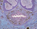
File:Hunter1935 text-fig06.jpg ==Text-Fig. 6. C.S. Glans region of penis from human foetus 100 mm. C.R. length==(731 × 1,153 (167 KB)) - 16:31, 4 January 2017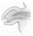
File:Hunter1935 text-fig04.jpg ==Text-fig. 4. L.S. Penis of human foetus 170 mm C.R. length==(677 × 736 (108 KB)) - 12:33, 4 January 2017- ...ion, looks more male than female, with an empty scrotum and a normal-sized penis-like phallus, however this structure is not quite as free from the perineum | Complete male virilisation, a normally-formed penis is present. Urethral opening at or near the tip, and the scrotum formed, bu2 KB (284 words) - 16:05, 8 June 2017
- | valign=top|prepubertal (testis volume < 1.5 ml<br>small penis (3 cm or less) <br>(age 9 and younger) ...estis volume 1.6 to 6 ml<br>skin on scrotum thins, reddens and enlarges<br>penis length unchanged<br>(age 9–11)5 KB (708 words) - 16:30, 2 June 2015
- ...rY}} chromosome | {{testis}} | {{spermatozoa}} | {{meiosis}} | {{AMH}} | {{penis}} | {{prostate}} | {{seminal vesicle}} | {{genital abnormalities}} | {{hypo5 members (0 subcategories, 2 files) - 11:10, 2 May 2019
File:Hypospadia classifications.jpg '''A''' Anterior - on inferior surface of glans penis. Hypospadias most common penis abnormality (1 in 300) from a failure of male urogenital folds to fuse in v(500 × 410 (47 KB)) - 13:29, 25 March 2019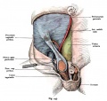
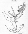
File:Kollmann445.jpg Skin and muscles, penis, and part of the scrotum are removed and in the area of the abdomen leaving Haut und Muskulatur, Penis und ein Teil des Scrotums sind entfernt und im Gebiet des Unterbauches nur(776 × 738 (112 KB)) - 11:46, 8 June 2017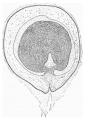
File:Hunter1935 text-fig07.jpg ==Text-Fig. 7. C.S. Glans region of penis from human foetus 170 mm. C.R. length==(814 × 1,153 (255 KB)) - 16:31, 4 January 2017- ...ference]][[Category:Genital]][[Category:Pig]][[Category:Female]][[Category:Penis]][[Category:Historic Embryology]][[Category:1910's]]</noinclude>447 bytes (59 words) - 22:24, 3 March 2020
- ...e|Testis Descent Movie]] - [[Media:Testis Descent_001.mp4|MP4 movie]] | [[Penis Development]] | [[Testis Development]] | [[Developmental Signals - Anti-Mul948 bytes (111 words) - 09:50, 8 March 2018
- ...of the testis, epididymis, vas deferens, seminal vesicle, prostate gland, penis, and sperm. The functional significance of the various histological structu ===Penis===3 KB (465 words) - 13:55, 27 May 2013
- * Q55.5 Congenital absence and aplasia of penis ...nital malformation of penis NOS Curvature of penis (lateral) Hypoplasia of penis5 KB (648 words) - 09:01, 13 October 2016
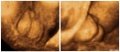
File:Hypospadia 3D ultrasound 01.jpg Ultrasonography in rendering mode, at {{GA}} 33 weeks, with short penis and with evidence of testicles inside a bifid scrotum.(1,150 × 497 (87 KB)) - 08:36, 8 June 2017- ...trunks which accompany the femoral vessels, the lymphatics from the glans penis vel clitoridis, and also some of the efferents from the superficial subingu734 bytes (120 words) - 23:07, 14 February 2013
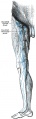
File:Gray0610.jpg ...nt. They receive as afferents lymphatic vessels from the integument of the penis, scrotum, perineum, buttock, and abdominal wall below the level of the umbi ...ut they also receive some of the vessels which drain the integument of the penis, scrotum, perineum, and buttock.(303 × 1,000 (81 KB)) - 12:56, 15 February 2013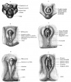
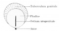
File:Gray1119.jpg * The first rudiment of the penis (or clitoris) is a structure termed the phallus * penis is developed from the phallus(700 × 807 (115 KB)) - 14:37, 28 May 2011- ...rY}} chromosome | {{testis}} | {{spermatozoa}} | {{meiosis}} | {{AMH}} | {{penis}} | {{prostate}} | {{seminal vesicle}} | {{genital abnormalities}} | {{hypo440 members (46 subcategories, 233 files) - 09:01, 23 April 2020
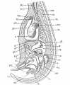
File:DeBeer1928 fig127.jpg ...kidney ; oe, oesophagus ; p, pancreas ; pi, pleural coelomic cavity ; pn, penis ; pr, pericardial coelomic cavity ; r, rectum ; sc, spinal cord ; stn, ster(1,055 × 1,284 (288 KB)) - 17:30, 26 April 2015- ...menstrual cycle}} | {{male}} | {{ChrY}} | {{testis}} | {{spermatozoa}} | {{penis}} | {{prostate}} | {{endocrine gonad}} | [[Movies#Genital|Genital Movies]]1 KB (130 words) - 14:52, 18 May 2019
File:Foster140.jpg ...tum ; ug. urinogenital sinus ; cp. elevation which becomes the clitoris or penis ; Is. ridge from which the labia majora or scrotum are developed.(679 × 824 (82 KB)) - 18:36, 12 January 2011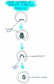
File:Cross section of genital tubercle male.jpg ...nital tubercle in a male fetus, as it forms the tubular urethra within the penis.(576 × 886 (84 KB)) - 22:52, 8 November 2014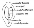
File:Keith1902 fig098.jpg ...inent tubercle — the genital tubercle, the apex of which becomes the glans penis or glans clitoris, according to sex. It is bounded by lateral folds, the ge(560 × 460 (40 KB)) - 11:39, 8 June 2017


