Search results
From Embryology
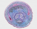
File:Male histology 002.jpg ==Male Histology - Penis== * cross-section of developing penis (Trichrome stain).(1,280 × 1,024 (490 KB)) - 00:21, 23 May 2012
File:Prepuce.jpeg ...1935. The drawings resemble and illustrate the observations of the foetal penis at different gestational age, depicting that the prepuce is a gradually gro The three images (A, B, C) show the overall continuous change of the penis in terms of the prepuce.(2,293 × 767 (382 KB)) - 17:27, 24 October 2014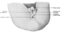
File:Keibel Mall 2 642.jpg The phallus has become the glans penis, the scrotal swellings are fully developed and the unpaired scrotal area is ...egory:Genital]][[Category:Male]][[Category:Historic Embryology]][[Category:Penis]](1,200 × 644 (95 KB)) - 12:57, 5 May 2019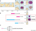
File:Mouse external genital development.jpg ...b, baculum (os penis); EB, estradiol benzoate; Flut, flutamide; gp, glans penis; p, prepuce; u, urethra.(800 × 701 (75 KB)) - 15:46, 8 June 2017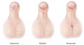
File:Hypospadias.jpg ...l''': The opening of the urethra is located somewhere near the head of the penis. ...'Midshaft''': The opening of the urethra is located along the shaft of the penis.(600 × 325 (53 KB)) - 22:57, 8 November 2014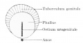
File:Keibel Mall 2 639.jpg [[Category:Penis]](700 × 367 (25 KB)) - 12:57, 5 May 2019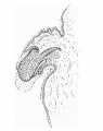
File:Hunter1935 text-fig02.jpg ==Text-fig. 2. L.S. Penis of human foetus 70 mm C.R. length==(483 × 612 (41 KB)) - 12:32, 4 January 2017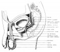
File:Nelsen1953 fig002.jpg [[Category:Male]][[Category:Genital]][[Category:Testis]][[Category:Penis]](1,200 × 1,037 (249 KB)) - 12:05, 7 September 2018
File:Fetal corpus cavernosum and corpus spongiosum 01.jpg ==Fetal Penis Development== ...lagen in the corpus cavernosum and corpus spongiosum (blue color) of fetal penis at different ages in weeks post-conception (WPC).(1,795 × 2,082 (919 KB)) - 11:42, 7 September 2014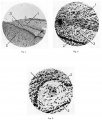
File:Hunter1935 plate01.jpg ...of the layer between the prepuee and glans. Pointers: 1, prepuce; 2, glans penis; 3, cell nest.(1,790 × 2,107 (711 KB)) - 16:27, 4 January 2017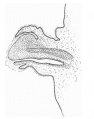
File:Hunter1935 text-fig03.jpg ==Text-fig. 3. L.S. Penis of human foetus 100 mm C.R. length==(666 × 845 (77 KB)) - 12:32, 4 January 2017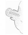
File:Hunter1935 text-fig01.jpg ==Text-fig. 1. L.S. Penis of human foetus 40 mm C.R. length==(488 × 612 (31 KB)) - 12:31, 4 January 2017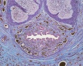
File:Male histology 003.jpg * cross-section of developing penis(1,280 × 1,024 (703 KB)) - 08:36, 29 August 2011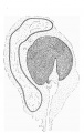
File:Hunter1935 text-fig06.jpg ==Text-Fig. 6. C.S. Glans region of penis from human foetus 100 mm. C.R. length==(731 × 1,153 (167 KB)) - 16:31, 4 January 2017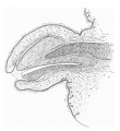
File:Hunter1935 text-fig04.jpg ==Text-fig. 4. L.S. Penis of human foetus 170 mm C.R. length==(677 × 736 (108 KB)) - 12:33, 4 January 2017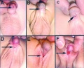
File:Hypospadia classifications.jpg '''A''' Anterior - on inferior surface of glans penis. Hypospadias most common penis abnormality (1 in 300) from a failure of male urogenital folds to fuse in v(500 × 410 (47 KB)) - 13:29, 25 March 2019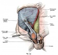
File:Kollmann445.jpg Skin and muscles, penis, and part of the scrotum are removed and in the area of the abdomen leaving Haut und Muskulatur, Penis und ein Teil des Scrotums sind entfernt und im Gebiet des Unterbauches nur(776 × 738 (112 KB)) - 11:46, 8 June 2017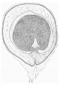
File:Hunter1935 text-fig07.jpg ==Text-Fig. 7. C.S. Glans region of penis from human foetus 170 mm. C.R. length==(814 × 1,153 (255 KB)) - 16:31, 4 January 2017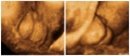
File:Hypospadia 3D ultrasound 01.jpg Ultrasonography in rendering mode, at {{GA}} 33 weeks, with short penis and with evidence of testicles inside a bifid scrotum.(1,150 × 497 (87 KB)) - 08:36, 8 June 2017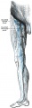
File:Gray0610.jpg ...nt. They receive as afferents lymphatic vessels from the integument of the penis, scrotum, perineum, buttock, and abdominal wall below the level of the umbi ...ut they also receive some of the vessels which drain the integument of the penis, scrotum, perineum, and buttock.(303 × 1,000 (81 KB)) - 12:56, 15 February 2013


