Uploads by Z5076158
From Embryology
This special page shows all uploaded files.
| Date | Name | Thumbnail | Size | Description | Versions |
|---|---|---|---|---|---|
| 10:08, 26 October 2017 | Adaptation Cerebellum.png (file) |  |
2.71 MB | In real stimulation, a considerable increase in pre-stimulus low-frequency activity is only found in the right cerebellar hemisphere (near to the stimulation site). But in sham stimulation, pre-stimulus low-frequency activity increases in both the righ... | 1 |
| 09:59, 26 October 2017 | Addiction Cerebellum.png (file) |  |
928 KB | Nissl staining showing the dopamine receptor expression within the cerebellum of a songbird <ref name=”PMIDPMC2904815”><pubmed>PMC2904815</pubmed></ref>. ==Reference== <references/> ==Copyright== Re-use of this article is permitted in accordance... | 1 |
| 09:27, 26 October 2017 | Cerebellum Dystonia.png (file) | 379 KB | 1 | ||
| 09:21, 26 October 2017 | Screen Shot 2017-10-26 at 10.20.18 am.png (file) |  |
1.02 MB | 1 | |
| 09:54, 25 October 2017 | Primary Brain.png (file) |  |
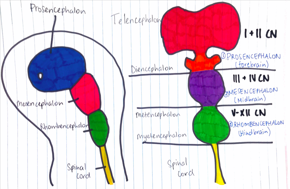
681 KB | In week 4, 3 primary brain vesicles are formed; forebrain (prosencephalon) midbrain (mesencephalon), and hindbrain (rhombencephalon). These structures then differentiate into 5 secondary brain vesicles during week 5. Drawn by Student z5076158 {{Templ... | 1 |
| 09:52, 25 October 2017 | Primary Brain Vesciles.pdf (file) | 453 KB | In week 4, 3 primary brain vesicles are formed; forebrain (prosencephalon) midbrain (mesencephalon), and hindbrain (rhombencephalon). These structures then differentiate into 5 secondary brain vesicles during week 5. Drawn by Student z5076158 {{Templ... | 1 | |
| 20:49, 23 October 2017 | Isthmic Organiser.png (file) |  |
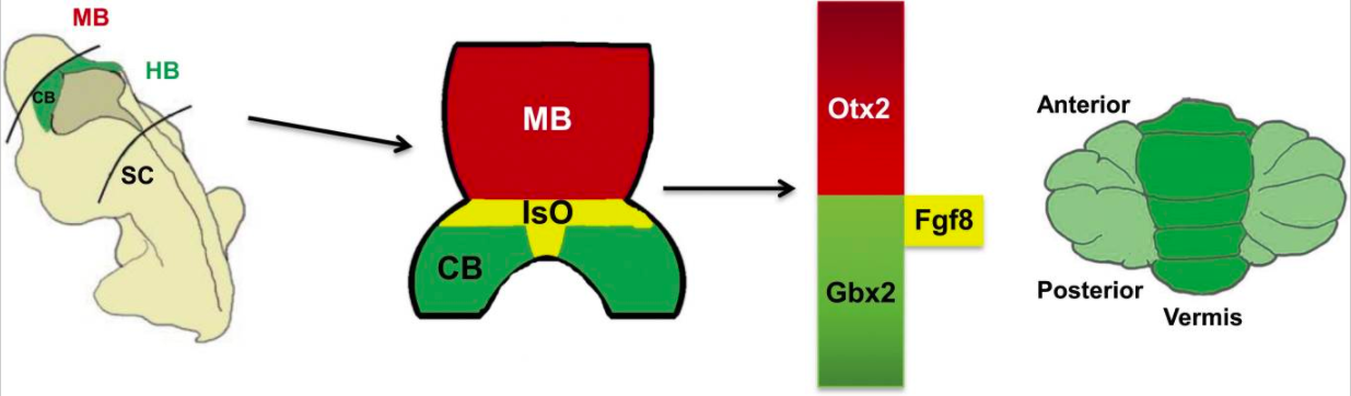
272 KB | The role of the Isthmic Organiser. "The establishment of gene expression domains along the anterior-posterior axis helps to segment the developing brain into the forebrain, midbrain (MB), hindbrain (HB) and spinal cord (SC). The Isthmic Organizer (IsO;... | 1 |
| 14:04, 16 October 2017 | Screen Shot 2017-10-16 at 3.01.32 pm.png (file) |  |
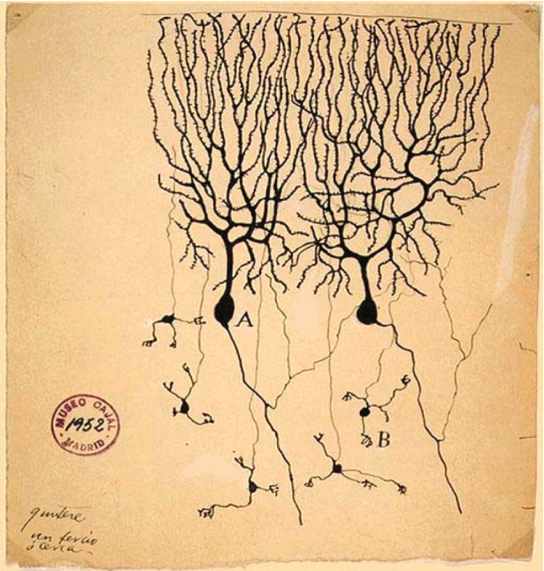
756 KB | Drawing of Purkinje cells (A) and granule cells (B) from pigeon cerebellum. Drawn by Santiago Ramón y Cajal, 1899. <ref><pubmed>4308982</pubmed></ref> =Copyright= <references/> © 2015 The Authors. Published by the Royal Society under the terms of th... | 1 |
| 12:52, 16 October 2017 | Screen Shot 2017-10-16 at 1.43.55 pm.png (file) |  |
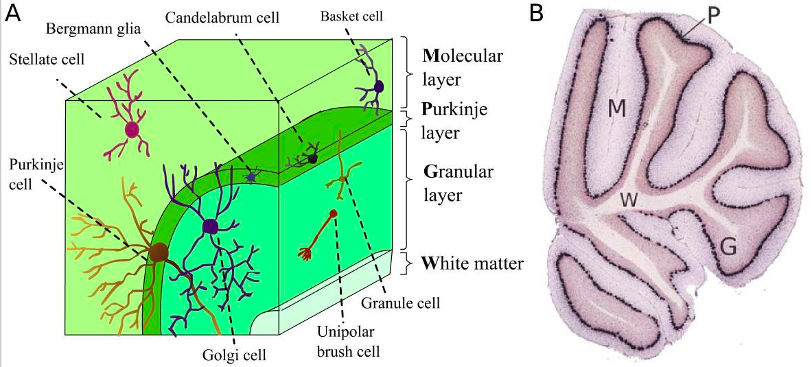
704 KB | (A). Cell types and where they are found in the cerebellar cortical layers. (B) Shows how the different layers of the cerebellum can be easily determined. P - the Purkinje layer; G - the granular layer; M - the molecular layer; W - the white matter. <r... | 1 |
| 12:47, 16 October 2017 | Cerebellum Layers and Cell Types.png (file) |  |
103 KB | (A). Cell types and where they are found in the cerebellar cortical layers. (B) Shows how the different layers of the cerebellum can be easily determined. P - the Purkinje layer; G - the granular layer; M - the molecular layer; W - the white matter. <r... | 1 |
| 14:35, 5 October 2017 | Screen Shot 2017-10-05 at 3.29.59 pm.png (file) |  |
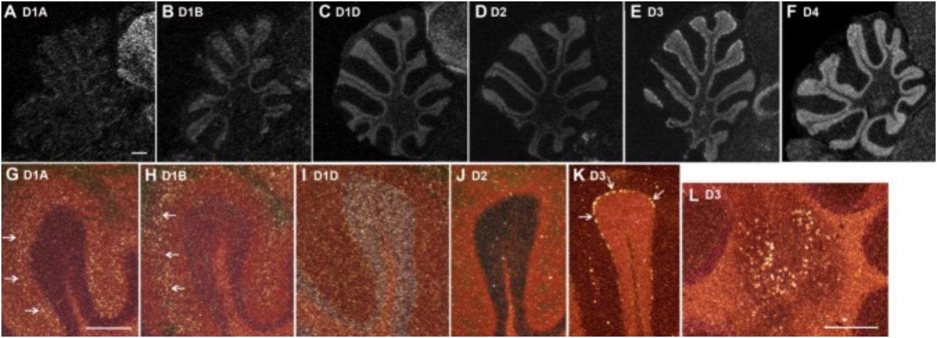
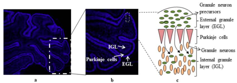
243 KB | ==Cerebellum development of cells== A 7 day old rat cerebellum that has been sectioned to show the 3 layers of cells - external granule layer, purkinje cells and internal granular layer (a). Showing granule cell precursors proliferating to differentiat... | 1 |
| 16:48, 3 October 2017 | Neural Circuit in the Cerebellum.jpg (file) |  |
55 KB | Source is cited from <ref><pubmed>21309081</pubmed></ref>. | 1 |
| 16:27, 14 September 2017 | Cerebellum Structure.jpg (file) |  |
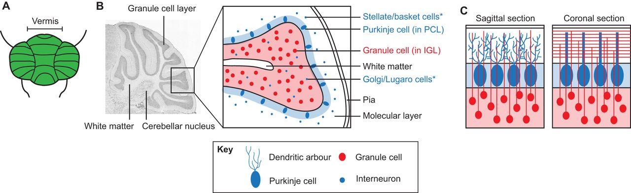
87 KB | <ref> PMID25336734 </ref> | 1 |