Uploads by Z3291423
From Embryology
This special page shows all uploaded files.
| Date | Name | Thumbnail | Size | Description | Versions |
|---|---|---|---|---|---|
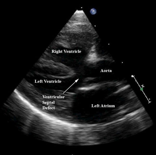
| 21:36, 17 August 2011 | Some common effects for patients with Tetralogy of Fallot.jpg (file) |  |
44 KB | This still frame of a modified parasternal long axis view from the same patient as imaged for Figures 6 and 7 demonstrates the large ventricular septal defect, aortic override, and right ventricular hypertrophy charactistic of patients with tetralogy of F | 1 |
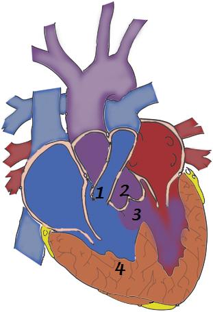
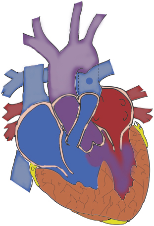
| 12:39, 11 September 2011 | Tetralogy of fallot-the 4 defects.jpg (file) |  |
42 KB | This picture demonstrates the four defects (numbers 1-4) found in Tetralogy of Fallot all the 4 defects contribute to poor oxygenation of blood. 1. Pulmonary Stenosis: The small pulmonary vessel causes less blood to enter to the lungs. 2. Displacement o | 1 |
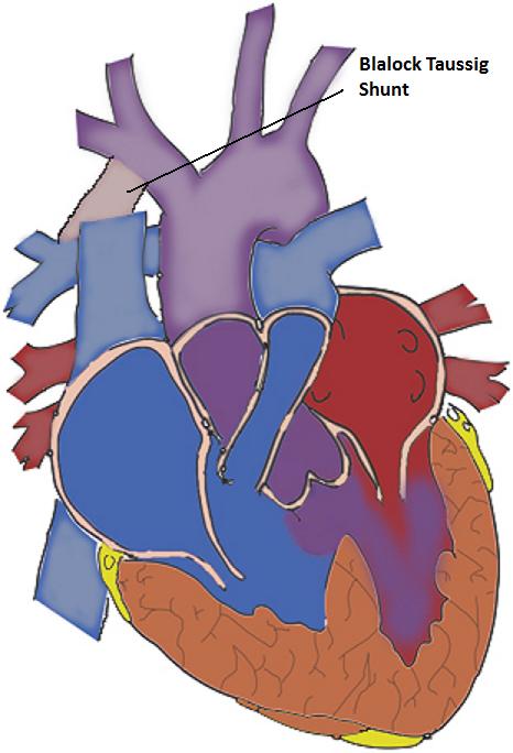
| 13:07, 11 September 2011 | The Blalock Taussig Shunt.jpg (file) |  |
60 KB | The Blalock Taussig Shunt is an artificial tubing that is sewn from the subclavian or Carotid arteries to the corresponding pulmonary artery. This shunt directs blood to the lungs for oxygenation and provides some cyanotic relief. This is a drawing base | 1 |
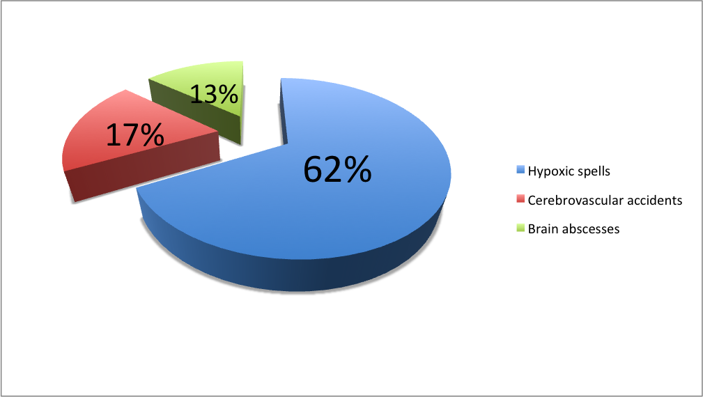
| 22:38, 4 October 2011 | Major causes of death in surgically untreated TOF patients.png (file) |  |
127 KB | The picture represents a graphical view of the major causes of death for patients who do not undergo surgery for TOF. 62% of patients died from Hypoxic spells 17% of patients died from Cerebrovascular accidents 13% of patients died from Brain abscesses | 1 |
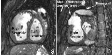
| 23:04, 5 October 2011 | Magnetic Resonance Imaging in an adult patient with tetralogy of Fallot.jpg (file) |  |
17 KB | The MRI shows a) Shows Dilation and hypertrophy of right ventricle b) Shows the stenotic and tortuous homograft conduit responsible for the dilated and hypertrophied right ventricle. © 2009 Bailliard and Anderson; licensee BioMed Central Ltd. This is | 1 |
| 18:24, 12 October 2011 | Potts Shunt.jpg (file) |  |
117 KB | The Potts shunt is a connection that is established between the descending portion of the aorta (on the left side of chest) to the left branch of the pulmonary artery. This Drawing is inspired by the following site (last image): http://www.med.nus.edu.s | 1 |
| 18:26, 12 October 2011 | Waterston Shunt.jpg (file) |  |
119 KB | The Waterston shunt was placed between the back of the aorta, to the right branch of the pulmonary. This image was inspired from the site (last image):http://www.med.nus.edu.sg/paed/resources/cardiac_thumbnail/surgery/shunts.htm Beginning six months aft | 1 |
| 23:47, 12 October 2011 | The Glenn Shunt.jpg (file) |  |
125 KB | he following picture shows the modified Glenn Shunt, where the Superior Vena Cava is anastomosed to the right branch of the pulmonary artery, which is still connected to the main pulmonary artery. This image was inspired from the following website: http: | 1 |
| 12:27, 13 October 2011 | Cyanotic baby.jpg (file) |  |
43 KB | This picture portrays the classic cyanotic appearance of a baby with Tetralogy of Fallot. The image was obtained from: http://upload.wikimedia.org/wikipedia/commons/thumb/e/e1/Cyanotic_neonate.jpg/150px-Cyanotic_neonate.jpg The copyright holder of this | 1 |