Uploads by Z3370664
From Embryology
This special page shows all uploaded files.
| Date | Name | Thumbnail | Size | Description | Versions |
|---|---|---|---|---|---|
| 22:19, 7 August 2012 | Gene morula.JPG (file) |  |
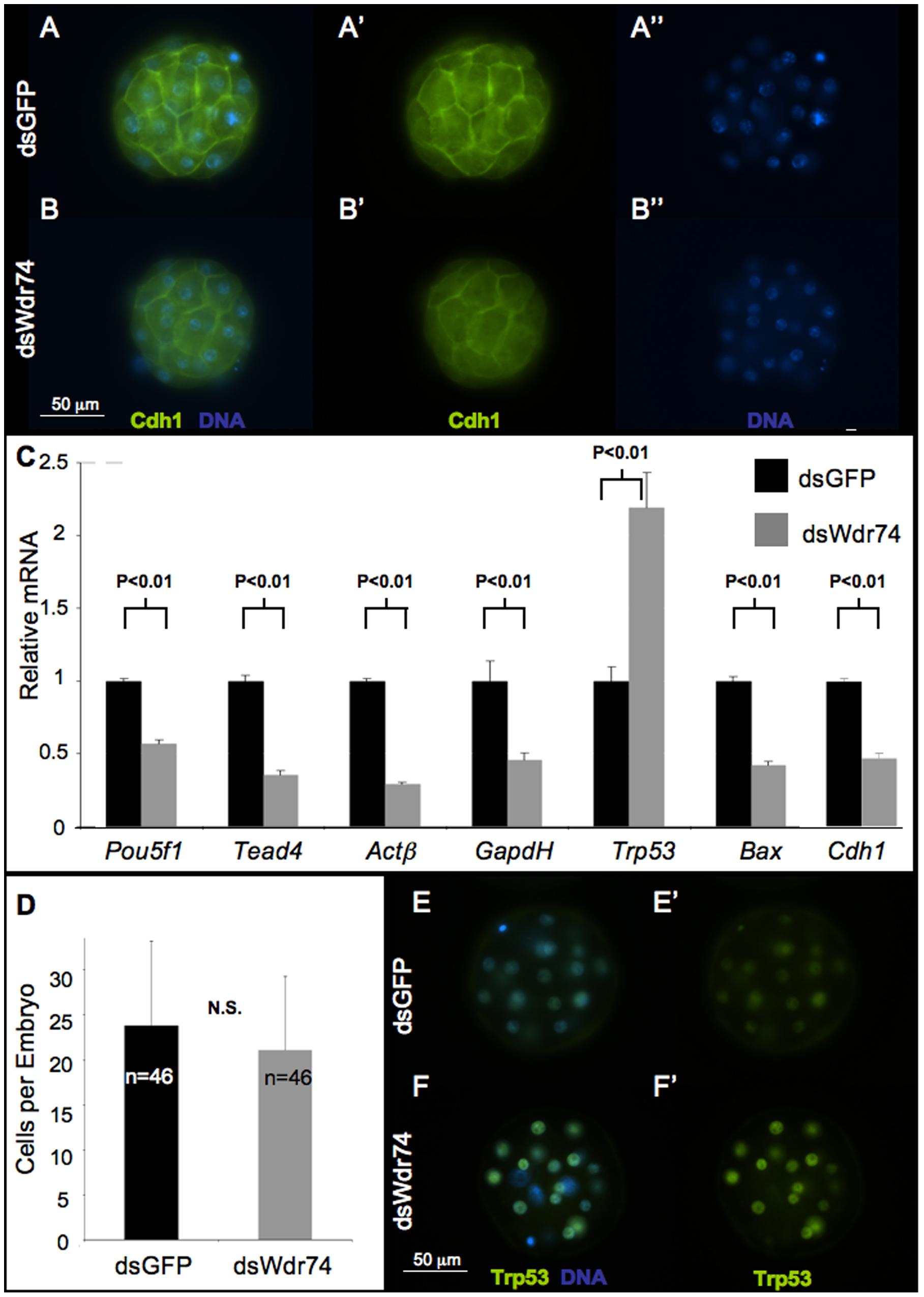
226 KB | == Gene_morula.JPG == Gene expression in dsWdr74 morula. A–B. E-cadherin (Cdh1) localization by immunofluorescence marks blastomere cell-cell adhesion as expected in dsGFP morula (A). E-Cadherin is appropriately localized but present at reduced in ds | 1 |
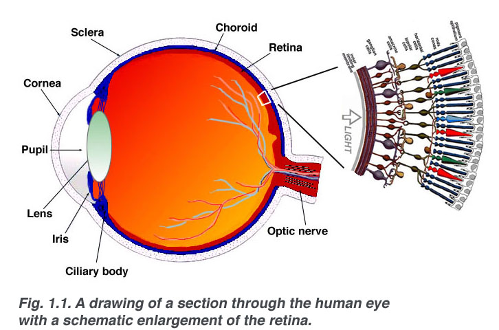
| 11:12, 19 September 2012 | Eyediagramcolour1.JPG (file) |  |
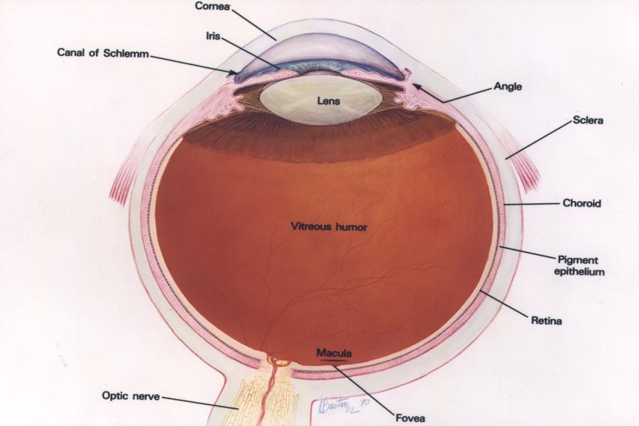
50 KB | Labeled diagram of a normal eye This image is courtesy of the US National Eye Institute, National Institutes of Health (NEI/NIH) http://www.nei.nih.gov/photo/keyword.asp?conditions=Normal+Eye+Images&match=all Copyright Policy Unless otherwise noted, | 1 |
| 11:31, 19 September 2012 | JNK1.png (file) |  |
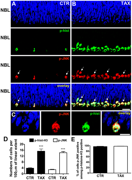
174 KB | Figure: JNK is phosphorylated during mitosis of retinal progenitor cells. (A, B) Representative confocal photomicrographs of immunohistochemistry for phospho-JNK (red) and phospho-histone-H3 (green) in sections of retinal tissue maintained for 3 hours in | 1 |
| 12:36, 28 September 2012 | Retina-cell-clusters.JPG (file) |  |
32 KB | "Endothelial cells form thick clusters in the LRP5 mutant retina." "Co-immunostaining was performed on retinal frozen sections of 3-week-old LRP5 littermates with endothelial cell specific Tie2 (red) and glial cell specific GFAP (green) antibodies. Cel | 3 |
| 17:52, 1 October 2012 | Aristotle-eye.jpg (file) |  |
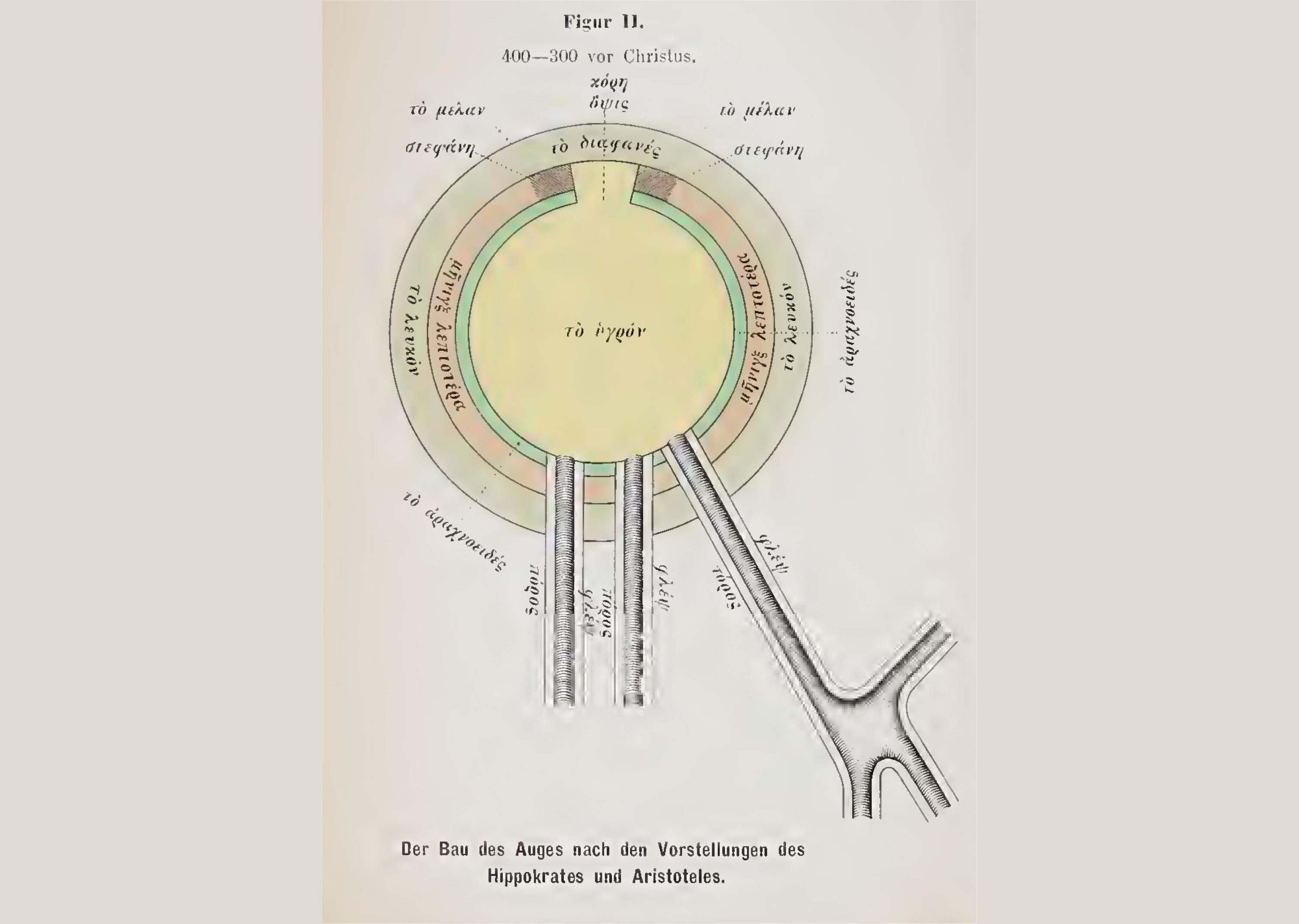
351 KB | 3 | |
| 18:03, 1 October 2012 | Galen-eye1.jpg (file) |  |
258 KB | 2 | |
| 18:05, 1 October 2012 | Rufus-eye.jpg (file) |  |
200 KB | The eye according to Rufus of Ephesus. Image Source: Magnus,H., (1901), Die Augenheilkunde der Alten, Breslau. This book was published in 1901, hence copyright has expired and it is now in the public domain. http://archive.org/details/dieaugenheilku | 2 |
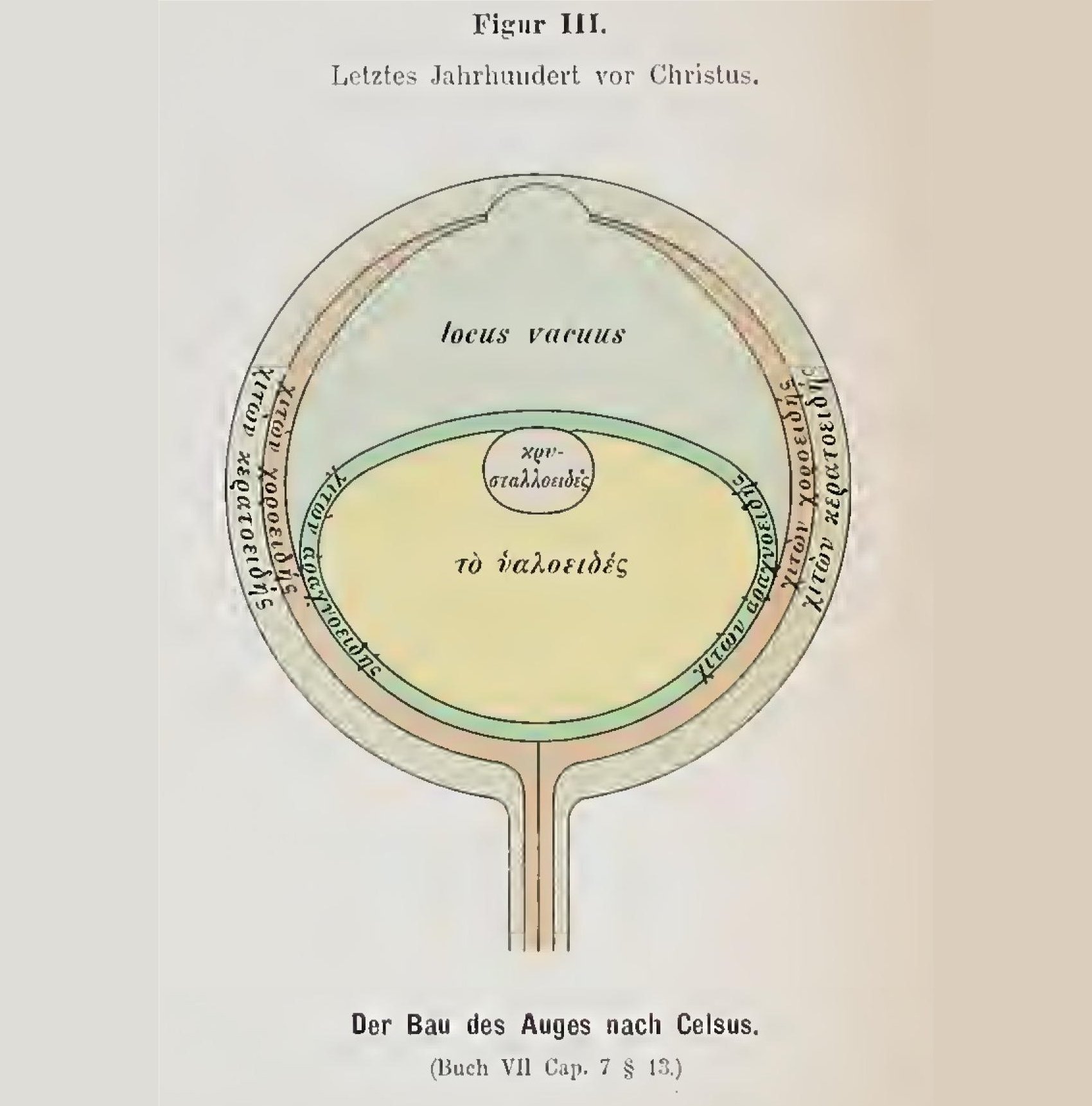
| 18:07, 1 October 2012 | Celsus-eye.jpg (file) |  |
204 KB | The eye according to Celsus. Image Source: Magnus,H., (1901), Die Augenheilkunde der Alten, Breslau. This book was published in 1901, hence copyright has expired and it is now in the public domain. http://archive.org/details/dieaugenheilkund00magn | 2 |
| 12:10, 2 October 2012 | Mip1-expression-in-pitx3.jpg (file) |  |
63 KB | "Analysis of mip1 expression in pitx3-mo and control embryos via in situ hybridization and RT-PCR." "A, D, F-H. Normal mip1 expression in control-injected embryos at 29-, 34- and 48-hpf. B, C, E, I–K. Altered mip1 expression is observed in pitx3 morph | 1 |
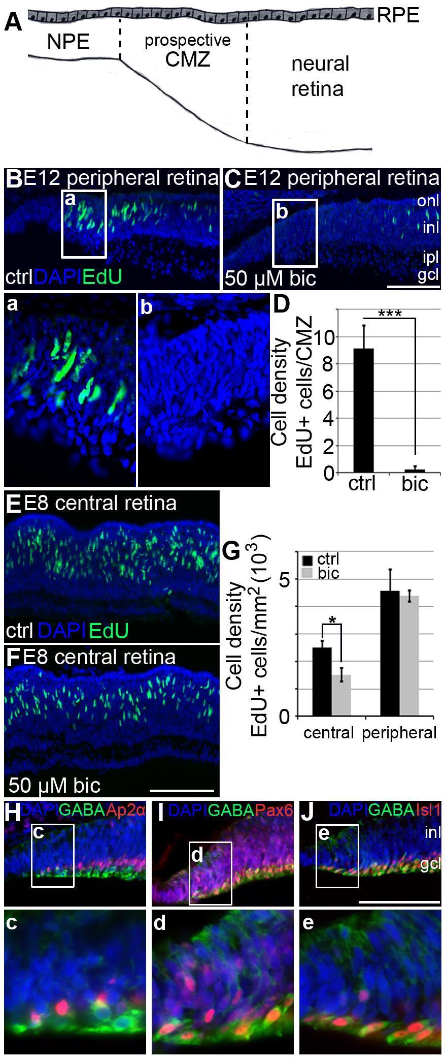
| 20:43, 2 October 2012 | Gaba-effects-retina.JPG (file) |  |
240 KB | "GABAA receptor mediated effects on retinal progenitor cell proliferation." "(A) Schematic diagram of the E12 peripheral retina. Cells between the dashed lines in the prospective ciliary marginal zone (CMZ) on the dorsal side of the retina were counted. | 1 |
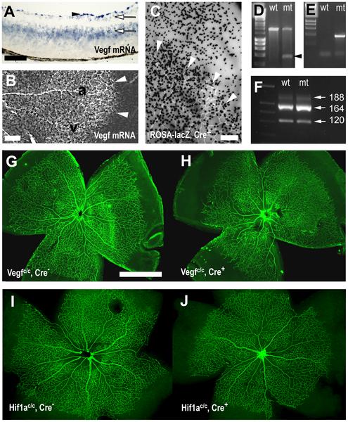
| 06:00, 3 October 2012 | Astrocyte-vegf-deletion.JPG (file) |  |
75 KB | "Astrocyte specific deletion of VEGF." "(A, B) In situ hybridisation showed Vegf mRNA expression in a retinal cross-section (A) and a retinal whole mount (B) at P5. (A) Vegf mRNA was most strongly expressed in retinal astrocytes (arrowhead), and weakly i | 1 |
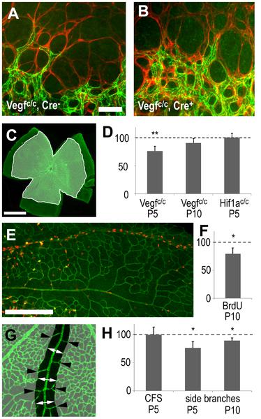
| 06:01, 3 October 2012 | Effect-of-vegf-on-retinal-vasculature.JPG (file) |  |
50 KB | "Effects of astrocyte-derived VEGF on retinal vascular development." "Immunohistochemistry, visualizing endothelial cells (claudin 5, green in A, B, C, E, G) and retinal astrocytes (GFAP, red in A, B), shows that angiogenic sprouting at the leading edge | 1 |
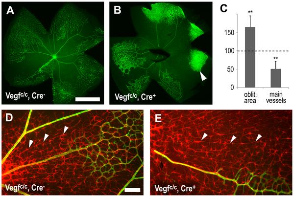
| 06:03, 3 October 2012 | Vegf-protects-vessels.JPG (file) |  |
50 KB | Astrocyte-derived VEGF protects vessels from hyperoxia. After hyperoxia exposure from P7–12 immunohistochemistry was used to visualize vessels with isolectin B4 (green, A, B), collagen IV (green, D, E) and retinal astrocytes (GFAP, red, D, E). (A–C) | 1 |
| 10:42, 3 October 2012 | Eye-pupil-sclera-iris.jpg (file) |  |
52 KB | Illustration of the front of the eye, showing the sclera, iris and pupil. Source: http://webvision.med.utah.edu/imageswv/pupil.jpeg Citation: <pubmed>21413389</pubmed> Copyright © 2012 Webvision: Attribution, Noncommercial, No Derivative Works Creati | 1 |
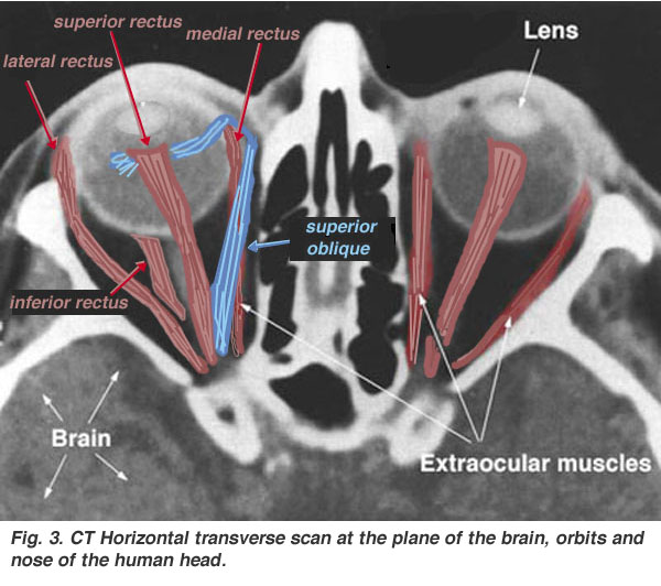
| 12:50, 5 October 2012 | Extraocular-muscles-scan.jpg (file) |  |
76 KB | Source: hhttp://webvision.med.utah.edu/imageswv/scan.jpeg Citation: <pubmed>21413389</pubmed> Copyright © 2012 Webvision: Attribution, Noncommercial, No Derivative Works Creative Commons license. Original copyright information from webvision: “Q: C | 1 |
| 13:21, 5 October 2012 | 5months-gestation-retina.jpg (file) |  |
173 KB | '''The layers of the retina in the fifth month of development.''' Source: http://webvision.med.utah.edu/imageswv/5months.jpeg Citation: Kolb H, Fernandez E, Nelson R. '''The Organization of the Retina and Visual System ''' (Online Book). PMID:[http://w | 1 |
| 13:26, 5 October 2012 | Eye-retina-layers.jpg (file) |  |
91 KB | The layers of the retina magnified, showing the direction of the layers of the retina in the back of the eye. Source: http://webvision.med.utah.edu/imageswv/Sagschem.jpeg Citation: Kolb H, Fernandez E, Nelson R. '''The Organization of the Retina and Vis | 1 |
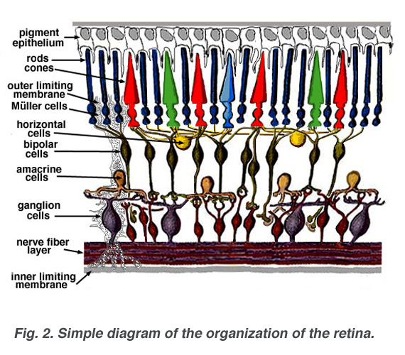
| 13:28, 5 October 2012 | Retina-layers-diagram.jpg (file) |  |
156 KB | '''A diagram of the layers of the retina.''' Source: http://webvision.med.utah.edu/imageswv/schem.jpeg Citation: Kolb H, Fernandez E, Nelson R. '''The Organization of the Retina and Visual System ''' (Online Book). PMID:[http://www.ncbi.nlm.nih.gov/pubm | 1 |
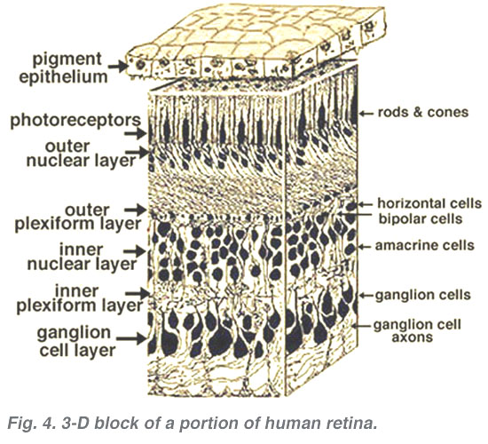
| 13:35, 5 October 2012 | Retina-layers-diagram2.jpg (file) |  |
96 KB | '''A diagram of the layers of the retina.''' Source: http://webvision.med.utah.edu/wp-content/uploads/2011/01/3dlabel.jpeg Citation: Kolb H, Fernandez E, Nelson R. '''The Organization of the Retina and Visual System ''' (Online Book). PMID:[http://www.n | 1 |