Uploads by Z3332629
From Embryology
This special page shows all uploaded files.
| Date | Name | Thumbnail | Size | Description | Versions |
|---|---|---|---|---|---|
| 14:02, 11 August 2011 | 1532-429X-12-14-3.jpg (file) |  |
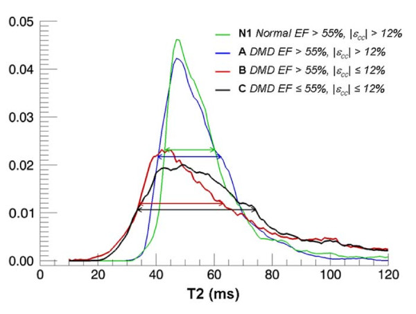
44 KB | T2 histograms. Examples of normalized (i.e. area under curve = 1) T2 histograms of DMD (Group A, B and C) and Normal control (N1) subjects show that DMD patients with normal EF but impaired εcc has higher heterogeneity in T2 compared to other groups. | 1 |
| 10:52, 18 August 2011 | DMD patients and causes of higher heterogeneity.jpg (file) |  |
44 KB | T2 histograms. Examples of normalized (i.e. area under curve = 1) T2 histograms of DMD (Group A, B and C) and Normal control (N1) subjects show that DMD patients with normal EF but impaired εcc has higher heterogeneity in T2 compared to other groups. | 1 |
| 08:58, 4 October 2011 | Dystrophin in the muscle fibre membrane.jpg (file) |  |
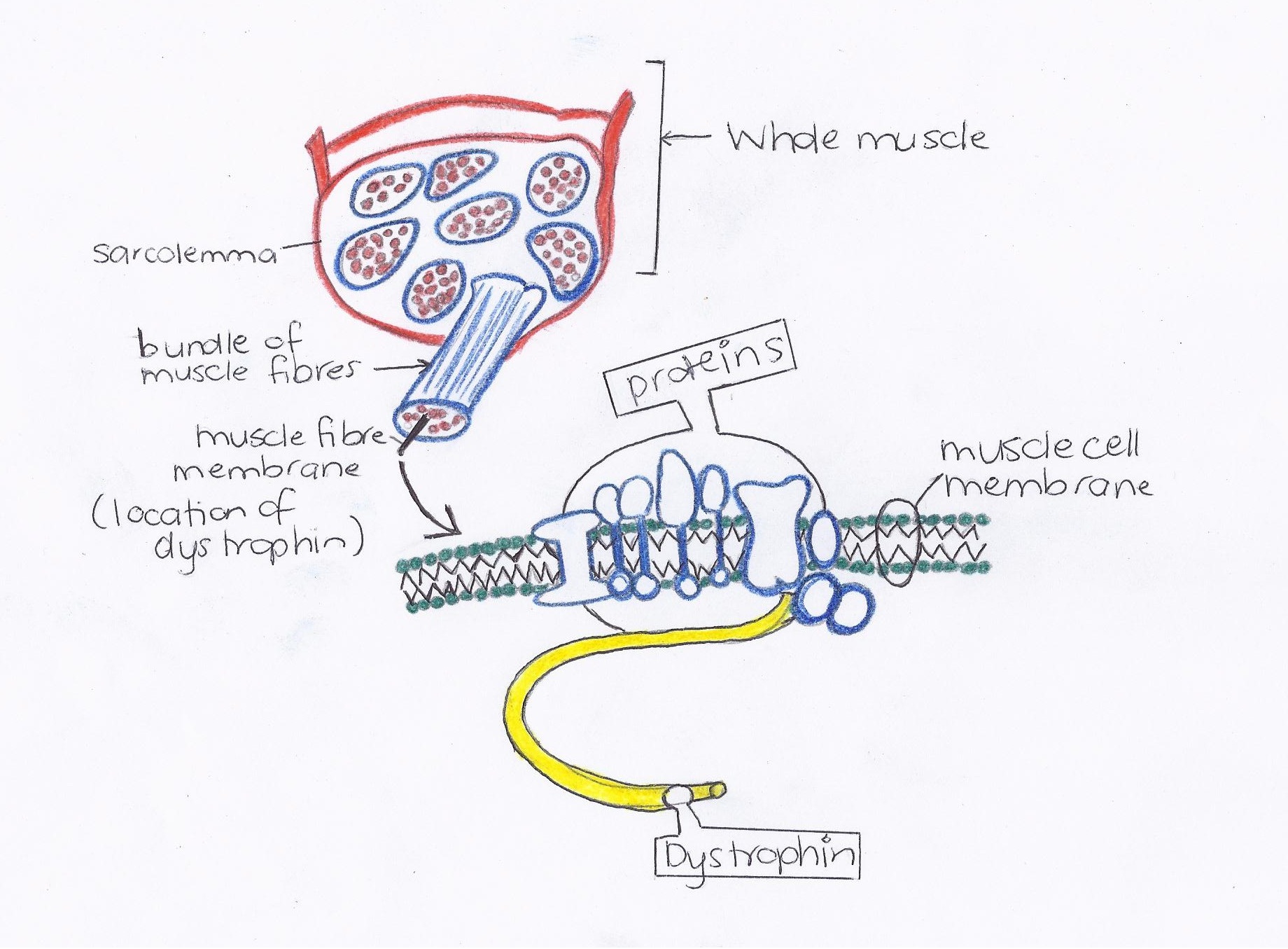
415 KB | This image is a visual display of the muscle fibre membrane and the location of dystrophin within the sarcolemma surrounding each muscle fibre. {{Template:2011 Student Image}}. Image creator: Ashleigh Pontifex (z3332629) Student image constructed bas | 1 |
| 17:16, 10 October 2011 | Dystrophin within the plasma membrane of muscle fibres.jpg (file) |  |
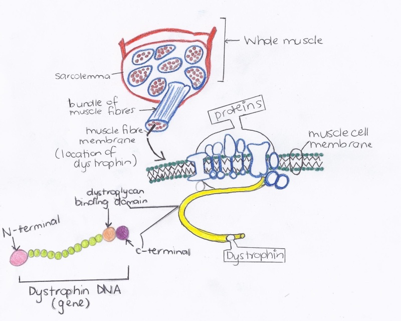
112 KB | This image is a visual display of the muscle fibre membrane and the location of dystrophin within the sarcolemma surrounding each muscle fibre. There are 4 main transmembrane proteins that compose the dystrophin-glycoprotein complex (DGP), also referred | 1 |
| 10:27, 22 September 2011 | Normal control muscle (a) vs. Duchennes muscular dystrophy muscle (b).jpg (file) | _vs._Duchennes_muscular_dystrophy_muscle_(b).jpg) |
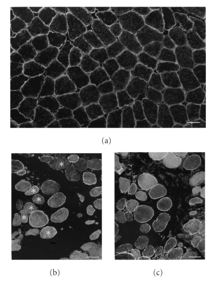
131 KB | Immunofluorescence with anti-AQP4 antibody of normal control muscle (a) and Duchenne muscular dystrophy muscle (DMD) (b), and that with anti-spectrin antibody of serial muscle section of DMD (c). Positive immunoreactivity with anti-AQP4 antibody is seen i | 1 |
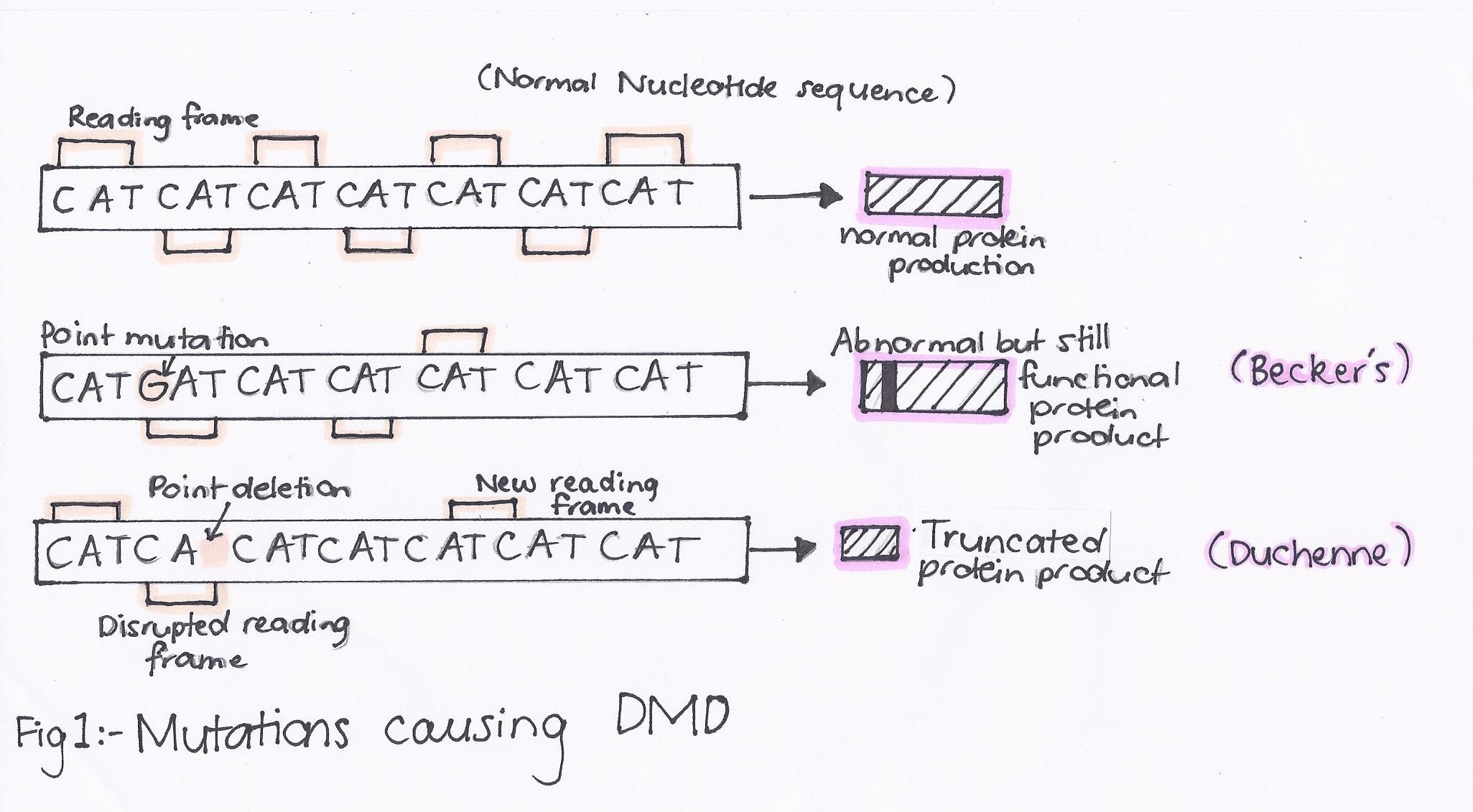
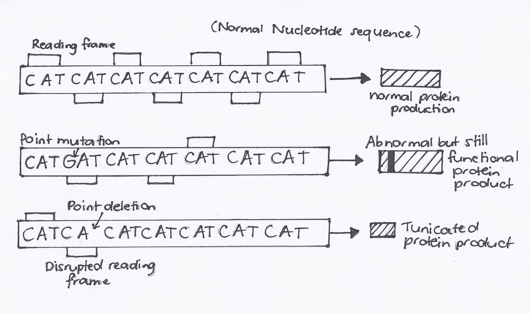
| 08:42, 4 October 2011 | Point mutations resulting in DMD.jpg (file) |  |
412 KB | This image is a visual display of the different types of mutations of the dystrophin gene that result in different forms of musuclar dystrophy. The first reading frame is that of a normal dystrophin gene and can be compared to the second reading frame tha | 1 |
| 19:25, 18 September 2011 | Point vs frameshift mutation of DMD gene.png (file) |  |
874 KB | Student image constructed based on the image present on http://emedicine.medscape.com/article/1173204-overview Point vs frameshift mutations. In contrast to most point mutations, which generally preserve the reading frame, frameshift mutations often le | 1 |
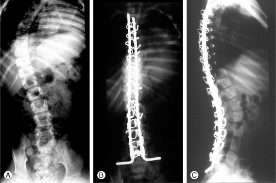
| 14:38, 16 August 2011 | Surgical correction of a 11-year old patient suffering from Duchenne muscular dystrophy.jpg (file) |  |
127 KB | This is an Open Access article distributed under the terms of the Creative Commons Attribution Non-Commercial License (http://creativecommons.org/licenses/by-nc/3.0) which permits unrestricted non-commercial use, distribution, and reproduction in any medi | 1 |
| 08:53, 4 October 2011 | X chromosome location of the dystrophin gene.jpg (file) |  |
188 KB | This image is a visual display of the location of the dystrophin gene on an X chromosome. Its noted location is on the short arm of the X chromosome at position 21.2, from base pair 31,137,344 to base pair 33,357,725. {{Template:2011 Student Image}}. | 1 |