File list
From Embryology
This special page shows all uploaded files.
| Date | Name | Thumbnail | Size | User | Description | Versions |
|---|---|---|---|---|---|---|
| 16:02, 16 April 2019 | Duodenal atresia 01.jpg (file) |  |
56 KB | Z8600021 | Upper gastrointestinal series showing complete obstruction to the flow of contrast at the second portion of the duodenum. There is also contrast filling of the biliary tree above the duodenal bulb noted (arrow). {{#pmid:30046504}} Copyright © 2018... | 1 |
| 15:58, 16 April 2019 | Placenta cord arteries abnormality 02.jpg (file) |  |
47 KB | Z8600021 | 1 | |
| 15:56, 16 April 2019 | Placenta cord arteries abnormality 01.jpg (file) |  |
72 KB | Z8600021 | ==Placenta cord arteries abnormality== Gross photograph of clamped segment of umbilical cord demonstrating segments of exposed spiraling umbilical arteries. {{#pmid:30906606}} The umbilical cord overall length was 29.5 cm and there were 13, 0.5- 1 cm... | 1 |
| 14:11, 13 April 2019 | VACTERL - esophageal atresia 01.jpg (file) |  |
135 KB | Z8600021 | == Figure 2: Infantogram showing Esophageal atresia as evidenced by coiling of nasogastric tube (yellow arrow) and absent right radial ray bones (red arrow). :'''Links:''' {{VACTERL}} ===Reference=== Chauhan S et al. Int J Contemp Pediatr. 2017 J... | 1 |
| 13:55, 13 April 2019 | VACTERL - vertebra 02.jpg (file) |  |
189 KB | Z8600021 | ==VACTERL - Vertebral Defect== Last menstrual period ((GA}} was 20 weeks 5 days. Measured fetal parameters of biparietal diameter such as head circumference, humerus, ulna, and radius showed foetus corresponding to 18–19 weeks. Hemi-vertebra at auto... | 1 |
| 13:48, 13 April 2019 | VACTERL - vertebra 01.jpg (file) |  |
74 KB | Z8600021 | ==VACTERL - Endocardial cushion defect== Gestational age according to her last menstrual period was 20 weeks 5 days. Measured fetal parameters of biparietal diameter such as head circumference, humerus, ulna, and radius showed foetus corresponding to 1... | 1 |
| 13:40, 13 April 2019 | VACTERL - cardiac 01.jpg (file) |  |
45 KB | Z8600021 | (Left) Endocardial cushion defect in form of absent crux. (arrow). (Right) Tricuspid regurgitation noted ===Reference=== {{#pmid:30662209}} ====Copyright==== https://creativecommons.org/licenses/by-nc-sa/4.0/ IJRI-28-452-g001.jpg | 1 |
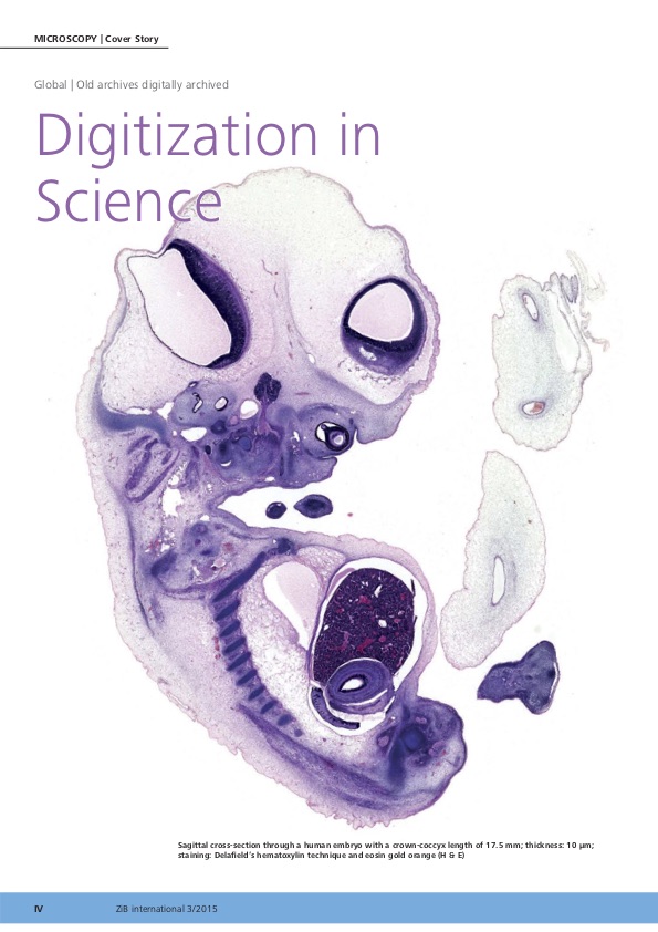
| 12:00, 13 April 2019 | EN Micro III-2015 Hill.jpg (file) |  |
109 KB | Z8600021 | 1 | |
| 10:31, 12 April 2019 | WNT3A transfer via filopodia.png (file) |  |
1.73 MB | Z8600021 | ==WNT3A transfer via filopodia== (a-c) Fluorescence photomicrographs of adjacent HeLa cells expressing RUSH-eGFP-WNT3A at indicated time points after biotin addition. From S7 video. (a) WNT3A is in the Golgi after biotin treatment. Inset showing WNT3A... | 1 |
| 09:55, 12 April 2019 | Acute otitis media infection age graph01.png (file) |  |
77 KB | Z8600021 | Fig 3. Age range of AOM infections. Each line represents an individual patient. N = 162 represents all patients who had at least one AOM infection. https://doi.org/10.1371/journal.pone.0212777.g003 acute otitis media (AOM) infection Copyright: © 201... | 1 |
| 09:44, 12 April 2019 | Third trimester maternal womb sounds 01.mp3 (file) | 6.93 MB | Z8600021 | ==Maternal externally recorded womb sound recording== ===Reference=== {{#pmid:29746604}} ====Copyright==== © 2018 Parga et al. This is an open access article distributed under the terms of the Creative Commons Attribution License, which permits unr... | 1 | |
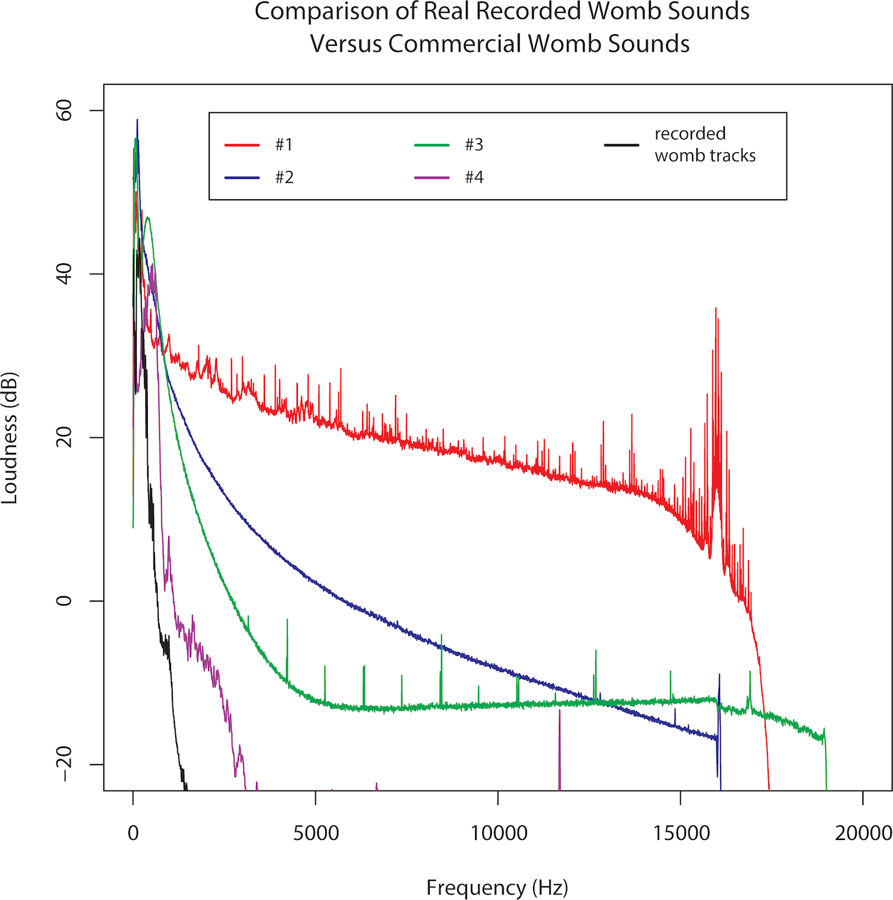
| 09:40, 12 April 2019 | Third trimester sounds 01.png (file) |  |
335 KB | Z8600021 | 2 | |
| 13:16, 11 April 2019 | Murine Cereblon in complex with Thalidomide.jpg (file) |  |
120 KB | Z8600021 | ==Crystal Structure of Murine Cereblon in Complex with Thalidomide== ===Reference=== Cite images created with the PDB ID and associated publication, NGL Viewer (AS Rose et al. (2018) NGL viewer: web-based molecular graphics for large complexes. Bioinf... | 1 |
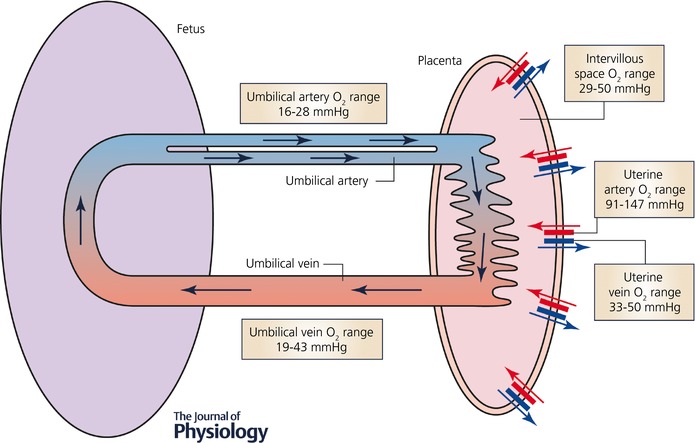
| 11:31, 10 April 2019 | Placental imaging 01.jpg (file) |  |
59 KB | Z8600021 | ==Placental Oxygen Levels== Simplified schematic diagram of the maternal and fetal placental circulations, showing the major compartments and published attributed in vivo oxygen values. :'''Links:''' {{placenta}} | {{trophoblast}} ===Reference===... | 1 |
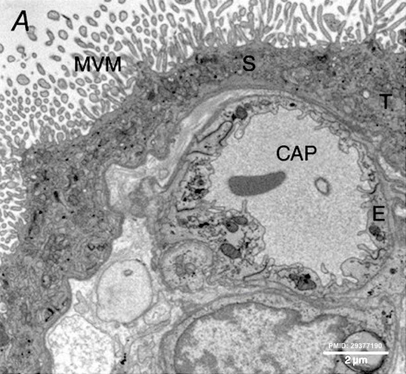
| 11:18, 10 April 2019 | Placental imaging 03A.jpg (file) |  |
181 KB | Z8600021 | ==Placental Imaging== A transmission electron micrograph of terminal villi showing microvillous membrane (MVM), an underlying capillary (CAP), a syncytiotrophoblast (S), a trophoblast (T) and endothelium (E). :'''Links:''' {{placenta}} | [[:File:Pl... | 1 |
| 11:11, 10 April 2019 | Placental imaging 03.jpg (file) |  |
227 KB | Z8600021 | ==Placental Imaging== A, a transmission electron micrograph of terminal villi showing microvillous membrane (MVM), an underlying capillary (CAP), a syncytiotrophoblast (S), a trophoblast (T) and endothelium (E). B, projection of an imaged stack (whole... | 1 |
| 09:39, 9 April 2019 | Prentiss1906 fig07.jpg (file) |  |
324 KB | Z8600021 | 1 | |
| 09:38, 9 April 2019 | Prentiss1906 fig06.jpg (file) |  |
138 KB | Z8600021 | 1 | |
| 09:37, 9 April 2019 | Prentiss1906 fig05.jpg (file) |  |
988 KB | Z8600021 | 1 | |
| 09:36, 9 April 2019 | Prentiss1906 fig04.jpg (file) |  |
490 KB | Z8600021 | 1 | |
| 09:35, 9 April 2019 | Prentiss1906 fig03.jpg (file) |  |
168 KB | Z8600021 | 1 | |
| 09:35, 9 April 2019 | Prentiss1906 fig02.jpg (file) |  |
159 KB | Z8600021 | 1 | |
| 09:34, 9 April 2019 | Prentiss1906 fig01.jpg (file) |  |
417 KB | Z8600021 | {{Ref-Prentiss1906}} | 1 |
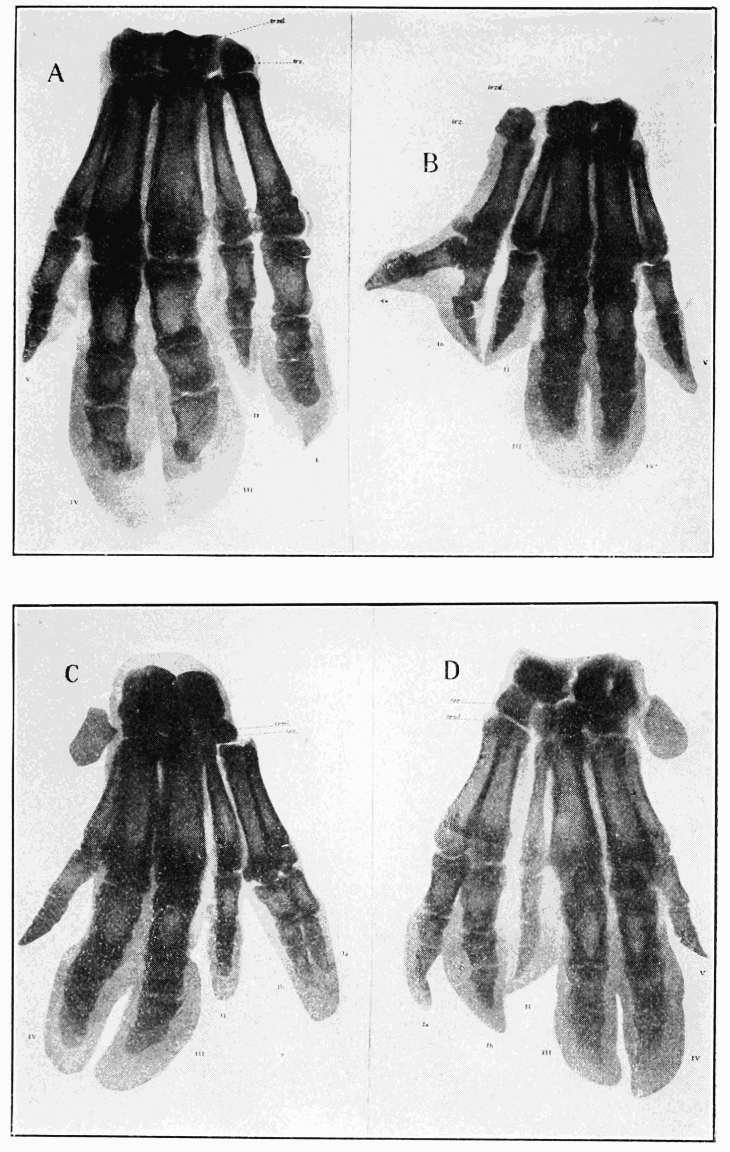
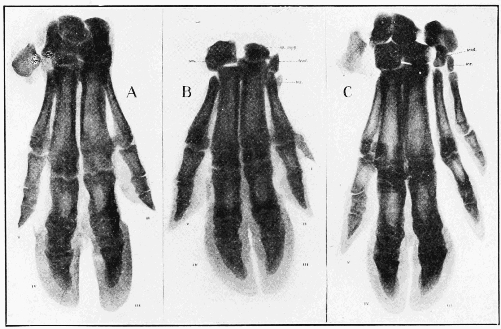
| 20:28, 1 April 2019 | Wood-Jones1934 fig05.jpg (file) |  |
270 KB | Z8600021 | {{Ref-Wood-Jones1934}} | 1 |
| 20:27, 1 April 2019 | Wood-Jones1934 fig04.jpg (file) |  |
94 KB | Z8600021 | {{Ref-Wood-Jones1934}} | 1 |
| 20:24, 1 April 2019 | Wood-Jones1934 fig03.jpg (file) |  |
41 KB | Z8600021 | {{Ref-Wood-Jones1934}} | 1 |
| 20:23, 1 April 2019 | Wood-Jones1934 fig02.jpg (file) |  |
149 KB | Z8600021 | {{Ref-Wood-Jones1934}} | 1 |
| 20:20, 1 April 2019 | Wood-Jones1934 fig01.jpg (file) |  |
76 KB | Z8600021 | {{Ref-Wood-Jones1934}} | 1 |
| 13:16, 30 March 2019 | 2019 Foundations Lecture - Introduction to Human Development.pdf (file) | 4.31 MB | Z8600021 | Category:PDFCategory:MedicineCategory:2019 | 1 | |
| 09:59, 29 March 2019 | Waldo Shumway.jpg (file) |  |
616 KB | Z8600021 | ==Waldo Shumway (1891-1956)== Born - 8 May 1891 New Brunswick, Middlesex County, New Jersey, USA Death - 8 Mar 1956 (aged 64) Manhattan, New York County (Manhattan), New York, USA {{Footer}} | 1 |
| 12:31, 27 March 2019 | Hair stem cells 01.jpg (file) |  |
123 KB | Z8600021 | ==Hair Stem Cells Niche== The hair follicle is a prominent example of how cell biology can help understand tissue homeostasis. Each hair follicle requires a niche of stem cells to undergo cyclical bouts of hair growth. The schematic (left) depicts t... | 1 |
| 15:50, 19 March 2019 | Trisomy 10 mosaicism cardiovascular abnormalities.jpg (file) |  |
147 KB | Z8600021 | ==Trisomy 10 mosaicism Cardiovascular Abnormalities== Echocardiogram showing: a - a patent ductus arteriosus b - patent foramen ovale :'''Links:''' {{Trisomy mosaicism}} ===Reference=== {{#pmid:30081864}} ====Copyright==== Open Access This art... | 1 |
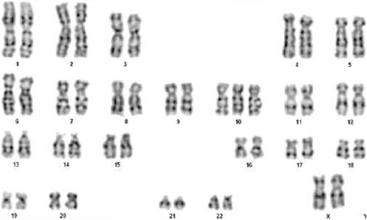
| 15:45, 19 March 2019 | Trisomy 10 mosaicism karyotype.jpg (file) |  |
76 KB | Z8600021 | Karyotype analysis: analysis of 100 split phase, 42 mitotic phase, karyotype of 47, XX ===Reference=== ====Copyright==== Open Access This article is distributed under the terms of the Creative Commons Attribution 4.0 International License (http://cr... | 1 |
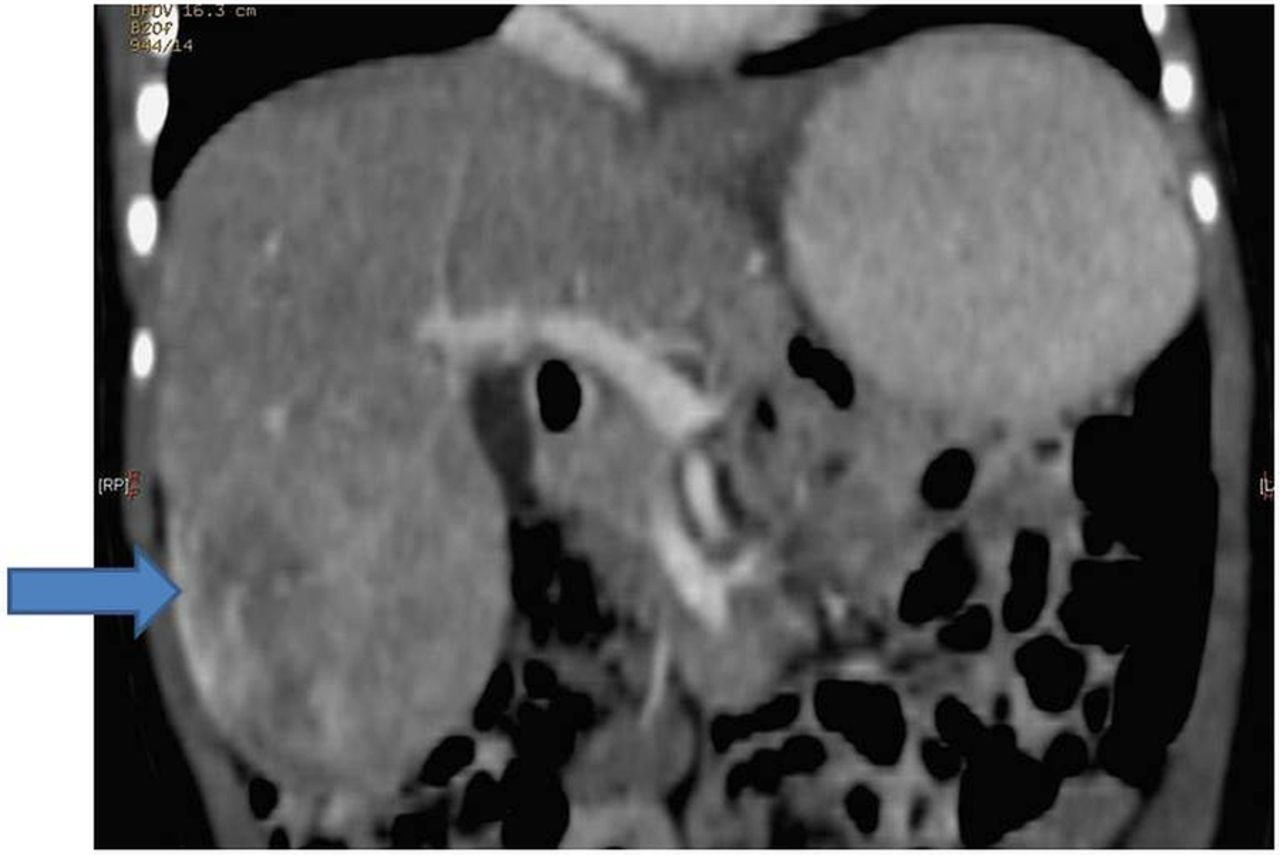
| 14:48, 19 March 2019 | Mosaic Trisomy 18 hepatoblastoma CT.jpg (file) |  |
79 KB | Z8600021 | ==Mosaic Trisomy 18 Hepatoblastoma== Diagnostic CT of the abdomen. {{#pmid:26795740}} Casereports-2016-January-2016---F2.large.jpg | 1 |
| 10:01, 19 March 2019 | Terminologia Embryologica User Guide.pdf (file) | 269 KB | Z8600021 | Category:PDF | 1 | |
| 15:15, 18 March 2019 | Angle1918 plate17.jpg (file) |  |
348 KB | Z8600021 | Angle1918 | 1 |
| 10:32, 15 March 2019 | Fetal week 12 head 14.jpg (file) |  |
95 KB | Z8600021 | 2 | |
| 10:31, 15 March 2019 | Fetal week 12 head 13.jpg (file) |  |
77 KB | Z8600021 | 2 | |
| 10:31, 15 March 2019 | Fetal week 12 head 12.jpg (file) |  |
47 KB | Z8600021 | 2 | |
| 09:50, 15 March 2019 | Fetal week 12 head 11.jpg (file) |  |
37 KB | Z8600021 | 1 | |
| 09:46, 15 March 2019 | Fetal week 12 head 1 icon.jpg (file) | 9 KB | Z8600021 | 1 | ||
| 09:42, 15 March 2019 | Fetal week 12 head 01.jpg (file) |  |
48 KB | Z8600021 | 12wk-6605-lat-Trans-Diewert-UBC.avi | 1 |
| 09:07, 15 March 2019 | Fetal week 12 head 01.mp4 (file) | 1,004 KB | Z8600021 | 12wk_6605_lat_Trans_Diewert_UBC.avi (398Wx398H) converted with handbrake 12wk_6605_lat_Trans_Diewert_UBC.mp4 | 1 | |
| 10:14, 13 March 2019 | Catshark egg case stage 1.jpg (file) |  |
81 KB | Z8600021 | 1 | |
| 10:14, 13 March 2019 | Catshark egg case stage 2.jpg (file) |  |
58 KB | Z8600021 | 1 | |
| 10:13, 13 March 2019 | Catshark egg case stage 3.jpg (file) |  |
76 KB | Z8600021 | 1 | |
| 10:13, 13 March 2019 | Catshark egg case stage 4.jpg (file) |  |
98 KB | Z8600021 | 1 | |
| 10:13, 13 March 2019 | Catshark egg case stage 5.jpg (file) |  |
97 KB | Z8600021 | 1 | |
| 10:13, 13 March 2019 | Catshark egg case stage 6.jpg (file) |  |
106 KB | Z8600021 | 1 | |
| 10:12, 13 March 2019 | Catshark egg case stage 7.jpg (file) |  |
95 KB | Z8600021 | Fig 7. The inside of the S. stellaris egg case at stage 7. A: Position of placoid denticles and banded patterns on the body (dorsal view). B: Fully developed eye and greater pigmentation on skin (lateral view). C: The magnified head and fully consumed... | 1 |