File list
From Embryology
This special page shows all uploaded files.
| Date | Name | Thumbnail | Size | User | Description | Versions |
|---|---|---|---|---|---|---|
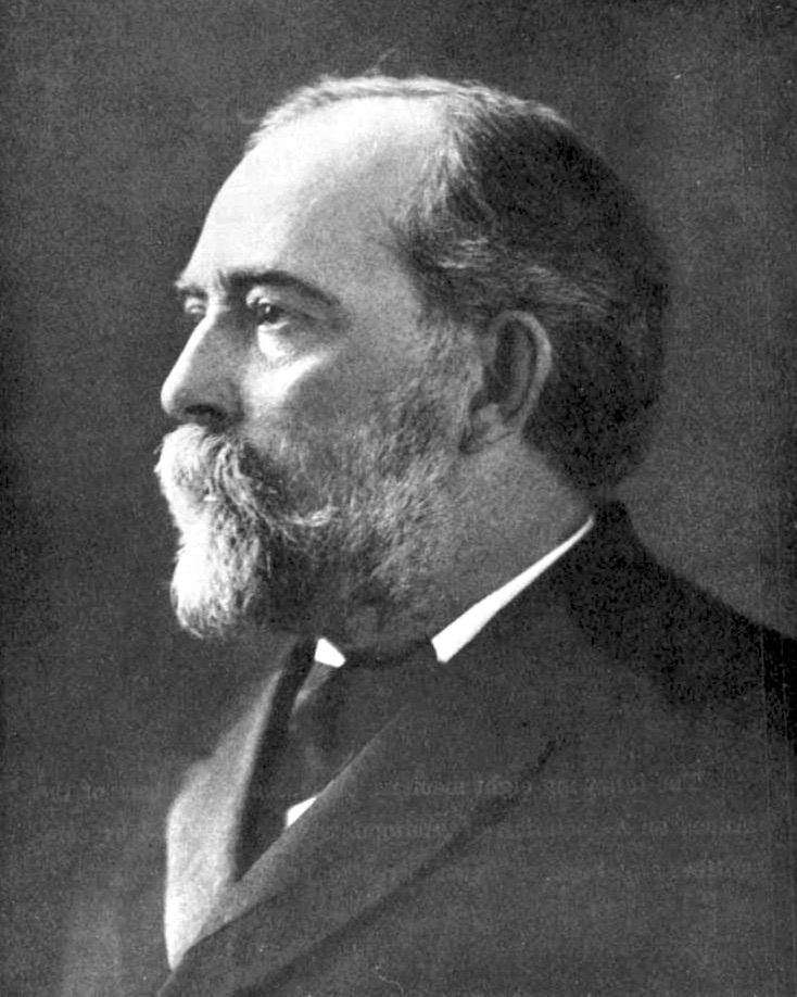
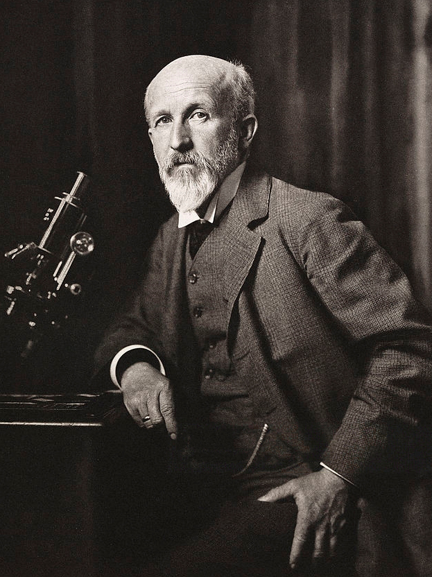
| 14:09, 12 August 2019 | Simon H. Gage.jpg (file) |  |
170 KB | Z8600021 | 1 | |
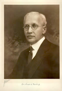
| 16:05, 10 August 2019 | Fred. J. Taussig.jpg (file) |  |
15 KB | Z8600021 | 2 | |
| 15:54, 8 August 2019 | PdP.gif (file) |  |
5.65 MB | Z8600021 | 1 | |
| 14:40, 8 August 2019 | 2019-ANAT2341-course outline-Final.pdf (file) | 395 KB | Z3485617 | 1 | ||
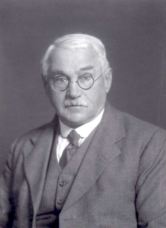
| 01:01, 6 August 2019 | Edward Fawcett.jpg (file) |  |
85 KB | Z8600021 | 3 | |
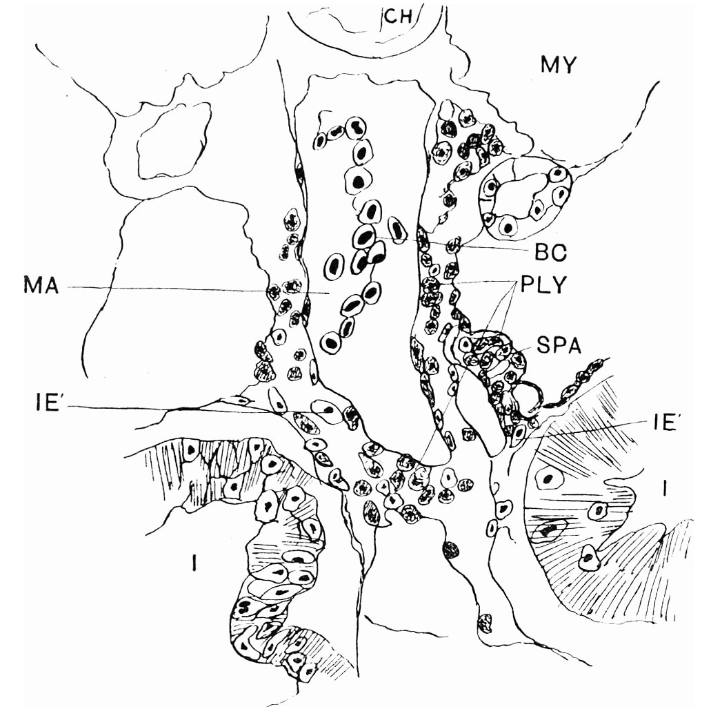
| 15:31, 2 August 2019 | Pearce1903 fig02.jpg (file) |  |
377 KB | Z8600021 | 2 | |
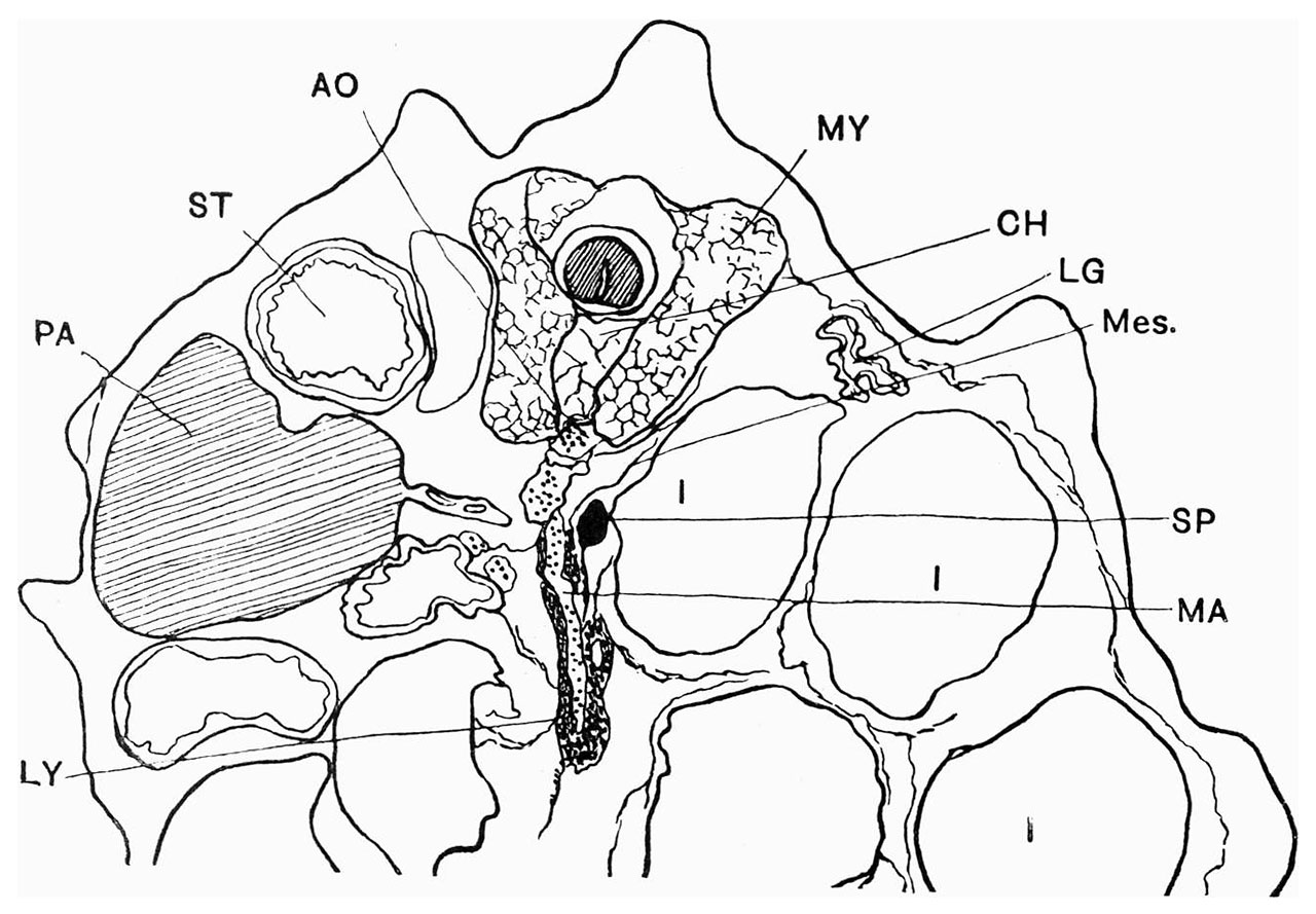
| 15:23, 2 August 2019 | Pearce1903 fig01.jpg (file) |  |
335 KB | Z8600021 | 2 | |
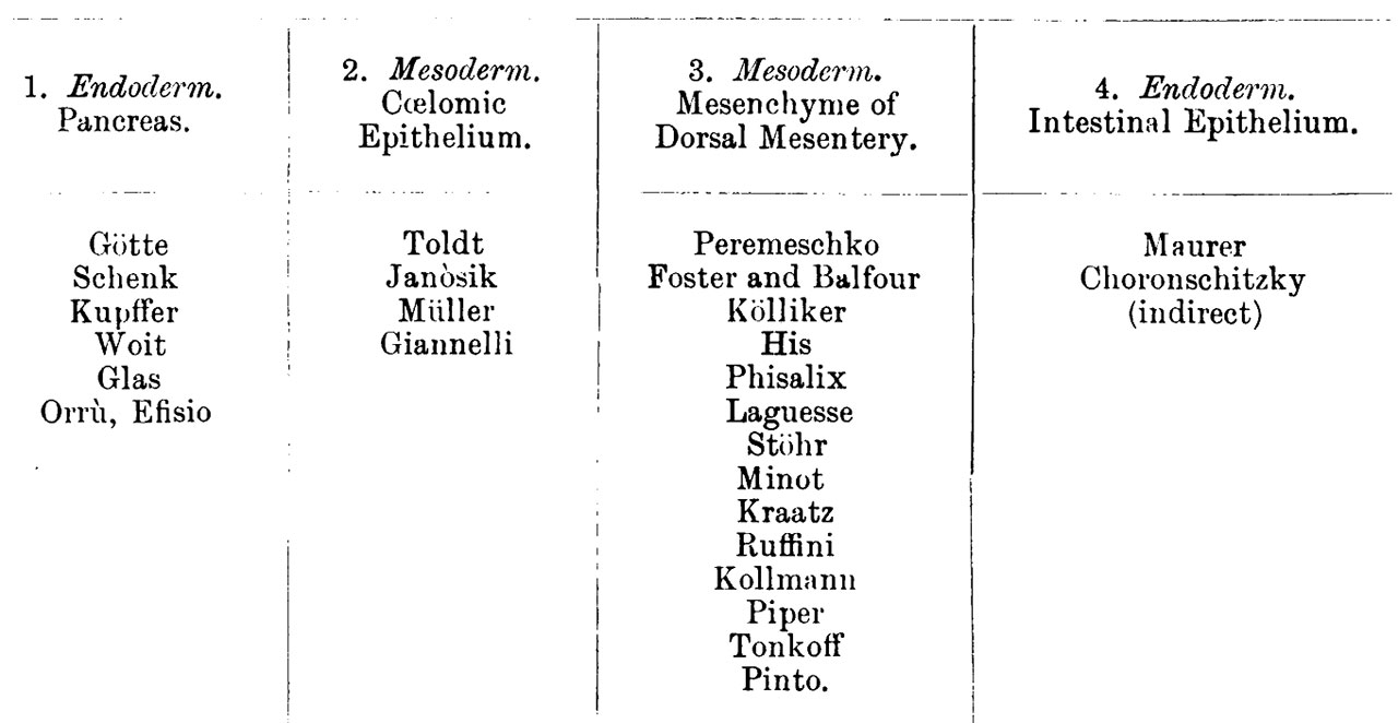
| 15:15, 2 August 2019 | Pearce1903 table1.jpg (file) |  |
332 KB | Z8600021 | {{Ref-Pearce1903}} | 1 |
| 13:47, 2 August 2019 | Lewis1903 plate04L.jpg (file) |  |
386 KB | Z8600021 | 2 | |
| 13:44, 2 August 2019 | Lewis1903 plate03L.jpg (file) |  |
354 KB | Z8600021 | 2 | |
| 13:39, 2 August 2019 | Lewis1903 plate02L.jpg (file) |  |
287 KB | Z8600021 | 3 | |
| 13:24, 2 August 2019 | Lewis1903 plate01L.jpg (file) |  |
246 KB | Z8600021 | 3 | |
| 13:16, 2 August 2019 | Lewis1903 plate04.jpg (file) |  |
403 KB | Z8600021 | reduce image size | 3 |
| 13:11, 2 August 2019 | Lewis1903 plate03.jpg (file) |  |
442 KB | Z8600021 | reduce image size | 3 |
| 13:05, 2 August 2019 | Lewis1903 plate02.jpg (file) |  |
258 KB | Z8600021 | reduce image size | 3 |
| 13:00, 2 August 2019 | Lewis1903 plate01.jpg (file) |  |
328 KB | Z8600021 | reduce image size | 3 |
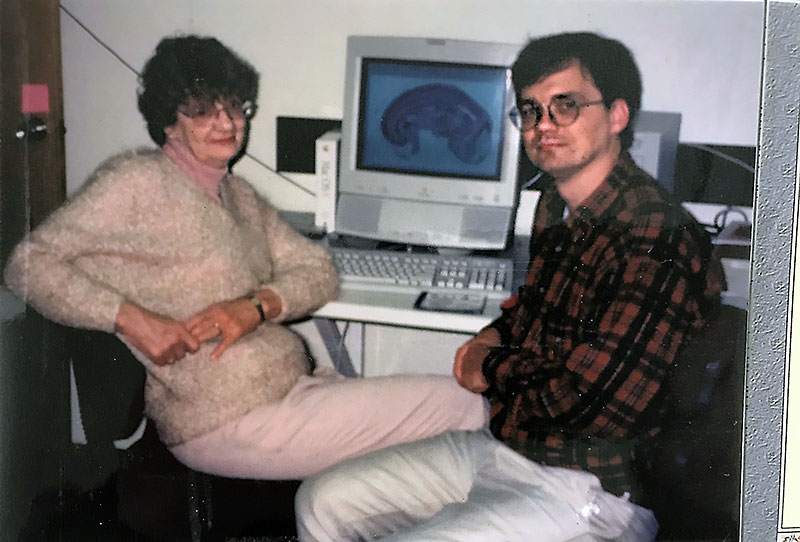
| 14:54, 31 July 2019 | Beverley and Mark 1997.jpg (file) |  |
88 KB | Z8600021 | 1 | |
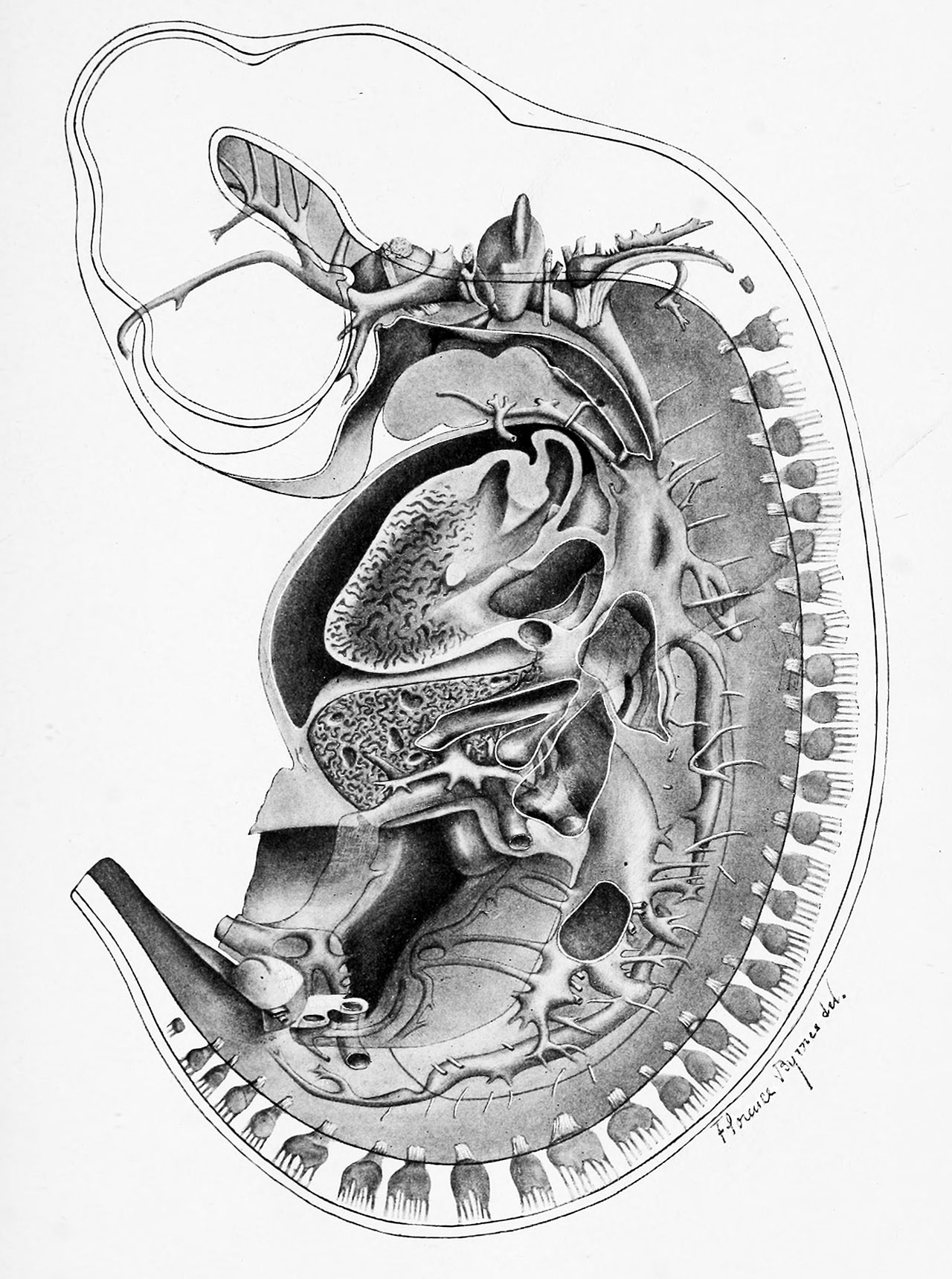
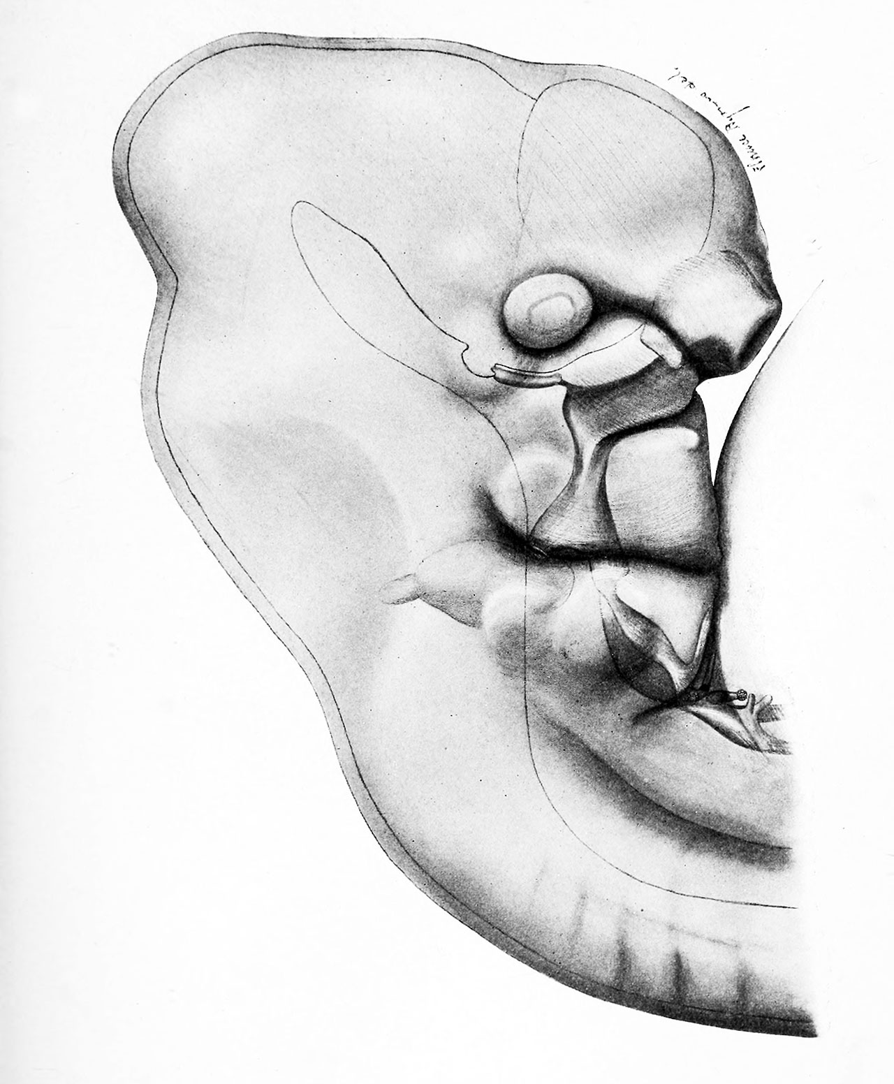
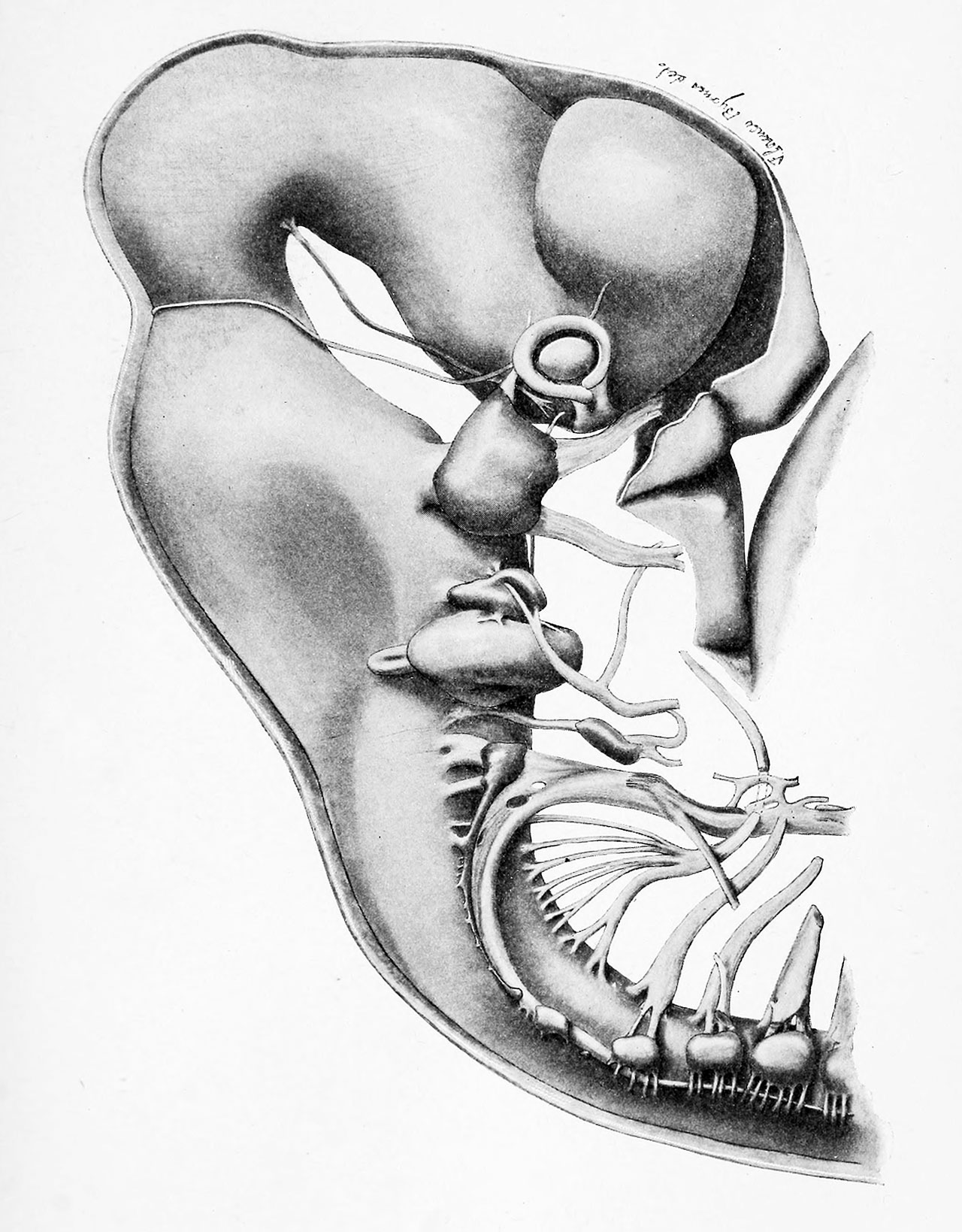
| 14:38, 30 July 2019 | Sabin1915 plate07.jpg (file) |  |
1.16 MB | Z8600021 | from original scan | 2 |
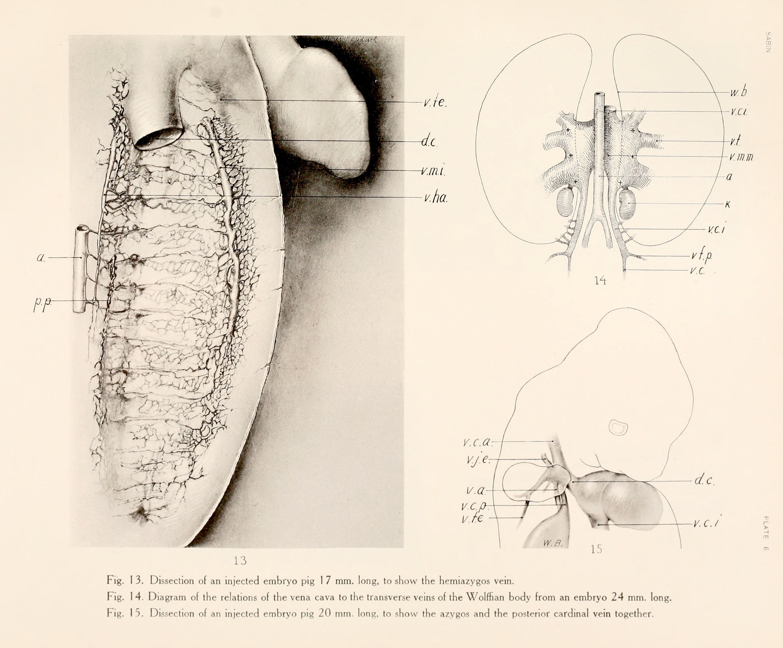
| 14:37, 30 July 2019 | Sabin1915 plate06.jpg (file) |  |
926 KB | Z8600021 | from original scan | 2 |
| 14:37, 30 July 2019 | Sabin1915 plate05.jpg (file) |  |
1,013 KB | Z8600021 | from original scan | 2 |
| 14:37, 30 July 2019 | Sabin1915 plate03.jpg (file) |  |
1.08 MB | Z8600021 | from original scan | 2 |
| 14:37, 30 July 2019 | Sabin1915 plate04.jpg (file) |  |
797 KB | Z8600021 | from original scan | 2 |
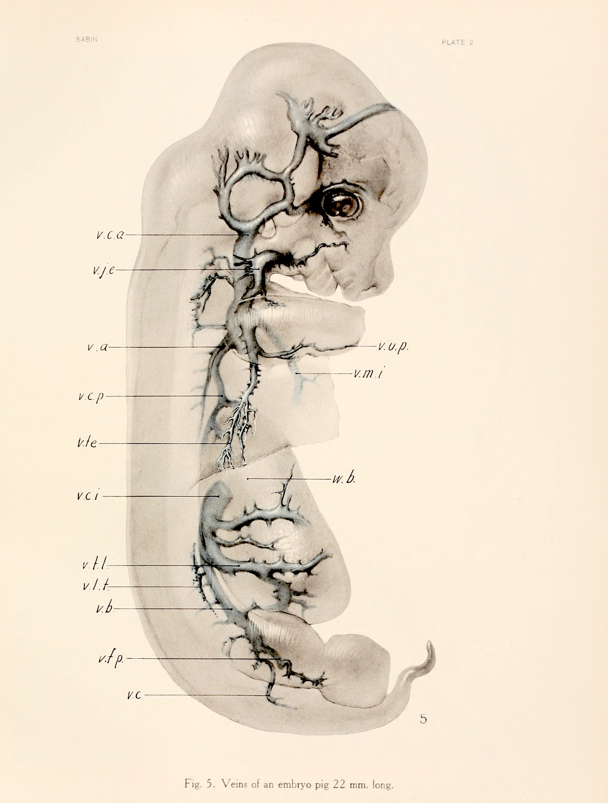
| 14:36, 30 July 2019 | Sabin1915 plate02.jpg (file) |  |
609 KB | Z8600021 | from original scan | 2 |
| 14:36, 30 July 2019 | Sabin1915 plate01.jpg (file) |  |
922 KB | Z8600021 | from original scan | 2 |
| 12:45, 30 July 2019 | Sabin1915.pdf (file) | 6.25 MB | Z8600021 | {{Ref-Sabin1915}} {| class="wikitable mw-collapsible mw-collapsed" ! Online Editor |- | 90px|left This 1915 paper by Florence Rena Sabin (1871 - 1953) describes early venous vascular... | 1 | |
| 06:32, 30 July 2019 | Site edits May-Jul 2019 graph.jpg (file) |  |
68 KB | Z8600021 | 1 | |
| 06:16, 30 July 2019 | Organoids - inner ear 01.jpg (file) |  |
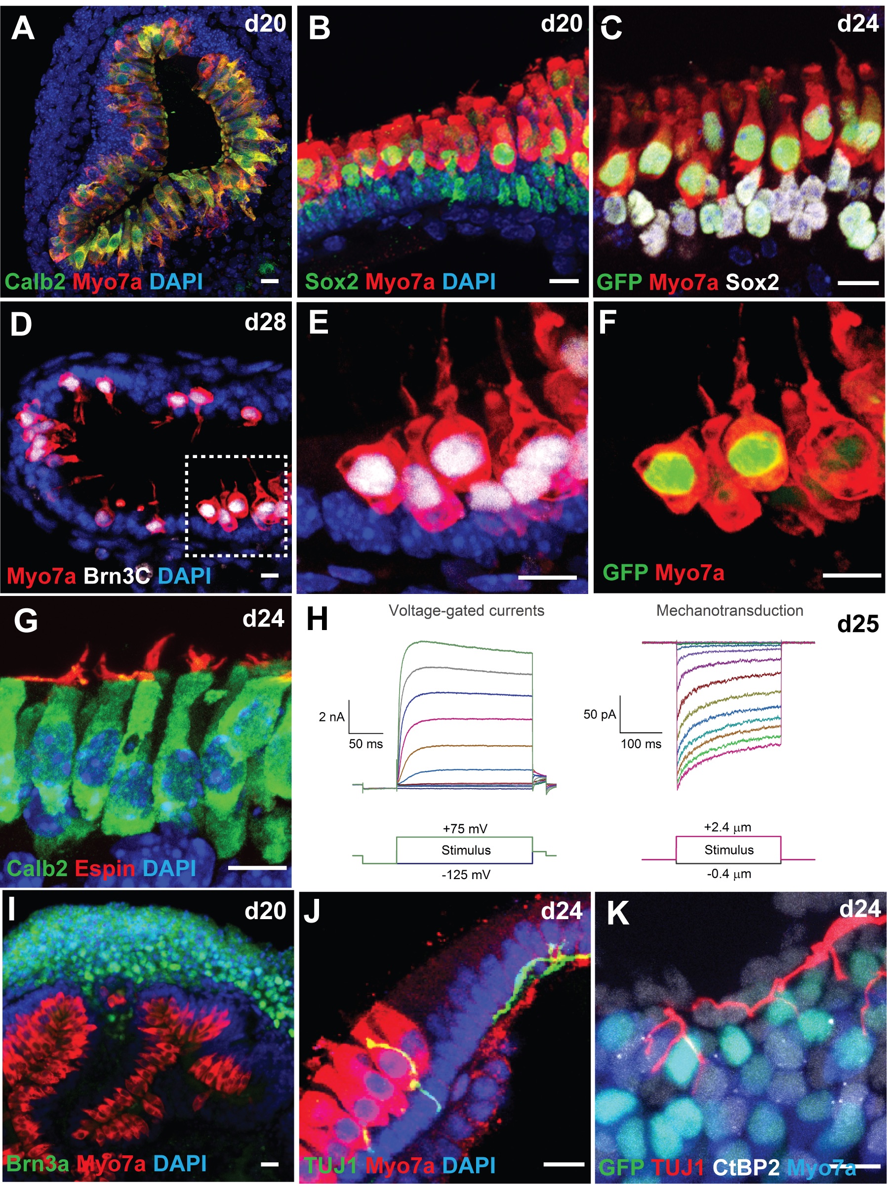
1.07 MB | Z8600021 | ==CHIR-treated aggregates give rise to inner ear organoids harboring mechanosensitive hair cells== (A-B) Co-localization of two hair cell markers Calb2 and Myo7a (A) or Sox2 and Myo7a (B) in cells lining the luminal surface of a vesicle. (C) Atoh1/nG... | 1 |
| 05:59, 30 July 2019 | Organoids overview 01.jpg (file) |  |
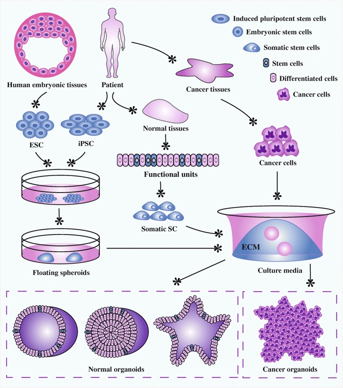
146 KB | Z8600021 | Organoid establishment from stem cells and cancer cells. Embryonic stem cells from human embryonic tissues and induced pluripotent stem cells from adult tissues firstly experience directed differentiation, generate floating spheroids, and subsequently... | 1 |
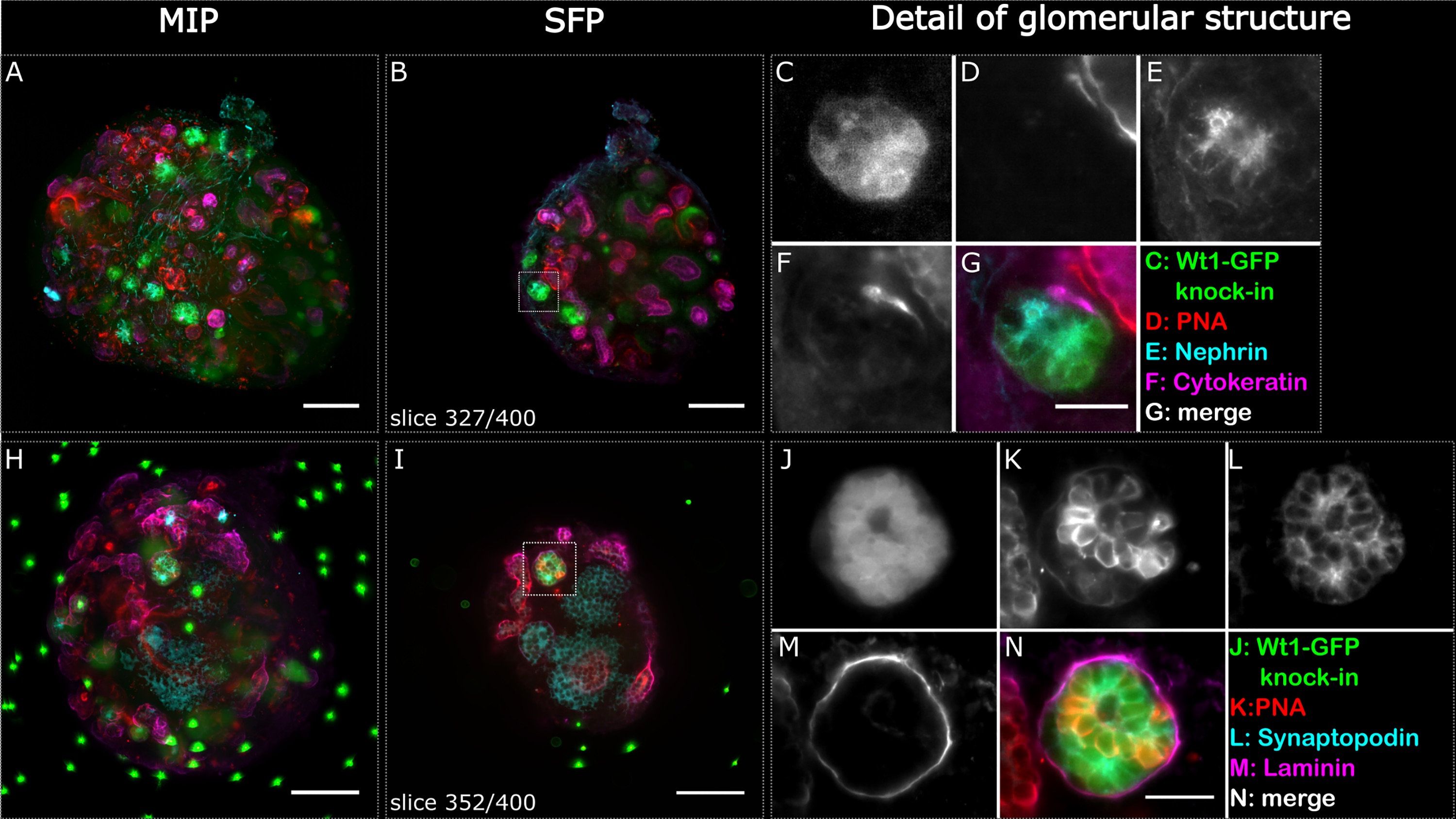
| 05:51, 30 July 2019 | Organoids - renal glomerulus.jpg (file) |  |
700 KB | Z8600021 | ==Renal Glomerular Organoids== Fig 2. Glomerular structures stained positively for podocyte markers indicating phenotypic maturity in re-aggregated embryonic renal organoids after 6 days of culture. Two whole organoids (A-G and H-N) were imaged in the... | 1 |
| 12:24, 29 July 2019 | Goat and kids.jpg (file) |  |
85 KB | Z8600021 | Goat kid in char of Sirajganj, Bangladesh. ====Copyright==== This file is licensed under the Creative Commons Attribution-Share Alike 3.0 Unported license. Goat_kid_in_char_of_Sirajganj,_Bangladesh_05.jpg {{Footer}} Category:Goat | 1 |
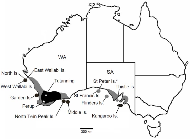
| 11:56, 29 July 2019 | Map tammar wallaby distribution.jpg (file) |  |
82 KB | Z8600021 | Fig 1 Former and current distribution of the tammar wallaby (Notamacropus eugenii) in Southern Australia. Collection localities and sites mentioned in the text are indicated. Dark shading represents extant distribution; light shading represents areas... | 1 |
| 11:54, 29 July 2019 | 2019-ANAT2341-course manual (FINAL).pdf (file) | 2.08 MB | Z3485617 | 4 | ||
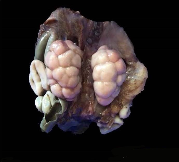
| 04:06, 29 July 2019 | Fetal kidney.jpg (file) |  |
43 KB | Z8600021 | Fetal kidney Normal human fetal kidney will show prominent lobulations which disappear postnatally to form a smooth surfaced kidney. ===Reference=== Dr. Roshan Nasimudeen, Department of Pathology, Calicut Medical College ====Copyright==== This work... | 1 |
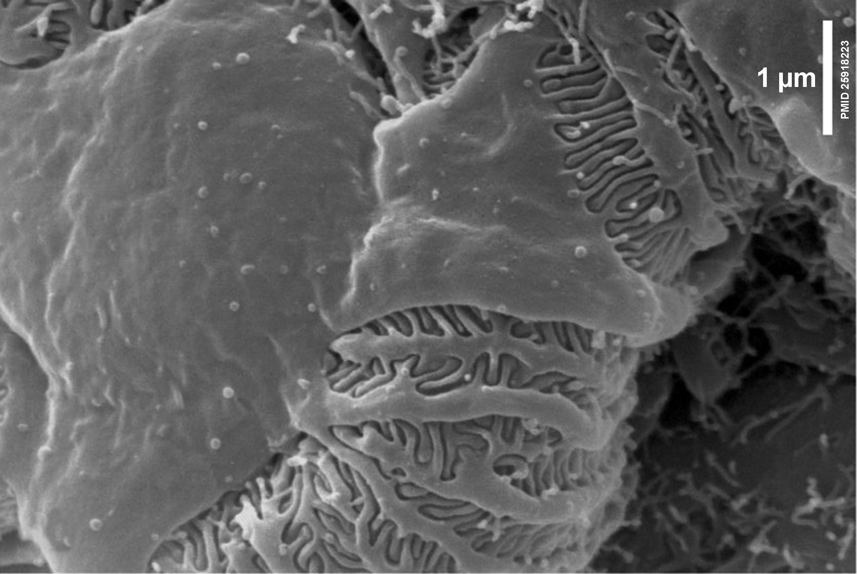
| 14:19, 24 July 2019 | Liver histology EM02.jpg (file) |  |
154 KB | Z8600021 | Reverted to version as of 20:47, 26 April 2018 (AEST) | 4 |
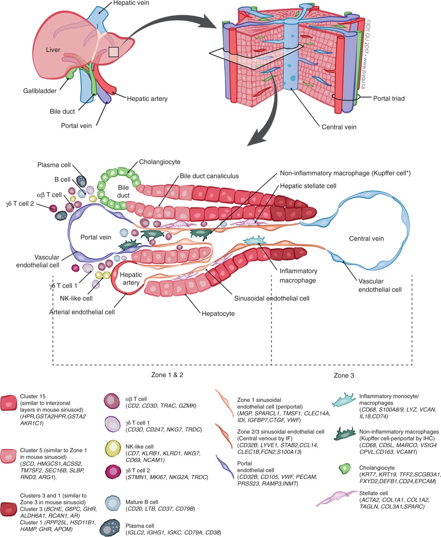
| 13:45, 24 July 2019 | Adult human liver cells.jpg (file) |  |
245 KB | Z8600021 | Summary map of the human liver. The main “building block” of the liver is the hepatic lobule, which includes a portal triad, hepatocytes aligned between a capillary network, and a central vein. The portal triad is made up of the hepatic artery, the... | 1 |
| 13:12, 24 July 2019 | Nephron EM11.jpg (file) |  |
398 KB | Z8600021 | 2 | |
| 09:42, 23 July 2019 | Frank R. Lillie.jpg (file) |  |
74 KB | Z8600021 | reduce image size | 2 |
| 13:55, 19 July 2019 | Bloom1926 fig01.jpg (file) |  |
170 KB | Z8600021 | 2 | |
| 13:04, 19 July 2019 | Radford1908 fig05.jpg (file) |  |
48 KB | Z8600021 | 2 | |
| 12:58, 19 July 2019 | Radford1908 fig04.jpg (file) |  |
224 KB | Z8600021 | 2 | |
| 12:54, 19 July 2019 | Radford1908 fig03.jpg (file) |  |
46 KB | Z8600021 | 2 | |
| 12:50, 19 July 2019 | Radford1908 fig02.jpg (file) |  |
168 KB | Z8600021 | 2 | |
| 12:46, 19 July 2019 | Radford1908 fig01.jpg (file) |  |
186 KB | Z8600021 | resize and crop legend | 2 |
| 12:40, 19 July 2019 | Radford1908-table1.jpg (file) |  |
91 KB | Z8600021 | {{Ref-Radford1908}} | 1 |
| 13:14, 18 July 2019 | Bennet Mills Allen.jpg (file) |  |
85 KB | Z8600021 | Adjusted in size and contrast. | 2 |
| 12:35, 18 July 2019 | Allen1904 plate7.jpg (file) |  |
704 KB | Z8600021 | 2 | |
| 11:26, 16 July 2019 | Oscar Hertwig.jpg (file) |  |
351 KB | Z8600021 | 2 | |
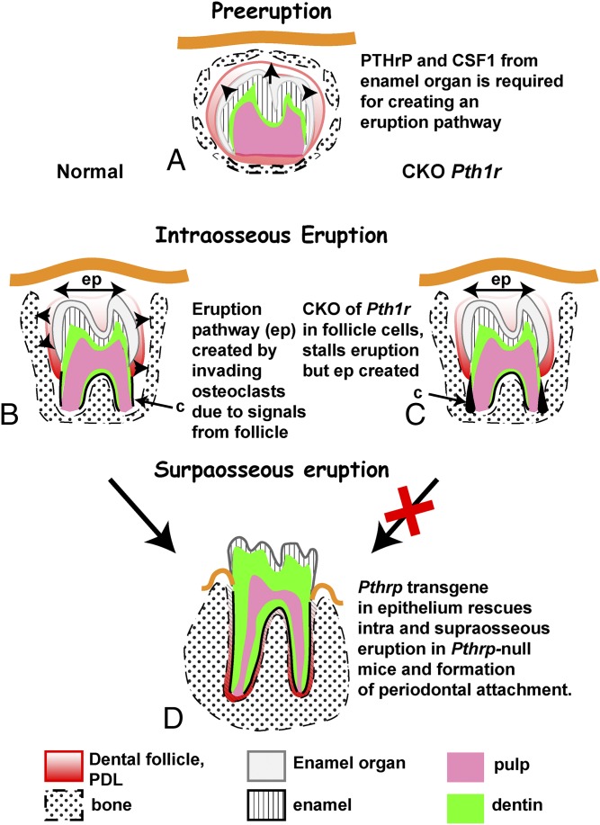
| 11:14, 16 July 2019 | Tooth eruption signaling.jpg (file) |  |
166 KB | Z8600021 | ==Tooth Eruption Signaling== Three phases of tooth eruption, preeruptive, intraosseous, and supraosseous, require signaling in the PTHrP/PTH1R pathway. (A) In the preeruptive phase, teeth are fully surrounded by a bony crypt. The enamel organ produces... | 1 |
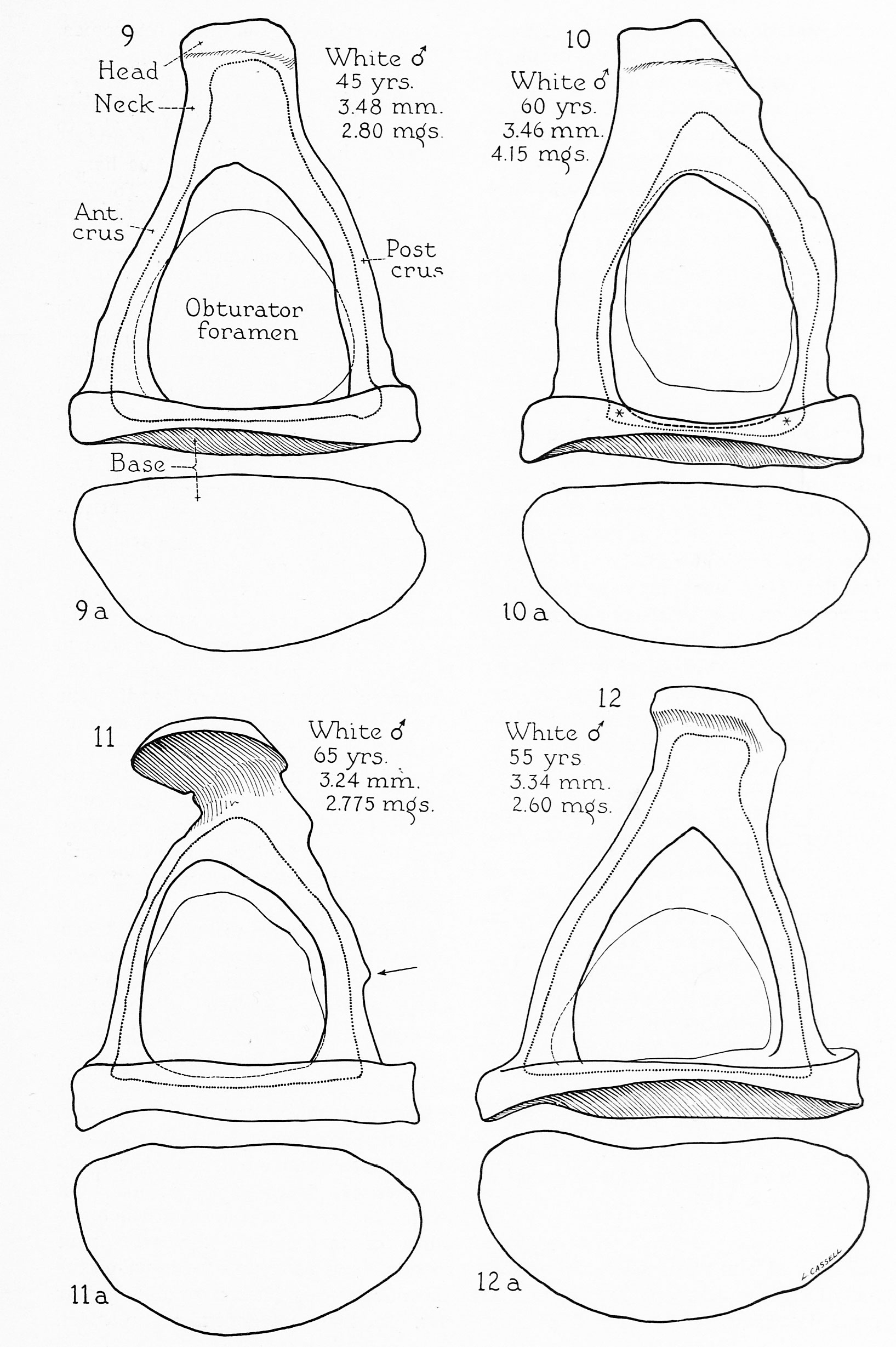
| 11:42, 12 July 2019 | BeatonAnson1940 fig09-12.jpg (file) |  |
422 KB | Z8600021 | 3 | |
| 11:27, 12 July 2019 | BeatonAnson1940 fig05-8.jpg (file) |  |
383 KB | Z8600021 | 3 |