File list
From Embryology
This special page shows all uploaded files.
| Date | Name | Thumbnail | Size | User | Description | Versions |
|---|---|---|---|---|---|---|
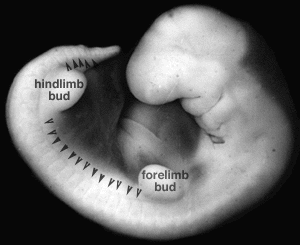
| 10:49, 10 August 2009 | Stage14 somites limbbuds.png (file) |  |
24 KB | MarkHill | Image source: UNSW Embryology http://embryology.med.unsw.edu.au/Notes/skmus.htm#Somite1 Category:Mesoderm Category:Somite | 1 |
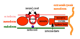
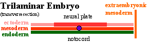
| 11:00, 9 August 2009 | Mesoderm cartoon4.gif (file) |  |
4 KB | S8600021 | Mesoderm Development cartoon Fourth of a set of 4 simple images to describe mesoderm development from trilaminar embryo onward. Original file name: Image_004.gif New Name: mesoderm cartoon4.gif Image Source: UNSW Embryology [http://embryology.med.unsw. | 1 |
| 10:59, 9 August 2009 | Image 004.gif (file) |  |
4 KB | S8600021 | 1 | |
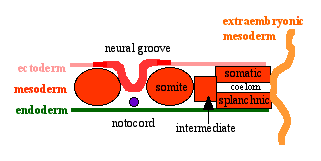
| 10:58, 9 August 2009 | Mesoderm cartoon3.gif (file) |  |
4 KB | S8600021 | Mesoderm Development cartoon Third of a set of 4 simple images to describe mesoderm development from trilaminar embryo onward. Original file name: Image_003.gif New Name: mesoderm cartoon3.gif Image Source: UNSW Embryology [http://embryology.med.unsw.e | 1 |
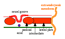
| 10:58, 9 August 2009 | Mesoderm cartoon2.gif (file) |  |
3 KB | S8600021 | Mesoderm Development cartoon First of a set of 4 simple images to describe mesoderm development from trilaminar embryo onward. Original file name: Image_002.gif New Name: mesoderm cartoon2.gif Image Source: UNSW Embryology [http://embryology.med.unsw.e | 1 |
| 10:57, 9 August 2009 | Mesoderm cartoon1.gif (file) |  |
3 KB | S8600021 | Mesoderm Development cartoon First of a set of 4 simple images to describe mesoderm development from trilaminar embryo onward. Original file name: Image_001.gif New Name: mesoderm cartoon1.gif Image Source: UNSW Embryology [http://embryology.med.unsw.e | 1 |
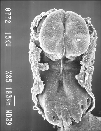
| 10:51, 9 August 2009 | Stage 9 SEM1.jpg (file) |  |
42 KB | S8600021 | Carnegie Stages 9 Features: embryonic disc, primitive node, primative streak, primative groove, somites, neural groove, brain plate region, connecting stalk, cut edge of amnion Facts: Week 3, 19 - 21 days, 1.5 - 2.5 mm, Somite Number 1 - 3 View 1: emb | 1 |
| 10:43, 9 August 2009 | Stage9sm.jpg (file) |  |
6 KB | S8600021 | Carnegie Stages 9 Features: embryonic disc, primitive node, primative streak, primative groove, somites, neural groove, brain plate region, connecting stalk, cut edge of amnion Facts: Week 3, 19 - 21 days, 1.5 - 2.5 mm, Somite Number 1 - 3 View 1: emb | 1 |
| 09:27, 9 August 2009 | Tooth molecular development.jpg (file) |  |
169 KB | S8600021 | Diagram showing stages of teeth development, a few genetic factors affecting phenotypes, and some signalling molecules and growth factors expressed in the epithelial and mesenchymal components of developing teeth. While most teeth-related genes exhibit, | 1 |
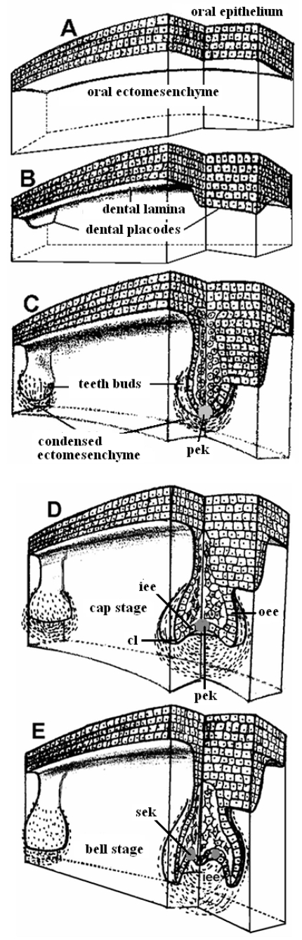
| 09:25, 9 August 2009 | Tooth development stage.jpg (file) |  |
303 KB | S8600021 | Stages in teeth development (A) Pre-patterned oral ectoderm is in close contact with cranial, neural crest ectomesenchyme. At this stage (ED 10) the odontogenic potential resides in the epithelium. (B) The epithelial cells secrete specific signals in d | 1 |
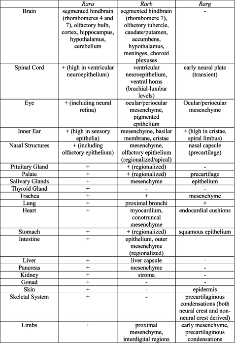
| 09:18, 9 August 2009 | Rar gene expression.jpg (file) |  |
153 KB | S8600021 | Summary of Rar gene expression patterns in the main developing organ systems. Data were essentially obtained in the mouse. See main text for references. + = ubiquitous (diffuse) expression. - = no expression detected. Link: http://www.pubmedcentral.n | 1 |
| 09:08, 9 August 2009 | Uterine teratoma.jpg (file) |  |
73 KB | S8600021 | Radiological and surgical specimen appearances of uterine teratoma. :"Teratomas are the commonest germ cell tumours and are most frequently found in the testes and ovary. Extragonadal teratomas are rare and mainly occur in midline structures. Uterine ter | 1 |
| 08:47, 9 August 2009 | Proboscis histology.jpg (file) |  |
166 KB | S8600021 | Histology of proboscis. a) Overview of a histological section of the proboscis showing the closed circular cartilaginous wall (arrow 1) with a central canal (arrow 2) with an epithelial lining (arrow 3) supported by loose connective tissue (arrow 4) whi | 1 |
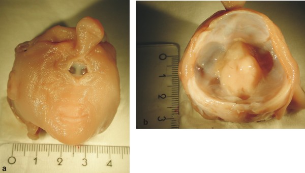
| 08:44, 9 August 2009 | Human holoprosencephaly cyclopia dissection.jpg (file) |  |
37 KB | S8600021 | Photographs of the macroscopic appearance of the head. a) Frontal view of the investigated head. b) View of the opened cranium with remnants of brain. Link: http://www.pubmedcentral.nih.gov/articlerender.fcgi?artid=2709107&rendertype=figure&id=F1 Ori | 1 |
| 15:37, 6 August 2009 | Connotea 32x32.png (file) |  |
1 KB | MarkHill | 1 | |
| 15:36, 6 August 2009 | Icon citeulike 16x16.gif (file) |  |
79 bytes | MarkHill | 1 | |
| 15:36, 6 August 2009 | Citeulike 16x16.png (file) |  |
413 bytes | MarkHill | 1 | |
| 15:35, 6 August 2009 | Delicious 32x32.png (file) |  |
888 bytes | MarkHill | 1 | |
| 15:35, 6 August 2009 | Reddit 32x32.png (file) |  |
2 KB | MarkHill | 1 | |
| 15:35, 6 August 2009 | Stumbleupon 32x32.png (file) |  |
2 KB | MarkHill | 1 | |
| 15:34, 6 August 2009 | Stumbleupon 16x16.png (file) |  |
901 bytes | MarkHill | 1 | |
| 15:34, 6 August 2009 | Digg 32x32.png (file) |  |
2 KB | MarkHill | 1 | |
| 15:34, 6 August 2009 | Facebook 32x32.png (file) |  |
2 KB | MarkHill | 1 | |
| 15:33, 6 August 2009 | Facebook 16x16.png (file) |  |
1 KB | MarkHill | 1 | |
| 09:35, 6 August 2009 | Unsw60.gif (file) | 2 KB | S8600021 | UNSW 60 years in 2009 http://www.unsw.edu.au/ | 1 | |
| 09:31, 6 August 2009 | Galletti1770 birth wax model.jpg (file) |  |
31 KB | S8600021 | One of a series of models commissioned by Giuseppe Galletti (? - 1819) currently held in the Institute and Museum of the History of Science (Italy) Istituto e Museo di Storia della Scienza (IMSS). Giuseppe Galletti and others used terracotta and wax model | 1 |
| 09:30, 6 August 2009 | Galletti1770 birth2.jpg (file) |  |
34 KB | S8600021 | One of a series of models commissioned by Giuseppe Galletti (? - 1819) currently held in the Institute and Museum of the History of Science (Italy) Istituto e Museo di Storia della Scienza (IMSS). Giuseppe Galletti and others used terracotta and wax model | 1 |
| 09:16, 6 August 2009 | Newborn.jpg (file) |  |
23 KB | S8600021 | Newborn infant Image source: UNSW Embryology http://embryology.med.unsw.edu.au/Child/birth1.htm | 1 |
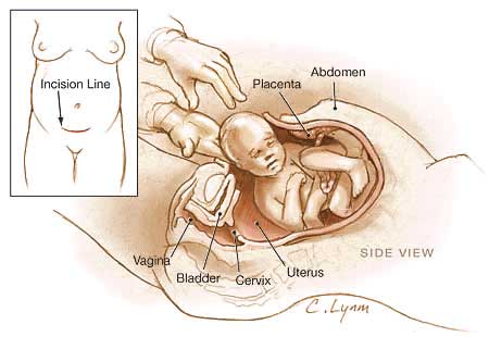
| 09:13, 6 August 2009 | Birth caesarean.jpg (file) |  |
18 KB | S8600021 | Birth by caesarean section The term "caesarean" comes from the historic description of Julius Ceasar's birth, though probably ficticious as his mother Aurelia survived his birth. The procedure involves surgically cutting skin, abdominal wall and uterus t | 1 |
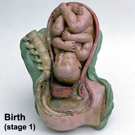
| 09:06, 6 August 2009 | Galletti1770 birth.jpg (file) |  |
34 KB | S8600021 | One of a series of models commissioned by Giuseppe Galletti (? - 1819) currently held in the Institute and Museum of the History of Science (Italy) Istituto e Museo di Storia della Scienza (IMSS). Giuseppe Galletti and others used terracotta and wax model | 1 |
| 07:59, 6 August 2009 | Mark Hill 08.jpg (file) |  |
20 KB | S8600021 | Dr Mark Hill, School of Medical Sciences, UNSW, Sydney Australia | 1 |
| 22:14, 5 August 2009 | Stage9 ventral.jpg (file) |  |
5 KB | S8600021 | Features: embryonic disc, primitive node, primative streak, primative groove, somites, neural groove, brain plate region, connecting stalk, cut edge of amnion Facts: Week 3, 19 - 21 days, 1.5 - 2.5 mm, Somite Number 1 - 3 View 1: embryonic disc, showin | 1 |
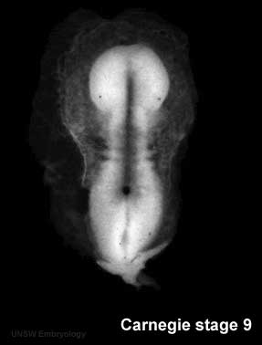
| 22:12, 5 August 2009 | Stage9 dorsal.jpg (file) |  |
5 KB | S8600021 | Carnegie Stage 9 Features: embryonic disc, primitive node, primative streak, primative groove, somites, neural groove, brain plate region, connecting stalk, cut edge of amnion Facts: Week 3, 19 - 21 days, 1.5 - 2.5 mm, Somite Number 1 - 3 View 1: embryo | 2 |
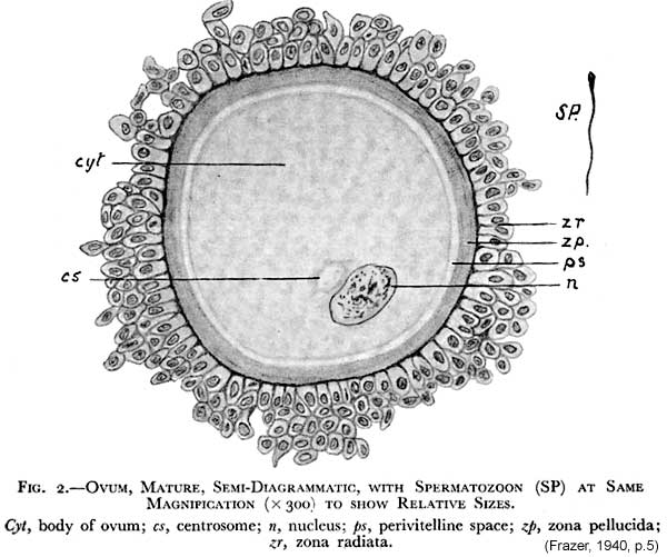
| 18:01, 5 August 2009 | Frazer043a 600.jpg (file) |  |
58 KB | S8600021 | 1 | |
| 18:00, 5 August 2009 | Frazer006 bw600.jpg (file) |  |
47 KB | S8600021 | 1 | |
| 18:00, 5 August 2009 | Frazer002 bw600.jpg (file) |  |
45 KB | S8600021 | 1 | |
| 17:59, 5 August 2009 | Frazer1940 titlepage 450.jpg (file) |  |
13 KB | S8600021 | 1 | |
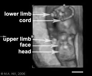
| 16:44, 5 August 2009 | Ultrasound12wk 3D image.jpg (file) |  |
8 KB | S8600021 | A 3D ultrasound static image of the 12 week fetus shows a ventral view with the fetus upside down, with the head down and cord to the top. | 1 |
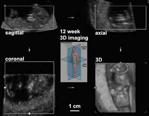
| 16:31, 5 August 2009 | Ultrasound12wk 3D.jpg (file) |  |
18 KB | S8600021 | Ultrasound image 12 week fetus, showing 3 dimensional (3d) axes Original file name: 12wk2_3D.jpg Image source: UNSW Embryology http://embryology.med.unsw.edu.au/Movies/usound/Hum3D.htm | 1 |
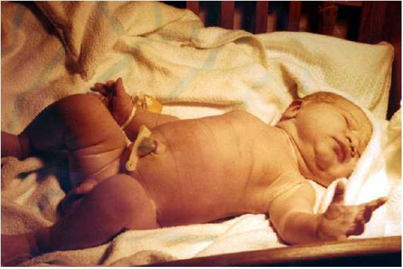
| 14:37, 5 August 2009 | Trisomy 21 newborn.jpg (file) |  |
16 KB | S8600021 | Trisomy 21 (Down Syndrome) Newborn Down syndrome or trisomy 21 is caused by nondisjunction of chromosome 21 in a parent who is chromosomally normal and is one of the most common chromosomal abnormalities in liveborn children. The frequency of trisom | 1 |
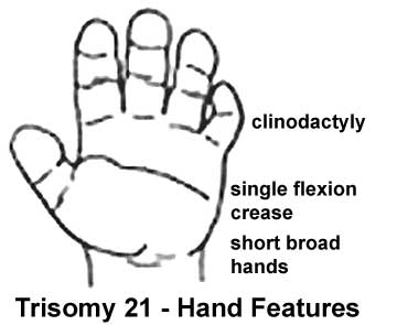
| 14:00, 5 August 2009 | Trisomy21 hand.jpg (file) |  |
12 KB | S8600021 | Trisomy 21 (Down Syndrome) Hand Down syndrome or trisomy 21 is caused by nondisjunction of chromosome 21 in a parent who is chromosomally normal and is one of the most common chromosomal abnormalities in liveborn children. The frequency of trisomy 21 | 1 |
| 13:39, 4 August 2009 | Human Embryo 17.8mm a CNS GIT.jpg (file) |  |
130 KB | MarkHill | Human Embryo (17.8mm) Gastrointestinal Tract Brain and Digestive System This plate consists of two reconstructions. The upper shows a left lateral view of the brain and cervical cord with the nerves in situ, the aortic arch, and other arteries of the le | 1 |
| 13:36, 4 August 2009 | Human Embryo 17.8mmCNS GIT.jpg (file) |  |
105 KB | MarkHill | Human Embryo (17.8mm) Gastrointestinal Tract Brain and Digestive System Reconstruction to illustrate chiefly the interior of the brain and the spinal cord; the digestive system and its appendages; thc arterial system; the left atrium and ventricle of th | 1 |
| 13:29, 4 August 2009 | Human Embryo 17.8mm GIT.jpg (file) |  |
38 KB | MarkHill | Human Embryo (17.8mm) Gastrointestinal Tract Wax-plate reconstruction of the stomach, the duodenum and the pancreas. The model is represented somewhat ventrally, from the right side A. du., antrum duodenale; C., corpus gastri; D. chol., ductus choledoch | 1 |
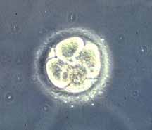
| 23:33, 3 August 2009 | Stage2.jpg (file) |  |
4 KB | S8600021 | Carnegie Stage 2 4 Cell Human Embryo: during each mitotic division the embryo does not increased in size and divides the existing cytoplasm. '''Image Source:''' UNSW Embryology, no reproduction without permission. Image reproduced with permission from D | 1 |
| 23:30, 3 August 2009 | Human oocyte.jpg (file) |  |
6 KB | S8600021 | Human oocyte with surrounding granulosa cells Image Source: UNSW Embryology, no reproduction without permission. http://embryology.med.unsw.edu.au/Notes/week1.htm Category:Human Embryo | 1 |
| 22:47, 3 August 2009 | Stage14 SEM.jpg (file) |  |
36 KB | S8600021 | Human embryo (Carnegie stage 14) scanning electron micrograph (original file name Stage14lateralsem.jpg) Facts: Week 5, 31 - 35 days, 5 - 7 mm View: Lateral view. Amniotic membrane removed. Features: midbrain, nasal placode, lens pit, 1,2,3 pharyngeal | 1 |
| 22:42, 3 August 2009 | Stage14 human scale.jpg (file) |  |
40 KB | S8600021 | Human embryo (Carnegie stage 14) Light microscope image of human embryo equivalent to SEM stage images (original file name Stage14lateralscale.jpg) '''Image Source:''' Prof Kathy Sulik scanning electron micrographs of the Carnegie stages of the early h | 1 |
| 22:40, 3 August 2009 | Stage14 human.jpg (file) |  |
32 KB | S8600021 | Human embryo (Carnegie stage 14) Light microscope image of human embryo equivalent to SEM stage images (original file name Stage14lateralbf.jpg) '''Image Source:''' Prof Kathy Sulik scanning electron micrographs of the Carnegie stages of the early huma | 1 |
| 22:35, 3 August 2009 | Stage8 human.jpg (file) |  |
14 KB | S8600021 | Human embryo (Carnegie stage 8) Light microscope image of human embryo equivalent to SEM stage images (original file name PresomiteSt8d18BFdorsal2.jpg) '''Image Source:''' Prof Kathy Sulik scanning electron micrographs of the Carnegie stages of the ear | 1 |