Somatic Cell Nuclear Transfer: Difference between revisions

From Embryology
(Created page with "==Introduction== Page Under Development thumb|300px|Early Human Zygote, with 2 pronuclei The first diploid cell that forms following [[fertilization...")
 
No edit summary
Line 1: Line 1:
==Introduction==
==Introduction==
Page Under Development
'''''Page Under Development'''''


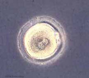
[[File:Early zygote.jpg|thumb|300px|Early Human Zygote, with 2 pronuclei]]
[[File:Early zygote.jpg|thumb|300px|Early Human Zygote, with 2 pronuclei]]
The first diploid cell that forms following [[fertilization]] by fusion of the haploid [[oocyte]] (egg) and [[spermatozoa]] (sperm) resulting in the combination of their separate genomes. The first image shows the cell with the 2 pronuclei still present before fusion enclosed within the zona pellucida. In humans, this cell then undergoes a series of mitotic cell divisions, still enclosed within the zona pellucida, during the first few days of development to form 2 blastomeres then a solid cell mass called the [[morula]].


:'''Zygote Links:''' [[Fertilization]] | [[Week 1]] | [[Morula]] | [[Blastocyst]] | [[Assisted Reproductive Technology|ART]] | [[Somatic Cell Nuclear Transfer|SCNT]] | [[:Category:Zygote|Category:Zygote]]
:[[Somatic Cell Nuclear Transfer|'''SCNT Links''': [[Somatic Cell Nuclear Transfer|Introduction]] | [[Stem_Cells_-_Adult#Somatic_Cell_Nuclear_Transfer|Stem Cells - SCNT]] | [[Assisted Reproductive Technology|ART]] | [[Fertilization]] | [[Week 1]] | [[Morula]] | [[Blastocyst]] | [[Assisted Reproductive Technology|ART]] | [[:Category:Zygote|Category:Zygote]]


== Some Recent Findings ==
Line 11: Line 12:
|-bgcolor="F5FAFF"  
|-bgcolor="F5FAFF"  
|  
|  
* '''cyclic AMP in the maturation of Ciona intestinalis oocytes'''<ref><pubmed>20810008</pubmed></ref>"Immature oocytes are arrested at prophase I of the meiotic process and maturation onset is indicated by oocyte nuclear disassembly (germinal vesicle breakdown or GVBD). Signaling pathways that elevate intracellular cyclic AMP (cAMP) may either prevent or induce oocyte maturation depending on the species. "
* '''Number of blastomeres and distribution of microvilli in cloned mouse embryos during compaction'''<ref><pubmed>20735894</pubmed></ref> "We concluded that: (i) the cleavage of blastomeres in cloned embryos was slow at least before compaction; (ii) the distribution of microvilli in cloned, normal, parthenogenetic, and tetraploid embryos was coherent before and after compaction; and (iii) the initiation of compaction in somatic cell nuclear transfer (SCNT) embryos was delayed compared with that of intracytoplasmic sperm injection (ICSI) embryos."


|}
|}
==Human Zygote Size==
Actual Size [[File:5cm ruler.jpg]]
Enlarged Size [[File:Stage1 size with ruler.jpg]]
==Parental Genomes==
[[File:Parental_genome_mix_01_icon.jpg|thumb|Labeled parental genomes in an early zygote (mouse)]]
Animation based upon individual images of mouse maternal and paternal genomes.
{| border='0px'
|-
| [[File:Parental_genome_mix_01_icon.jpg|120px|link=Movie_-_Parental_genomes‎‎]]
|-bgcolor="a3bfb1"
| &nbsp;‎‎'''Mouse Genome'''
|-bgcolor="F5FFFA"
| [[Quicktime_Movie_-_Parental_genomes|Quicktime]] | [[Movie_-_Parental_genomes|Flash]]
|}
[[File:Mouse zygote pronuclei 01.jpg]]
'''Mouse zygote pronuclei'''<ref name=PMID21321204><pubmed>21321204</pubmed>| [http://www.ncbi.nlm.nih.gov/pmc/articles/PMC2132672 PMC2132672] | [http://www.pnas.org/content/108/9/3642.abstract PNAS]</ref>
==Formation of Zygote ==
* male and female pronuclei, 2 nuclei approach each other and nuclear membranes break down
* DNA replicates, first mitotic division
* sperm contributes centriole which organizes mitotic spindle
[[File:Human_zygote_two_pronuclei_22.jpg|300px]] [[File:Parental_genome_mix_01_icon.jpg|300px]]
[[Development_Animation_-_Pronuclear_Fusion|Movie - Pronuclear Fusion]] | [[Movie_-_Parental_genomes|Movie - Parental Genomes]]
'''Conceptus''' - term refers to all material derived from this fertilized zygote and includes both the embryo and the non-embryonic tissues (placenta, fetal membranes).
==Zygote Protein Expression==
[[File:Mouse- zygote protein expression.jpg]]
The table above shows the pattern of protein expression (as percentages of total) in the mouse zygote according to 14 molecular function categories.<ref><pubmed>20876089</pubmed>| [http://www.pnas.org/content/107/41/17639.full PNAS]</ref>
:'''Links:''' [[:File:Mouse- germinal vesicle oocyte protein expression.jpg|Germinal vesicle oocyte protein expression]] | [[:File:Mouse-_MII_oocyte_protein_expression.jpg|MII oocyte protein expression]] | [[:File:Mouse- zygote protein expression.jpg|Zygote Protein Expression]] | [[Mouse Development]] | [[Oocyte Development]] | [[Zygote]]


==Male Pronucleus Reprogramming==
{|
| valign=top|'''Mouse zygote paternal genome reprogramming'''<ref name=PMID21321204><pubmed>21321204</pubmed>| [http://www.ncbi.nlm.nih.gov/pmc/articles/PMC2132672 PMC2132672] | [http://www.pnas.org/content/108/9/3642.abstract PNAS]</ref>
Mouse zygotes male pronucleus contains 5-hydroxymethylcytosine (5hmC) thought to be formed by enzymatic oxidation of 5-methylcytosine (5mC).
* '''A''' - Mouse zygote double-stained with anti-5hmC antibody (green) and anti-5mC antibody (red). The smaller maternal pronucleus is closer to the polar body (pb). A bright-field image is shown on the far left.
* '''B''' - Additional zygotes double-stained with anti-5hmC antibody (green) and anti-5mC antibody (red). Merged images are shown.
* '''C''' - Zygotes obtained by in vitro fertilization were double-stained similarly. Two polyspermic zygotes (to the right) exhibit 5hmC staining in two paternal pronuclei.
* '''D''' - 5mC and 5hmC staining reveal two separate chromosome sets at metaphase of zygote division. A confocal image is shown.
* '''E''' - Individual chromosomes are largely stained for either 5mC (likely originated from the maternal pronucleus) or 5hmC (likely from the paternal pronucleus) at anaphase of zygote division. Two Z sections of the same zygote are shown.
:'''Links:''' [[Molecular_Development_-_Epigenetics|Epigenetics]]
| [[File:Mouse_zygote_paternal_genome_reprogramming_01.jpg|400px]]
|}


'''Mouse zygote mitosis'''<ref name=PMID21321204><pubmed>21321204</pubmed>| [http://www.ncbi.nlm.nih.gov/pmc/articles/PMC2132672 PMC2132672] | [http://www.pnas.org/content/108/9/3642.abstract PNAS]</ref>
[[Molecular_Development_-_Epigenetics|Epigenetics]]


[[File:Mouse_zygote_mitosis_metaphase.jpg]] [[File:Mouse_zygote_mitosis_anaphase.jpg]]


==References==
==References==

Revision as of 13:05, 22 August 2012

Introduction

Page Under Development

Early Human Zygote, with 2 pronuclei

The first diploid cell that forms following fertilization by fusion of the haploid oocyte (egg) and spermatozoa (sperm) resulting in the combination of their separate genomes. The first image shows the cell with the 2 pronuclei still present before fusion enclosed within the zona pellucida. In humans, this cell then undergoes a series of mitotic cell divisions, still enclosed within the zona pellucida, during the first few days of development to form 2 blastomeres then a solid cell mass called the morula.

[[Somatic Cell Nuclear Transfer|SCNT Links: Introduction | Stem Cells - SCNT | ART | Fertilization | Week 1 | Morula | Blastocyst | ART | Category:Zygote


Some Recent Findings

  • Number of blastomeres and distribution of microvilli in cloned mouse embryos during compaction[1] "We concluded that: (i) the cleavage of blastomeres in cloned embryos was slow at least before compaction; (ii) the distribution of microvilli in cloned, normal, parthenogenetic, and tetraploid embryos was coherent before and after compaction; and (iii) the initiation of compaction in somatic cell nuclear transfer (SCNT) embryos was delayed compared with that of intracytoplasmic sperm injection (ICSI) embryos."


Epigenetics


References

  1. <pubmed>20735894</pubmed>

Journals

  • Zygote An international journal dedicated to the rapid publication of original research in early embryology, Zygote covers interdisciplinary studies in animals and humans, from gametogenesis through fertilization to gastrulation.

Reviews

Articles

Search Pubmed

June 2010 "zygote" All (6213) Review (651) Free Full Text (1543)

Search Pubmed: zygote


Glossary Links

Glossary: A | B | C | D | E | F | G | H | I | J | K | L | M | N | O | P | Q | R | S | T | U | V | W | X | Y | Z | Numbers | Symbols | Term Link

Cite this page: Hill, M.A. (2024, April 18) Embryology Somatic Cell Nuclear Transfer. Retrieved from https://embryology.med.unsw.edu.au/embryology/index.php/Somatic_Cell_Nuclear_Transfer

What Links Here?
© Dr Mark Hill 2024, UNSW Embryology ISBN: 978 0 7334 2609 4 - UNSW CRICOS Provider Code No. 00098G